





| Research Article | ||
Open Vet. J.. 2026; 16(2): 788-801 Open Veterinary Journal, (2026), Vol. 16(2): 788-801 Research Article Emergence of novel highly pathogenic H5N1 avian influenza in slender-billed gulls (Chroicocephalus genei) and gull-billed terns (Gelochelidon nilotica) at Dukan Lake, IraqNasih Ali1*, Harem Mustafa1, Nadia Salih2, Pavel Qader3, Snur Hassan1 and Dashty Amin41Department of Anatomy and Histopathology, College of Veterinary Medicine, University of Sulaimani, Sulaymaniyah, Iraq 2Department of Basic Sciences, College of Veterinary Medicine, University of Sulaimani, Sulaymaniyah, Iraq 3Department of Surgery and Theriogenology, College of Veterinary Medicine, University of Sulaimani, Sulaymaniyah, Iraq 4Qaiwan International University, Department of Biomedical Engineering, Sulaymaniyah, Iraq *Corresponding Author: Nasih Ali. Department of Anatomy and Histopathology, College of Veterinary Medicine, University of Sulaimani, Sulaymaniyah, Iraq. Email:nasih.ali [at] univsul.edu.iq Submitted: 24/09/2025 Revised: 02/12/2025 Accepted: 17/12/2025 Published: 28/02/2026 © 2026 Open Veterinary Journal
ABSTARCTBackground: Highly pathogenic avian influenza (HPAI) H5N1 continues to cause severe mortality among wild birds globally. Aim: The present study aimed to investigate the mass mortality in Slender-billed Gulls (Chroicocephalus genei) and Gull-billed Terns (Gelochelidon nilotica) at Dukan Lake, Iraq, during the 2024 breeding season. Methods: Field monitoring was conducted at breeding colonies, and dead carcasses were collected for necropsy, histopathology, and molecular analysis. Virus detection and genetic characterization were performed by reverse transcription polymerase chain reaction and sequencing. In addition, the viral lineage was inferred by phylogenetic analysis of the hemagglutinin (HA) and neuraminidase (NA) genes. Results: More than 3,200 gulls and terns were found dead, exhibiting severe neurological signs and systemic illness. Necropsy and histopathological investigations indicated significant multi-organ damage, with gulls showing more pronounced visceral lesions than terns, while terns exhibited greater neurological damage. Molecular characterization confirmed the virus as HPAI H5N1, and analysis of the HA and NA genes revealed that the strain from Dukan Lake falls within clade 2.3.4.4b, displaying a high genetic resemblance (>98%) with the recently identified H5N1 strain isolated from Mute swans in Kazakhstan. The virus showed multibasic cleavage sites characteristic of high pathogenicity but maintained receptor-binding specificity for avian hosts. Moreover, the results demonstrate the recent introduction of the virus via migratory flyways and the significance of gulls and terns in the disease ecology of HPAI virus (HPAIV). Conclusion: This outbreak is the first recorded mass mortality of gulls and terns due to HPAI H5N1 in Iraq. The results emphasize the need for improved surveillance at migratory stopover and breeding sites to observe viral evolution, host susceptibility, and potential risks for cross-species transmission. Keywords: Highly pathogenic avian influenza, H5N1, Gulls and terns, Dukan Lake, Iraq. IntroductionThe highly pathogenic avian influenza (HPAI) virus (HPAIV) has significantly affected wild birds and mammals globally (Fusaro et al., 2024). Wild birds serve as a natural reservoir of IAV s with various combinations of 16 hemagglutinin (HA) and 9 neuraminidase (NA) subtypes (Hill et al., 2022). Notably, waterbirds from the orders Anseriformes (such as ducks, geese, and swans) and Charadriiformes (such as gulls, waders, and terns) play a crucial role in the spread of avian influenza viruses, particularly certain highly pathogenic strains like H5N1 (Haman et al., 2024). In the past, HPAIVs were rarely found in wild birds and generally did not cause clinical disease when present (Sonnberg et al., 2013). In late 2002, outbreaks of HPAIV H5N1 were reported in Hong Kong parks, resulting in illness and mortality among both local and migratory birds. Simultaneously, HPAIV H5N1 was detected in deceased chickens and humans (Ellis et al., 2004). The antigenic analysis demonstrated that the novel isolates of HPAIV H5N1 had a reactivity pattern distinct from that of HPAIV H5N1 isolated in 1997 and 2001, resulting in systemic infection in experimentally infected domestic ducks (Sturm-Ramirez et al., 2004). This change began with occasional incidents of H5N1 HPAIV-related deaths among wild birds, and it soon became apparent that wild birds were spreading the virus over great distances (Ramey et al., 2022). HPAIV caused by H5N1 radically altered the scope and diversity of hosts, virulence, transmission, and maintenance dynamics (Charostad et al., 2023; Webby and Uyeki, 2024). Therefore, the identification and characterization of novel subtypes of avian influenza viruses that adapt in migratory birds will trigger early warnings for cross-species infection and transmission (Wang et al., 2022). Since 2014, the HPAI H5 clade 2.3.4.4 has been the predominant lineage in Europe, with a new variant emerging in 2020 (King et al., 2021). This development triggered the largest documented global HPAI outbreak, along with a rising number of reports on viral transmission to mammalian species (Fusaro et al., 2024). The extensive spread of HPAI H5N1 viruses from clade 2.3.4.4b has had a significant impact on rare and protected bird species, leading to drastic population declines (Banyard et al., 2022). Particularly, between 2021 and 2022, several high-mortality outbreaks were observed among colony-nesting seabirds worldwide (Lane et al., 2024). Gulls and terns are the most closely related taxon; they share ecological characteristics and frequently coexist in the same habitat (Benkaroun et al., 2016). However, gulls and terns are known as clinical hosts of H5N1 HPAIV (Sharshov, 2015; Rijks et al., 2022). Several species, including the black-headed gull (Chroicocephalus ridibundus) (Ellis et al., 2004), common gull (Larus canus) (Sharshov et al., 2010), brown-headed gull (Larus brunnicephalus) (Liu et al., 2005), Sandwich tern (Thalasseus sandvicensis) (Rijks et al., 2022), and Cabot’s tern (Thalasseus acuflavidus) (Reischak et al., 2023) have been diagnosed with disease and mortality as a result of natural HPAIV H5N1 infection. In addition, laughing gulls (Larus atricilla) and herring gulls (Larus argentatus) have been shown to be highly susceptible to distinct HPAIV H5N1 isolates in experimental infections, with a drastic and brief clinical pathology characterized predominantly by neurologic signs, with a high mortality rate (Brown et al., 2008; Tarasiuk et al., 2022). Interestingly, gull-origin virus demonstrated a shared affinity for the central nervous system, pancreas, and adrenal gland as the primary replication target organs (Brown et al., 2006). Moreover, using an HPAIV H5N1 isolate from healthy common gulls (Larus canus), it was shown that the virus could be shed through the oropharynx and cloaca for up to 2 weeks post-infection (Zaĭkovskaia et al., 2012). The pathogenicity of H5N1 is associated with the presence of multiple basic amino acids at the cleavage site of the HA protein, resulting in the creation of a polybasic cleavage site. This alteration enables HA to have a broader range of activity beyond the gastrointestinal tract. Consequently, HPAI outbreaks are characterized by rapid disease progression and high mortality rates (Luczo et al., 2015; De Bruin et al., 2022). Dukan Lake is located in the northwest of Iraq, near the town of Dukan in Sulaymaniyah Province. Various gull species have been observed in the area, with the Slender-billed Gull (Chroicocephalus genei) and Gull-billed Tern (Gelochelidon nilotica) being the most notable species that reside or migrate through the region (Ararat, 2009). Their migratory behavior, social habits, and tendency to form mixed-species colonies enhance their capacity as reservoirs for avian influenza transmission. In May 2024, a significant emergence of H5N1 was observed within the gull and tern populations in that area. This study aims to document the outbreak and provide detailed pathological and molecular analysis of H5N1 HPAVI in the gull and tern populations in the region. Materials and MethodsField studyThe study was conducted at Dukan Lake, northwest of Sulaimani province, Iraq. The islands within this lake provide vital breeding habitats for various migratory bird species. Specifically, the Slender-billed Gull and Gull-billed Tern arrive at the lake in early April, lay their eggs in early May, and breed in large, dense colonies. In May 2024, following heavy rainfall, mass mortality was observed only on two islands in the lake (Fig. 1; S1, S2). Only these two species were found dead or moribund. During this active outbreak, field monitoring was conducted at the site for disease surveillance and sampling collection. Clinical signs were observed in both affected species. Carcasses were chosen to be broadly representative, covering various stages of death (fresh to moderate decay), both species, and various parts of the islands. Only carcasses with intact organs were sampled. A total of 16 samples (9 gulls, 7 terns) were sampled based on biosafety considerations using HPAI standards, as well as diagnostic availability at a level representative of wildlife outbreak investigations. The oropharynx and cloaca were collected using sterile handled swabs and preserved in a viral transport medium. The swab samples were then placed on ice packs and sent to a research center at the College of Veterinary Medicine at the University of Sulaimani for further analysis. Necropsy findingsFollowing aseptic collection, the carcasses were sent to the research center for necropsy and tissue sampling. Necropsy examinations were conducted in a biosafety cabinet located within the specialized high-containment area designated for particular animal pathogens. Carcasses were examined for gross lesions, and aseptic collection of tissue samples (liver, brain, and kidneys) was performed from 16 dead birds, comprising 9 gulls and 7 terns. The samples were then stored at −20°C for subsequent molecular analyses. Equivalent tissue samples were fixed in 10% neutral buffered formalin for histopathological assessment.
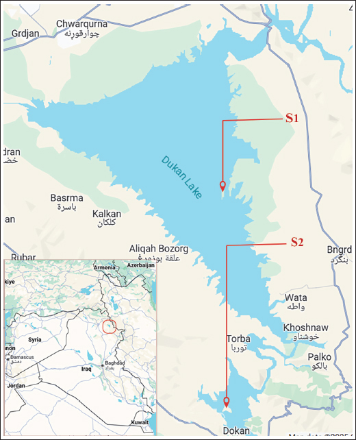
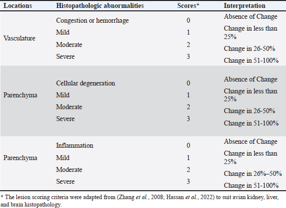
Fig. 1. Map of Dukan Lake, S1 and S2, showing the location of breeding colonies and the HPAI H5N1 outbreak. Samples were also collected at these two sites. Histopathological assessmentThe fixed tissue samples from the liver, brain, and kidneys were examined histopathologically. The samples were first preserved in 10% neutral buffered formalin for at least 48 hours before undergoing a series of dehydration treatments with ethanol. The sections were dehydrated, clarified in xylene, and embedded in paraffin. Thin tissue sections of 4 μm were produced from each specimen for histopathological examination and were stained with hematoxylin and eosin (H&E). The lesions were evaluated using a light microscope and computer-assisted image analysis software (Am ScopeTM, Japan). The following lesions were assessed (Table 1) to grade and score the pathologic alteration in the kidney: tubular degeneration, glomerular degeneration, vascular disturbance, and inflammation. The main liver abnormalities were estimated as degeneration, necrosis, vascular disturbance, and inflammatory reaction. Brain lesions include neuronal degeneration, vascular disturbance, and inflammation. The alterations were classified into the following categories: Grade 0: No significant histological findings (0%). Grade 1: Minor alteration encompassing up to 20% of change commencement. Grade 2: Significant alteration affecting 21%–50% of patients. Grade 3: Moderate to severe alterations affecting 51%–75% of patients. Grade 4: Severe change involving ≤75%; widespread changes identified. The lesion scoring criteria were adapted and modified from (Zhang et al., 2008; Hassan et al., 2022) to suit avian kidney, liver, and brain histopathology. Table 1. Assessment of the kidney, liver, and brain lesions in the study groups.
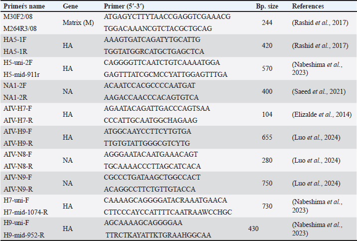
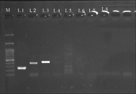
Nucleic acid extractionNucleic acid extractions from swabs and tissues were conducted using the AddBio® Viral Nucleic Acid Extraction Kit (AddBio Co., Korea) according to the manufacturer’s instructions. The eluted nucleic acid was quantified using UV spectrophotometry (260/280 nm), followed by storage at −20°C. Detection of influenza virusMultiplex reverse transcription polymerase chain reaction (PCR) (RT-PCR) was used to analyze samples of influenza A virus, specifically targeting the matrix (M) gene. The M, HA, and NA genes were amplified individually using the SuPrimeScript RT-PCR Premix from GeNet Bio, Korea. Additionally, the samples were screened for H7, H9, N7, N8, and N9 subtypes. A 20 μl one-step RT-PCR reaction was set up, comprising 10 μl of master mix from AddBio Co., Korea, 4 μl of RNA, and 1 μl (10 pmol) each of forward and reverse primers (Table 2). The reaction volume was adjusted by adding 4 μl of diethylpyrocarbonate-treated water. The amplification process was carried out in a thermocycler, which was initially incubated at 50°C for 30 minutes. The PCR was started with denaturation at 95°C for 10 minutes, followed by 40 cycles of denaturation (95°C for 35 seconds), annealing at 55°C for 35 seconds, and elongation at 72°C for 30 seconds. The final elongation was performed at 72°C for 10 minutes. Subsequently, PCR products were visualized (Fig. 2) using a 1.5% agarose gel and a UV Trans Illuminator (Ingenius, USA). The PCR products were then purified using the E.Z.N.A.® Kit (Omega Bio-tek, Inc. USA). Sequencing and phylogenetic analysisThe HA and NA RT-PCR products were purified before sequencing using the Sanger method with an ABI3730 XL genetic analyzer (Macrogen, Korea). The resulting nucleotide sequences were trimmed to remove ambiguous ends and aligned using the ApE software (A Plasmid Editor, Version 2.0.51) to generate consensus sequences with uniform start and termination sites. These sequences were then subjected to a Basic Local Alignment Search Tool analysis against the GenBank nucleotide database to identify homologous influenza virus genomic sequences. The phylogenetic tree was created using the maximum likelihood approach in MEGA 11 software with a neighbor-joining tool based on nucleotide sequences from the complete HA and NA coding regions of H5N1 AIV genotypes. Statistical analysisStatistical differences in mean pathologic lesions were assessed using GraphPad Prism 9. One-way analysis of variance was used to examine lesion scores in the liver, kidney, and brain. Statistically significant differences were established with p-values below 0.05. Table 2. List of primers used in this study.
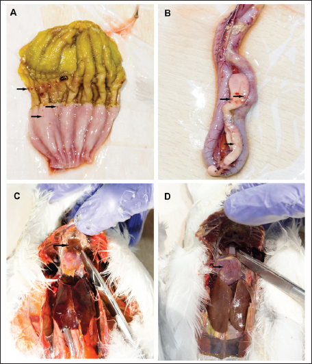
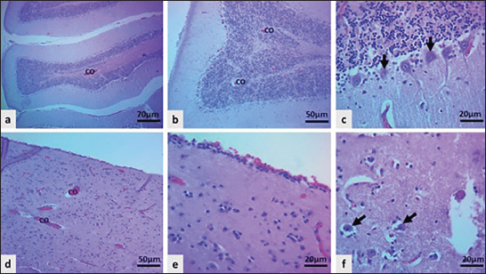
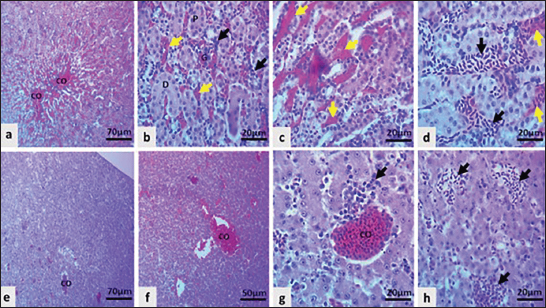
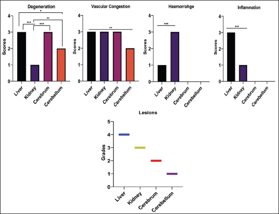
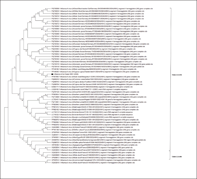
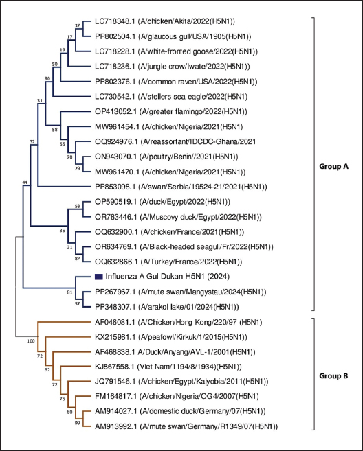
Fig. 2. Gel electrophoresis shows the PCR result of positive for amplification of H5N1, Lane M (left) DNA marker (100 bp), Lane 1 matrix gene (240 bp), Lane 2 H5 gene (400 bp), Lane 3 N1 gene (420 bp), Lane 4 negative result of N9, Lane 5 negative control, Lane 6 negative result of N8, lane 7 negative result of H7, lane 8 negative result of H9. Ethical approvalThe components of this study did not involve human participants, and ethical approval was not required for sampling carcasses and dying birds. ResultsHistory and clinical findingsMass mortality was observed among Slender-billed Gulls (Chroicocephalus genei) and Gull-billed Terns (Gelochelidon nilotica) at two sites (S1 and S2) in Dukan Lake (Fig. 1). A total of 2,458 dead adult and fledgling gulls, along with 783 dead adult and fledgling terns, were recorded (Table 3). In addition, numerous debilitated and near-death individuals were observed within both species. Interestingly, no mortality was noted among other co-nesting species, including various wading birds and shorebirds, as well as poultry and backyard chickens surrounding the lake. The clinical manifestations in the affected birds included elevated body temperature, greenish diarrhea, depression, lack of vocalization, and prominent neurological signs such as seizures, head tremors, torticollis, opisthotonus, and severe imbalance. In addition, some infected gulls showed mild respiratory symptoms. Pathological findingsNecropsy findings in the field included petechial hemorrhage in the gizzard, proventriculus (Fig. 3A), pancreas and intestine (Fig. 3B), hydropericardium, and flaccid heart (Fig. 3C and D), and congestion and enlargement of the liver and kidneys. Airsaculitis and pericarditis were observed in some cases. Overall, gross lesions appeared milder in terns than in gulls. In contrast, the multifocal congestion of brain tissue was more prominent in terns. HistopathologyThe histopathologic features of the infected terns and gulls revealed various lesions in different organs. The cerebellum had moderate vascular congestion with degeneration of Purkinje cells (Fig. 4a–c), whereas the cerebrum had marked congestion and degeneration of neuropil and neuron (Fig. 4d–f). The grade in the cerebellum was 1 versus the cerebrum was 2, (Fig. 6). The highly significant lesions seen in the kidney with high grades, including marked congestion and hemorrhage, tubular degeneration, and marked heterophil infiltration (Fig. 5a–c), were highly significant in the liver with high grades compared with all other organs (Fig. 5d–f). Quantitative evaluation of lesion scores revealed species-specific differences. Lesion scores in terns were significantly higher than those in gulls (p < 0.05), especially in the cerebrum. However, liver and kidney lesions were more severe in gulls than in terns (p < 0.01). Scores mean ± SD of different organs are shown in Figure 6. These findings are consistent with the observation that terns had a greater degree of neural involvement and gulls had more severe visceral pathology. Molecular diagnostic findingsOne-step RT-PCR detection of the M gene indicated an AIV. Further HA and NA subtyping confirmed H5 and N1 positivity, respectively (Fig. 2). The other results were negative for the N8, N9, H7, and H9 subtypes. Finally, specific HA and NA genes were sequenced for further analysis. Phylogenetic analysisThe phylogenetic tree based on the HA gene shows that the influenza A Gul Dukan H5N1 (2024) field virus (Fig. 7) is classified within the HPAI H5N1 subtype clade 2.3.4.4b. The Dukan Lake strain forms a cluster with isolates from mute swans in the Mangystau region and whooper swans in the Karakol lake in Kazakhstan, namely (A/mute swan/Mangystau/1-S24R-2/2024 and A/Cygnus cygnus/Karakol lake/01/2024), sharing identities of 99.35% and 98.86%, respectively, with a few mutations at the bases 1101 A changed to G, at 1174 A instead of T, at 1417 T was instead of A, at 1527 T instead of A, whereas genetic distances to the European and East Asian 2.3.4.4b viruses were slightly lower (97.2%–98.1%), suggesting a more recent introduction from Central Asia rather than Western Eurasia. Four point mutations were detected in the HA gene (1101A→G, 1174A→T, 1417T→A, 1527T→A); all occurred outside major receptor-binding or antigenic sites and did not alter known functional motifs. Overall, the phylogenetic position and mutational profile of the strains showed a close evolutionary relationship with Kazakhstan swan-derived strains and recent regional viral movement. Complementary analysis of the partial NA gene sequence indicates that the virus is in group A (Fig. 8), a finding consistent with the phylogenetic placement inferred from the HA gene. In contrast, the strain is clearly separated from Group B classical H5N1 lineages (Hong Kong 97-, Anyang 2001-, and Egypt/Kalyobia 2011-like viruses), confirming that it does not derive from older regional lineages. The amino acid sequences of the influenza A virus H5N1 (2024) from the Gul Dukan strain revealed multibasic cleavage sites with the PLRE KRRKR.GLF motif, a hallmark of HPAI. The receptor binding domains of the HA gene were identified by specific amino acid residues, including H119, K208, E201, N205, K293, G237, Q238, and G240, which serve as important molecular markers for HPAI. DiscussionIn this investigation, the HPAIV H5N1 was studied within the populations of Slender-billed Gull and Gull-billed Tern, which caused mass mortality during the 2024 breeding season in Dukan Lake, Iraq. The lake is a stopover site on the Central Asian Flyway, used by birds traveling between breeding grounds that migrate annually through Kazakhstan, the Caspian basin, and northern Iraq during spring migration (Schielzeth et al., 2010). In early 2024, H5N1 was detected in mute and whooper swans from Kazakhstan and neighboring Central Asia, which were genetically close to the Dukan strain (Sultankulova et al., 2024; Tabynov et al., 2024). The tight phylogenetic link between our isolate and these Central Asian viruses, together with the absence of concurrent poultry outbreaks locally, suggests that migratory waterbirds introduced H5N1 into Dukan Lake rather than in-country viral transmission; therefore, this outbreak highlights the importance of wild birds, especially migratory species, in the dissemination of HPAIVs throughout different geographic areas. The rapid spread and high mortality rates of H5N1 HPAIV align with those of prior studies, which have shown the susceptibility of gulls and terns to H5N1 HPAIV (Rijks et al., 2022; Kydyrmanov et al., 2024; Przymencki et al., 2024). Table 3. Mortality rate and population size of gulls and terns in impacted colonies of Dukan Lake in 2024.
Fig. 3. Gross lesions of HPAI H5N1 in gulls and terns. (A) Proventriculus and Gizzard (Gull): Petechial hemorrhages (black arrow) are present on the mucosa of the gizzard and proventriculus. (B) Pancreas (Gull): Petechial hemorrhages (black arrow) are present in the pancreas, with multifocal to coalescing red areas consistent with necrosis (arrowheads). (C) Pericardium and Liver (Tern): Hydropericardium with clear straw-colored fluid accumulation in the pericardial sac is evident. The liver appears congested, with a darkened, possibly necrotic appearance. (D) Flaccid heart (black arrow): Heart (Tern). Mortality rates were higher in the North Islands (Fig. 1,S1) than in the South Islands (Fig. 1, S2) due to higher colony population densities. Additionally, the highest mortality among gulls were recorded compared to terns. These findings suggest that gulls could serve as the primary vector for H5N1 HPAIV. This may be attributed to their higher population density and the fact that they are far more numerous than terns in the lack, with more aggressive scavenging behavior, which could increase exposure (Verhagen et al., 2021), and possibly different innate immune responses than terns, as shown by the high susceptibility and pathogenesis of HPAIV in gull species (Tarasiuk et al., 2022). Similarly, the higher mortality rates reported here are comparable to recent H5N1 outbreaks in gulls, such as European Herring gull (Larus argentatus), Black-headed gull (Chroicocephalus ridibundus) (Lean et al., 2024), Mediterranean gull (Ichthyaetus melanocephalus) (Przymencki et al., 2024), and Common gull (Larus canus) ((Sharshov et al., 2010). In contrast, Haman (2024) reported higher mortality in both adult and young terns. The mortality recorded between the two species indicates variations in susceptibility, highlighting that terns and gulls should not be considered to have similar responses to avian influenza, particularly H5N1 subtype.
Fig. 4. Light microscopic sections of brain revealed; a-c: Moderate congestion (CO) in the cerebellum with degeneration of Purkinje cells, as indicated by black arrows. d-f: Marked congestion of cerebral vasculature (CO) and severe degeneration of neuropil and neuron as indicated by black arrows, (H&E stain).
Fig. 5. Light microscopic sections of the kidney and liver presented; (a–c) Severe congestion and hemorrhage (CO, black arrows) of the renal vasculature and glomeruli (G) with mild degeneration of the proximal (P) and distal convoluted tubules (D) and inflammatory reaction (black arrows). (d–f) Marked congestion of the central vein and sinusoid (CO), severe hepatic swelling, and severe heterophil infiltration as indicated by black arrows (H&E stain). The clinical findings, particularly the predominant neurological disorders, align with findings reported by other studies regarding Sandwich terns (Thalasseus sandvicensis) (Rijks et al., 2022), Herring gulls (Larus argentatus) (Tarasiuk et al., 2022), and Caspian terns (Hydroprogne caspia) (Haman et al., 2024) infected with various clades of HPAIV.
Fig. 6. The chart revealed different pathologic scores and grades among the studied organs. At necropsy, the observed gross lesions in both species, such as hemorrhages in the gastrointestinal tract and myocardial changes, are similar to those described by other authors for Black-headed gull (Chroicocephalus ridibundus) (Lean et al., 2024) , Herring gulls (Larus argentatus) (Brown et al., 2008), and Caspian terns (Hydroprogne caspia) (Kydyrmanov et al., 2024). HPAIV showed a strong tropism for visceral organs, particularly the gastrointestinal tract in gulls, resulting in systemic endothelial and parenchymal damage. In contrast, the terns exhibited milder visceral lesions and more pronounced neurological changes. Brain congestion was frequently observed in terns but not in gulls. This pattern closely corresponds to recent outbreaks in Sandwich terns (Thalasseus Sandvicensis), for which encephalitis and neuronal degeneration characterized the disease presentation (Knief et al., 2024). Our histological findings support this, showing extensive neuronal necrosis and marked Purkinje cell degeneration in the brain tissues of the terns. Moreover, both species showed multiple organ necrosis, degeneration, and vascular congestion in histopathological results. Such lesions are a mirror of those documented in experimentally infected Common gulls (Larus canus) (Zaĭkovskaia et al., 2012) and Black-headed gulls (Chroicocephalus ridibundus) (Ramis et al., 2014) with HPAIV isolates.
Fig. 7. The phylogenetic tree was created using the entire coding sequence of the HA gene of H5N1 avian influenza virus genotypes. The strain used in this study is Influenza A gul Dukan 2024 (marked with a black node). According to phylogenetic analysis, the virus belongs to group B within clade 2.3.4.4b of H5N1, a lineage that has been prevalent in global transmission since 2020 and stands out as one of the most genetically and epidemiologically successful clades of H5Nx viruses (Graziosi et al., 2024; Van Borm et al., 2025). Genetic analysis of the HA gene sequence reveals that the H5N1 Dukan Lake strain is closely associated with strains previously identified in Kazakhstan, infecting both mute swans and whooper swans (Sultankulova et al., 2024; Tabynov et al., 2024). This finding is consistent with other studies showing that the spread of HPAIV is highly interconnected among the breeding, stopover, and wintering areas of the world’s migratory birds across Eurasia (Verhagen et al., 2021; Iancu et al., 2025). Moreover, the significant genetic similarity (>98%) with Central Asian isolates, along with a few point mutations in HA, indicates a recent introduction rather than prolonged local circulation. The impact of these alterations on antigenicity, host range, or pathogenicity requires further investigation using antigenic mapping and receptor-binding tests (Wang et al., 2022). NA gene analysis also demonstrated the close genetic similarity of the Dukan Lake strain with those of Central Asian isolates, and no obvious reassortments were identified. Nevertheless, surveillance must continue because reassortment with locally circulating LPAI viruses in gull and tern populations might produce new H5Nx variants with unknown phenotypic characteristics (Verhagen et al., 2021).
Fig. 8. The phylogenetic tree was generated using nucleotide sequences obtained from the whole NA coding areas of H5N1 avian influenza virus genotypes. Single amino acid substitutions at positions H119, K208, E201, N205, K293, G237, Q238, and G240 in the HA protein have been shown to enhance the affinity of avian influenza virus for α-2,6 SA (human) compared to α-2,3 SA (avian) (Boltz et al., 2010; Bialy and Shelton, 2020). In multiple sequence alignments, the influenza A strain H5N1 failed to establish binding to α-2,6 SA (human) in the HA assay, preferring instead classic avian α-2,3 SA specificities (Boltz et al., 2010). This observation indicates that no documented cases have occurred in animals throughout the pandemic. Although the current study provides important information about the first confirmed outbreak in Iraqi gulls and terns, this study is still limited by sequencing only HA and NA genes, as whole-genome sequencing would be necessary to exclude reassortment with local low-pathogenic avian influenza viruses. The absence of virus isolation and experimental infection studies limited the ability to directly confirm the identified strain’s pathogenicity and tissue tropism. Despite monitoring surrounding seabirds, poultry, and backyard chickens, screening was limited in scope and duration and may not exclude low-level viral circulation. These limitations emphasize the need for prolonged surveillance, comprehensive whole-genome sequencing, and regulated pathogenicity studies in future investigations. ConclusionThe outbreak of the HPAI H5N1, clade 2.3.4.4b, at Iraq’s Dukan Lake resulted in mass mortality among Slender-billed Gulls and Gull-billed Terns, causing necropsy and damage to multiple organs. Molecular and phylogenetic analyses confirmed a close genetic relationship between the virus and Central Asian isolates, suggesting a recent introduction through migratory pathways. The presence of multibasic cleavage sites in HA suggests ongoing adaptation to avian hosts. Regional and global surveillance is required to track viral evolution and minimize cross-species transmission. AcknowledgmentNot applicable. Conflicts of interestThe authors have no conflicts of interest to declare. FundingThis study received no funding. Authors’ contributionsNA: Conceptualization, Data curation, Formal analysis, Original draft writing, and Project administration. HM: conceptualization, investigation, methodology, and resources. NS: Conceptualization, supervision, validation, visualization, and project administration. PQ: Data curation, investigation, methodology, formal analysis, review, and editing. SH: Conceptualization, methodology, writing, review, and editing. DA: Methodology, formal analysis, and investigation. All authors have read and approved the published version of the manuscript. Data availabilityAll data were presented in the study. ReferencesArarat, K. 2009. Key biodiversity areas: rapid assessment of birds in Kurdistan, northern Iraq. BioRisk 3(2009), 187–203; doi:10.3897/biorisk.3.21 Banyard, A.C., Lean, F.Z.X., Robinson, C., Howie, F., Tyler, G., Nisbet, C., Seekings, J., Meyer, S., Whittard, E., Ashpitel, H.F., Bas, M., Byrne, A.M.P., Lewis, T., James, J., Stephan, L., Lewis, N.S., Brown, I.H., Hansen, R.D.E. and Reid, S.M. 2022. Detection of highly pathogenic avian influenza virus H5N1 clade 2.3.4.4b in great skuas: a species of conservation concern in Great Britain. Viruses 14(2), 212; doi:10.3390/v14020212 Benkaroun, J., Shoham, D., Kroyer, A.N.K., Whitney, H. and Lang, A.S. 2016. Analysis of influenza A viruses from gulls: an evaluation of inter-regional movements and interactions with other avian and mammalian influenza A viruses. Cogent. Biol. 2(1), 1234957; doi:10.1080/23312025.2016.1234957 Bialy, D. and Shelton, H. 2020. Functional neuraminidase inhibitor resistance motifs in avian influenza A (H5Nx) viruses. Antiviral Res. 182(2020), 104886; doi:10.1016/j.antiviral.2020.104886 Boltz, D.A., Seiler, P., Govorkova, E.A., Suzuki, Y., Webster, R.G., Obert, C., Mondry, R., Keating, R., Douangngeun, B., Hiramatsu, H., Sinthasak, S. and Phommachanh, P. 2010. Emergence of H5N1 avian influenza viruses with reduced sensitivity to neuraminidase inhibitors and novel reassortants in Lao People’s Democratic Republic. J. Gen. Virol. 91(4), 949–959; doi:10.1099/vir.0.017459-0 Brown, J.D., Stallknecht, D.E. and Swayne, D.E. 2008. Experimental infections of herring gulls (Larus argentatus) with H5N1 highly pathogenic avian influenza viruses by intranasal inoculation of virus and ingestion of virus-infected chicken meat. Avian. Pathol. 37(4), 393–397; doi:10.1080/03079450802216595 Brown, J.D., Stallknecht, D.E., Beck, J.R., Suarez, D.L. and Swayne, D.E. 2006. Susceptibility of North American ducks and gulls to H5N1 highly pathogenic avian influenza viruses. Emerg. Infect. Dis. 12(11), 1663; doi:10.3201/eid1211.060652 Charostad, J., Rezaei Zadeh Rukerd, M., Mahmoudvand, S., Bashash, D., Hashemi, S.M.A., Nakhaie, M. and Zandi, K. 2023. A comprehensive review of highly pathogenic avian influenza (HPAI) H5N1: an imminent threat at doorstep. Travel. Med. Infect. Dis. 55(2023), 102638; doi:10.1016/j.tmaid.2023.102638 De Bruin, A.C.M., Funk, M., Spronken, M.I., Gultyaev, A.P., Fouchier, R.A.M. and Richard, M. 2022. Hemagglutinin subtype specificity and mechanisms of highly pathogenic avian influenza virus genesis. Viruses 14(7), 1566; doi:10.3390/v14071566 Elizalde, M., Agüero, M., Buitrago, D., Yuste, M., Arias, M.L., Muñoz, M.J., Lelli, D., Pérez-Ramírez, E., Moreno-Martin, A.M. and Fernández-Pinero, J. 2014. Rapid molecular haemagglutinin subtyping of avian influenza isolates by specific real-time RT-PCR tests. J. Virological Methods 196(2014), 71–81; doi:10.1016/j.jviromet.2013.10.031 Ellis, T.M., Barry Bousfield, R., Bissett, L.A., Dyrting, K.C., Luk, G.S.M., Tsim, S.T., Sturm-Ramirez, K., Webster, R.G., Guan, Y. and Peiris, J.S.M. 2004. Investigation of outbreaks of highly pathogenic H5N1 avian influenza in waterfowl and wild birds in Hong Kong in late 2002. Avian. Pathol. 33(5), 492–505; doi:10.1080/03079450400003601 Fusaro, A., Gonzales, J.L., Kuiken, T., Mirinavičiūtė, G., Niqueux, E., Ståhl, K., Staubach, C., Svartström, O., Terregino, C., Willgert, K., Baldinelli, F., Delacourt, R., Georganas, A. and Kohnle, L. 2024. Avian influenza overview December 2023–March 2024. Efsa. J. 22(3), e8754; doi:10.2903/j.efsa.2024.8754 Graziosi, G., Lupini, C., Catelli, E. and Carnaccini, S. 2024. Highly pathogenic avian influenza (HPAI) H5 clade 2.3.4.4b virus infection in birds and mammals. Animals 14(9), 1372; doi:10.3390/ani14091372 Haman, K.H., Pearson, S.F., Brown, J., Frisbie, L.A., Penhallegon, S., Falghoush, A.M., Wolking, R.M., Torrevillas, B.K., Taylor, K.R., Snekvik, K.R., Tanedo, S.A., Keren, I.N., Ashley, E.A., Clark, C.T., Lambourn, D.M., Eckstrand, C.D., Edmonds, S.E., Rovani-Rhoades, E.R., Oltean, H., Wilkinson, K., Fauquier, D., Black, A. and Waltzek, T.B. 2024. A comprehensive epidemiological approach documenting an outbreak of H5N1 highly pathogenic avian influenza virus clade 2.3.4.4 b among gulls, terns, and harbor seals in the Northeastern Pacific. Front. Vet. Sci. 11(2024), 1483922; doi:10.3389/fvets.2024.1483922 Hassan, S.M.A., Saeed, A.K., Rahim, O.O. and Mahmood, S.A.F. 2022. Alleviation of cisplatin-induced hepatotoxicity and nephrotoxicity by L-carnitine. Iranian J. Basic Med. Sci. 25(7), 897; doi:10.22038/ijbms.2022.65427.14395 Hill, N.J., Bishop, M.A., Trovão, N.S., Ineson, K.M., Schaefer, A.L., Puryear, W.B., Zhou, K., Foss, A.D., Clark, D.E., Mackenzie, K.G., Gass, J.D., Borkenhagen, L.K., Hall, J.S. and Runstadler, J.A. 2022. Ecological divergence of wild birds drives avian influenza spillover and global spread. PLos Pathogens 18(5), e1010062; doi:10.1371/journal.ppat.1010062 Iancu, I., Bărbuceanu, F., Tîrziu, E., Pascu, C., Costinar, L., Degi, J., Badea, C., Gligor, A., Bucur, I., Popa, S.A., Gurau, M. and Herman, V. 2025. Determination of H5N1 avian influenza virus persistence following a 2024 backyard poultry outbreak in Romania. Vet. Sci. 12(10), 922; doi:10.3390/vetsci12100922 King, J., Harder, T., Conraths, F.J., Beer, M. and Pohlmann, A. 2021. The genetics of highly pathogenic avian influenza viruses of subtype H5 in Germany, 2006–2020. Transboundary. Emerg. Dis. 68(3), 1136–1150; doi:10.1111/tbed.13843 Knief, U., Bregnballe, T., Alfarwi, I., Ballmann, M.Z., Brenninkmeijer, A., Bzoma, S., Chabrolle, A., Dimmlich, J., Engel, E., Fijn, R., Fischer, K., Hälterlein, B., Haupt, M., Hennig, V., Herrmann, C., In ‘t Veld, R., Kirchhoff, E., Kristersson, M., Kühn, S., Larsson, K., Larsson, R., Lawton, N., Leopold, M., Lilipaly, S., Lock, L., Marty, R., Matheve, H., Meissner, W., Morrison, P., Newton, S., Olofsson, P., Packmor, F., Pedersen, K.T., Redfern, C., Scarton, F., Schenk, F., Scher, O., Serra, L., Sibille, A., Smith, J., Smith, W., Sterup, J., Stienen, E., Strassner, V., Valle, R.G., Van Bemmelen, R.S.A., Veen, J., Vervaeke, M., Weston, E., Wojcieszek, M. and Courtens, W. 2024. Highly pathogenic avian influenza causes mass mortality in Sandwich Tern Thalasseus sandvicensis breeding colonies across north-western Europe. Bird. Conservation. Int. 34(2024), e6; doi:10.1017/S0959270923000400 Kydyrmanov, A., Karamendin, K., Kassymbekov, Y., Daulbayeva, K., Sabyrzhan, T., Khan, Y., Nuralibekov, S., Baikara, B. and Fereidouni, S. 2024. Mass mortality in terns and gulls associated with highly pathogenic avian influenza viruses in Caspian Sea, Kazakhstan. Viruses 16(11), 1661; doi:10.3390/v16111661 Lane, J.V., Jeglinski, J.W., Avery., Gomm, S., Ballstaedt, E., Banyard, A.C., Barychka, T., Brown, I.H., Brugger, B., Burt, T.V. and Careen, N. 2024. High pathogenicity avian influenza (H5N1) in Northern Gannets (Morus bassanus): global spread, clinical signs and demographic consequences. Ibis 166(2), 633–650; doi:10.1111/ibi.13275 Lean, F.Z.X., Falchieri, M., Furman, N., Tyler, G., Robinson, C., Holmes, P., Reid, S.M., Banyard, A.C., Brown, I.H., Man, C. and Núñez, A. 2024. Highly pathogenic avian influenza virus H5N1 infection in skua and gulls in the United Kingdom, 2022. Vet. Pathol. 61(3), 421–431; doi:10.1177/03009858231217224 Liu, J., Xiao, H., Lei, F., Zhu, Q., Qin, K., Zhang, X.W., Zhang, X.L., Zhao, D., Wang, G., Feng, Y., Ma, J., Liu, W., Wang, J. and Gao, G.F. 2005. Highly pathogenic H5N1 influenza virus infection in migratory birds. Science 309(5738), 1206; doi:10.1126/science.1115273 Luczo, J.M., Stambas, J., Durr, P.A., Michalski, W.P. and Bingham, J. 2015. Molecular pathogenesis of H5 highly pathogenic avian influenza: the role of the haemagglutinin cleavage site motif. Rev. Med. Virol. 25(6), 406–430; doi:10.1002/rmv.1846 Luo, S., Xie, Z., Li, M., Li, D., Zhang, M., Ruan, Z., Xie, L., Wang, S., Fan, Q., Zhang, Y., Huang, J. and Zeng, T. 2024. Simultaneous differential detection of H5, H7, H9 and nine NA subtypes of avian influenza viruses via a GeXP assay. Microorganisms 12(1), 143; doi:10.3390/microorganisms12010143 Nabeshima, K., Asakura, S., Iwata, R., Honjo, H., Haga, A., Goka, K. and Onuma, M. 2023. Sequencing methods for HA and NA genes of avian influenza viruses from wild bird feces using Oxford Nanopore sequencing. Comparative Immunol. Microbiol. Infect. Dis. 102(2023), 102076; doi:10.1016/j.cimid.2023.102076 Przymencki, M., Beuch, S., Indykiewicz, P., Litwiniak, K., Bukaciński, D., Bukacińska, M., Zieliński, P., Betleja, J., Marchowski, D., Ledwoń, M., Bzoma, S., Buczyński, A., Wardecki, Ł., Bednarz, Ł., Flis, A., Łożyńska, H., Kusal, B. and Chodkiewicz, T. 2024. Mortality among scarce breeding gulls and terns during a highly pathogenic avian influenza (HPAI) H5N1 virus outbreak in Poland during 2023. Bird Study 71(4), 326–335; doi:10.1080/00063657.2024.2439131 Ramey, A.M., Hill, N.J., Deliberto, T.J., Gibbs, S.E.J., Camille Hopkins, M., Lang, A.S., Poulson, R.L., Prosser, D.J., Sleeman, J.M., Stallknecht, D.E. and Wan, X.F. 2022. Highly pathogenic avian influenza is an emerging disease threat to wild birds in North America. J. Wildlife Manage. 86(2), e22171; doi:10.1002/jwmg.22171 Ramis, A., Van Amerongen, G., Van De Bildt, M., Leijten, L., Vanderstichel, R., Osterhaus, A. and Kuiken, T. 2014. Experimental infection of highly pathogenic avian influenza virus H5N1 in black-headed gulls (Chroicocephalus ridibundus). Vet. Res. 45(1), 84; doi:10.1186/s13567-014-0084-9 Rashid, P.M.A., Saeed, N.M. and Dyary, H.O. 2017. Genetic characterization and phylogenic analysis of H5N1 avian influenza virus detected in peafowl in Kirkuk province, Iraq. J. Med. Virol. 89(7), 1179–1185; doi:10.1002/jmv.24762 Reischak, D., Rivetti, A.V., Otaka, J.N.P., Domingues, C.S., Freitas, T.D.L., Cardoso, F.G., Montesino, L.O., Da Silva, A.L.S., Malta, F., Amgarten, D., Goés-Neto, A., De Oliveira, A.F. and Camargos, M.F. 2023. First report and genetic characterization of the highly pathogenic avian influenza A (H5N1) virus in Cabot’s tern (Thalasseus acuflavidus), Brazil. Vet. Anim. Sci. 22(2023), 100319; doi:10.1016/j.vas.2023.100319 Rijks, J.M., Leopold, M.F., Kühn, S., In ‘t Veld, R., Schenk, F., Brenninkmeijer, A., Lilipaly, S.J., Ballmann, M.Z., Kelder, L., De Jong, J.W., Courtens, W., Slaterus, R., Kleyheeg, E., Vreman, S., Kik, M.J.L., Gröne, A., Fouchier, R.A.M., Engelsma, M., De Jong, M.C.M., Kuiken, T. and Beerens, N. 2022. Mass mortality caused by highly pathogenic influenza A (H5N1) virus in sandwich terns, the Netherlands, 2022. Emerg. Infect. Dis. 28(12), 2538; doi:10.3201/eid2812.221292 Saeed, N.M., Rashid, P.M.A. and Dyary, H.O. 2021. Genetic characterization of highly pathogenic avian influenza A (H5N8) virus isolated from domestic geese in Iraq, 2018. BMC. Vet. Res. 17(1), 124; doi:10.1186/s12917-021-02831-y Schielzeth, H., Kamp, J., Eichhorn, G., Heinicke, T., Koshkin, M.A., Lachmann, L., Sheldon, R.D. and Koshkin, A.V. 2010. Wader, gull and tern population estimates for a key breeding and stopover site in Central Kazakhstan. Bird Conservation Int. 20(2), 186–199; doi:10.1017/S0959270910000031 Sharshov, K. 2015. Neuropathological effect of clade 2.3. 2 H5N1 influenza virus isolated from wild birds. J. Emerg. Dis. Virol. 1(2015), N1–N4; doi:10.16966/2473-1846.102 Sharshov, K.A., Kurskaia, O.G., Zaĭkovskaia, A.V., Iurlov, A.K., Susloparov, I.M., Ternovoĭ, V.A., Durymanov, A.G., Zolotykh, S.I., Shestopalov, A.M. and Drozdov, I.G. 2010. Characteristic of high pathogenic avian influenza virus subtype H5N1 isolated from common gull (Larus canus). Zhurnal. Mikrobiologii. Epidemiologii. I. Immunobiologii. 1(1), 29–32; https://pubmed.ncbi.nlm.nih.gov/20218341/ Sonnberg, S., Webby, R.J. and Webster, R.G. 2013. Natural history of highly pathogenic avian influenza H5N1. Virus Res. 178(1), 63–77; doi:10.1016/j.virusres.2013.05.009 Sturm-Ramirez, K.M., Ellis, T., Bousfield, B., Bissett, L., Dyrting, K., Rehg, J.E., Poon, L., Guan, Y., Peiris, M. and Webster, R.G. 2004. Reemerging H5N1 influenza viruses in Hong Kong in 2002 are highly pathogenic to ducks. J. Virol. 78(9), 4892–4901; doi:10.1128/jvi.78.9.4892-4901.2004 Sultankulova, K.T., Argimbayeva, T.U., Aubakir, N.A., Bopi, A., Omarova, Z.D., Melisbek, A.M., Karamendin, K., Kydyrmanov, A., Chervyakova, O.V., Kerimbayev, A.A., Burashev, Y.D., Kasymbekov, Y.T. and Orynbayev, M.B. 2024. Reassortants of the highly pathogenic influenza virus A/H5N1 causing mass swan mortality in Kazakhstan from 2023 to 2024. Animals 14(22), 3211; doi:10.3390/ani14223211 Tabynov, K., Strochkov, V., Sandybayev, N., Karibayev, T., Berdikulov, M., Yelchibayeva, L., Zharmambet, K., Kuanyshbek, A., Zhumadilova, Z. and Tabynov, K. 2024. Detection and genomic characterization of an avian influenza virus A/mute swan/Mangystau/1-S24R-2/2024 (H5N1; clade 2.3. 4.4 b) strain isolated from the lung of a dead swan in Kazakhstan. Microbiol. Resource Announcements 13(8), e00260–24; doi:10.1128/mra.00260-24 Tarasiuk, K., Kycko, A., Knitter, M., Świętoń, E., Wyrostek, K., Domańska-Blicharz, K., Bocian, Ł., Meissner, W. and Śmietanka, K. 2022. Pathogenicity of highly pathogenic avian influenza H5N8 subtype for herring gulls (Larus argentatus): impact of homo-and heterosubtypic immunity on the outcome of infection. Vet. Res. 53(1), 108; doi:10.1186/s13567-022-01125-x Van Borm, S., Ahrens, A.K., Bachofen, C., Banyard, A.C., Bøe, C.A., Briand, F., -X.., Dirbakova, Z., Engelsma, M., Fusaro, A. and Germeraad, E. 2025. Genesis and Spread of Novel Highly Pathogenic Avian Influenza A (H5N1) Clade 2.3.4.b Virus Genotype EA-2023-DG Reassortant, Western Europe. Emerg. Infect. Dis. 31(6), 1100; doi:10.3201/eid3106.241870 Verhagen, J.H., Fouchier, R.A.M. and Lewis, N. 2021. Highly pathogenic avian influenza viruses at the wild–domestic bird interface in Europe: future directions for research and surveillance. Viruses 13(2), 212; doi:10.3390/v13020212 Wang, Y., Zhang, H., Wang, M., Guo, J., Zhao, C., Cui, P., Deng, G., Chu, D., Shen, J., Sun, X., Gao, X., Li, Y., Liu, W., Peng, P. and Li, X. 2022. Genetic analysis of a novel H16N3 virus isolated from a migratory gull in China in 2021 and animal studies of infection. Microbiol. Spectr. 10(6), 2484; doi:10.1128/spectrum.02484-22 Webby, R.J. and Uyeki, T.M. 2024. An update on highly pathogenic avian influenza A (H5N1) virus, clade 2.3.4.4b. J. Infect. Dis. 230(3), 533–542; doi:10.1093/infdis/jiaf085 Zaĭkovskaia, A.V., Sharshov, K.A., Sherstkov, E.A., Iurlov, A.K. and Shestopalov, A.M. 2012. Experimental infection caused by influenza A (H5N1) virus in common gull (Larus canus). Voprosy Virusologii. 57(5), 43–46; https://pubmed.ncbi.nlm.nih.gov/20218341/ Zhang, J., Brown, R.P., Shaw, M., Vaidya, V.S., Zhou, Y., Espandiari, P., Sadrieh, N., Stratmeyer, M., Keenan, J., Kilty, C.G., Bonventre, J.V. and Goering, P.L. 2008. Immunolocalization of Kim-1, RPA-1, and RPA-2 in kidney of gentamicin-, mercury-, or chromium-treated rats: relationship to renal distributions of iNOS and nitrotyrosine. Toxicologic Pathol. 36(3), 397–409; doi:10.1177/0192623308315832 | ||

| How to Cite this Article |
| Pubmed Style Ali N, Mustafa H, Salih N, Qader P, Hassan S, Amin D. Emergence of novel highly pathogenic H5N1 avian influenza in slender-billed gulls (Chroicocephalus genei) and gull-billed terns (Gelochelidon nilotica) at Dukan Lake, Iraq. Open Vet. J.. 2026; 16(2): 788-801. doi:10.5455/OVJ.2026.v16.i2.2 Web Style Ali N, Mustafa H, Salih N, Qader P, Hassan S, Amin D. Emergence of novel highly pathogenic H5N1 avian influenza in slender-billed gulls (Chroicocephalus genei) and gull-billed terns (Gelochelidon nilotica) at Dukan Lake, Iraq. https://www.openveterinaryjournal.com/?mno=286272 [Access: June 27, 2026]. doi:10.5455/OVJ.2026.v16.i2.2 AMA (American Medical Association) Style Ali N, Mustafa H, Salih N, Qader P, Hassan S, Amin D. Emergence of novel highly pathogenic H5N1 avian influenza in slender-billed gulls (Chroicocephalus genei) and gull-billed terns (Gelochelidon nilotica) at Dukan Lake, Iraq. Open Vet. J.. 2026; 16(2): 788-801. doi:10.5455/OVJ.2026.v16.i2.2 Vancouver/ICMJE Style Ali N, Mustafa H, Salih N, Qader P, Hassan S, Amin D. Emergence of novel highly pathogenic H5N1 avian influenza in slender-billed gulls (Chroicocephalus genei) and gull-billed terns (Gelochelidon nilotica) at Dukan Lake, Iraq. Open Vet. J.. (2026), [cited June 27, 2026]; 16(2): 788-801. doi:10.5455/OVJ.2026.v16.i2.2 Harvard Style Ali, N., Mustafa, . H., Salih, . N., Qader, . P., Hassan, . S. & Amin, . D. (2026) Emergence of novel highly pathogenic H5N1 avian influenza in slender-billed gulls (Chroicocephalus genei) and gull-billed terns (Gelochelidon nilotica) at Dukan Lake, Iraq. Open Vet. J., 16 (2), 788-801. doi:10.5455/OVJ.2026.v16.i2.2 Turabian Style Ali, Nasih, Harem Mustafa, Nadia Salih, Pavel Qader, Snur Hassan, and Dashty Amin. 2026. Emergence of novel highly pathogenic H5N1 avian influenza in slender-billed gulls (Chroicocephalus genei) and gull-billed terns (Gelochelidon nilotica) at Dukan Lake, Iraq. Open Veterinary Journal, 16 (2), 788-801. doi:10.5455/OVJ.2026.v16.i2.2 Chicago Style Ali, Nasih, Harem Mustafa, Nadia Salih, Pavel Qader, Snur Hassan, and Dashty Amin. "Emergence of novel highly pathogenic H5N1 avian influenza in slender-billed gulls (Chroicocephalus genei) and gull-billed terns (Gelochelidon nilotica) at Dukan Lake, Iraq." Open Veterinary Journal 16 (2026), 788-801. doi:10.5455/OVJ.2026.v16.i2.2 MLA (The Modern Language Association) Style Ali, Nasih, Harem Mustafa, Nadia Salih, Pavel Qader, Snur Hassan, and Dashty Amin. "Emergence of novel highly pathogenic H5N1 avian influenza in slender-billed gulls (Chroicocephalus genei) and gull-billed terns (Gelochelidon nilotica) at Dukan Lake, Iraq." Open Veterinary Journal 16.2 (2026), 788-801. Print. doi:10.5455/OVJ.2026.v16.i2.2 APA (American Psychological Association) Style Ali, N., Mustafa, . H., Salih, . N., Qader, . P., Hassan, . S. & Amin, . D. (2026) Emergence of novel highly pathogenic H5N1 avian influenza in slender-billed gulls (Chroicocephalus genei) and gull-billed terns (Gelochelidon nilotica) at Dukan Lake, Iraq. Open Veterinary Journal, 16 (2), 788-801. doi:10.5455/OVJ.2026.v16.i2.2 |


