Open Veterinary Journal, (2026), Vol. 16(6): 3575-3578
Case Report
10.5455/OVJ.2026.v16.i6.27
Presumptive lipemic uveitis with a fibrinous component after phacoemulsification in a hyperlipidemic dog
Fernanda F. Felippsen1*, Paula S. Hünning1, Celeste Blumenthal Guimarães Samará1,
Luciane Dubina Pinto2 and João Antonio Tadeu Pigatto3
1Self-Employed Veterinarian, Porto Alegre, Brazil
2College of Veterinary Medicine, Feevale University, Campo Bom, Brazil
3College of Veterinary Medicine, Federal University of Rio Grande do Sul, Porto Alegre, Brazil
*Corresponding Author: Fernanda F. Felippsen. Self-Employed Veterinarian, Porto Alegre, Brazil.
Email: fernandaf-felippsen [at] gmail.com
Submitted: 26/09/2025 Revised: 25/04/2026 Accepted: 08/05/2026 Published: 05/06/2026
© 2025 Open Veterinary Journal
This is an Open Access article distributed under the terms of the Creative Commons Attribution-Non Commercial-No Derivatives License (http://creativecommons.org/licenses/by-nc-nd/4.0/), which permits non-commercial re-use, distribution, and reproduction in any medium, provided the original work is properly cited, and is not altered, transformed, or built upon in any way.
ABSTRACT
Background: Lipemic uveitis (LU) is a rare ophthalmological condition characterized by the abnormal presence of lipoproteins in the anterior chamber of the eye, resulting in an opaque, whitish appearance of the aqueous humor. This report describes a case of a patient with postoperative anterior chamber opacity with a presumptive lipemic component and associated fibrinous reaction following phacoemulsification in a dog with concurrent metabolic disease.
Case Description: An 8-year-old Yorkshire Terrier diagnosed with diabetes mellitus and Cushing's syndrome developed opacity in the aqueous humor of the right eye, without any signs of discomfort, after phacoemulsification surgery. Laboratory tests revealed marked hypertriglyceridemia and hypercholesterolemia, supporting a presumptive diagnosis of LU. Treatment included topical anti-inflammatory, antibiotics, mydriatics, subconjunctival steroid administration, and intracameral tissue plasminogen activator. Clinical response was positive, with improved vision, reduced aqueous humor turbidity, and return of normal ocular reflexes.
Conclusion: This case highlights the importance of metabolic control and postoperative monitoring in dogs with endocrine comorbidities, reinforcing the importance of LU differential diagnosis and the need for early recognition and prompt intervention in predisposed patients.
Keywords: Cataract surgery, Cushing’s syndrome, Diabetes mellitus, Hypercholesterolemia, Hypertriglyceridemia.
Introduction
Lipemic uveitis (LU) is a condition of uveitis with the presence of lipoproteins in the anterior chamber of the eye. A relatively uncommon clinical finding during ophthalmological examination, as described in the human medical literature and in several canine case reports (Violette and Ledbetter, 2019). It is characterized by a whitish aqueous humor opacity caused by the breakdown of the blood-aqueous barrier due to systemic hyperlipidemia and inflammation, inducing the accumulation of lipids transported by lipoproteins such as low-density lipoproteins and chylomicrons, which are normally absent in this ocular compartment (Chan and Li, 1998; Maggs, 2009). LU is mostly associated with hyperlipidemia, which can be primary, hereditary, and common in breeds such as miniature Schnauzers and Beagles, or secondary to systemic disorders such as diabetes mellitus (DM), hypothyroidism, and hyperadrenocorticism. Violette and Ledbetter (2019)observed that LU can occur spontaneously or as a complication, especially after phacoemulsification. Understanding the physiopathological mechanisms is essential for the differential diagnosis and appropriate management of animals presenting with signs of intraocular inflammation and metabolic alterations. This study reports a case of presumptive LU after phacoemulsification in a canine patient.
Case Details
A male Yorkshire Terrier, approximately 8 years old and weighing 7 kg, was indicated for bilateral cataract surgery by phacoemulsification. The patient was diagnosed with Cushing’s syndrome and DM. The owners reported that the vision loss began 2 years ago, simultaneously with the diabetes diagnosis. The dog was being treated with insulin, trilostane, and bezafibrate and was under the care of a veterinary endocrinologist.
In the ophthalmic evaluation, intumescent cataracts were observed in both eyes, and no fundus reflex was detected using biomicroscopy with a slit-lamp. A decision to perform stop-and-chop phacoemulsification was performed, maintaining the aphakic eyes. Glucose levels were measured during the procedure, and the patient experienced hyperglycemia peaks of up to 442 mg/dl (normal levels are 60–120 mg/dl), thus requiring regular intravenous insulin administration.
The surgery was uneventful. The patient was discharged with an Elizabethan collar and postoperative topical regimen that included a combination of anti-inflammatory dexamethasone and moxifloxacin (Vigadexa®), an anti-inflammatory based on diclofenac sodium (Still®), mydriatic based on tropicamide hydrochloride (Mydriacyl®), and a hypotensive combination of brinzolamide hydrochloride and timolol hydrochloride. Additionally, it was also prescribed 10 mg of the oral nonsteroidal anti-inflammatory drug robenacoxib (Onsior®) for 7 days.
Within 24 hours postoperatively, the patient’s right eye presented with aqueous humor lipemia, consisting of cloudy fluid and fibrin filling the anterior chamber of the eye, and the intraocular pressure (IOP) was 12 mm Hg (Fig. 1A). The left eye exhibited expected postoperative edema with an IOP of 10 mm Hg. The patient received a subconjunctival injection of 0.1 ml of dexamethasone (4 mg/ml) and topical 1% atropine eye drops in both eyes. Complete blood count and biochemical analysis of cholesterol and triglycerides were requested.
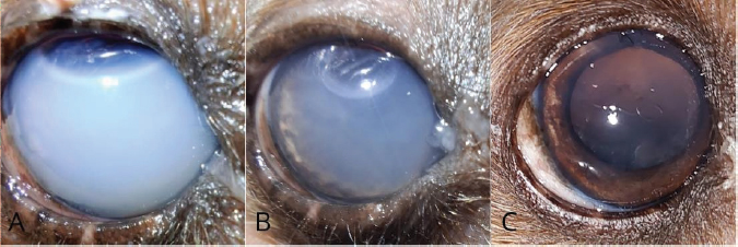
Fig. 1. The right eye of a Yorkshire Terrier after phacoemulsification (A) Lipemic aqueous humor 24 hours postoperatively. (B) Reduction of cloudiness 48 hours postoperatively. (C) Clinical improvement 7 days postoperatively.
Within 48 hours postoperatively, the right eye had satisfactory midriasis with positive flare and IOP of 13 mm Hg. The left eye presented with slight synechiae and fibrin in the anterior chamber, an IOP of 11 mmHg, reduced lipemic humor, and a positive menace response. Atropine 1% eye drops were administered again, and subconjunctival dexamethasone was administered to the right eye.
Biochemical analysis revealed hypertriglyceridemia (2,345.00 mg/dl, reference range : 21–116 mg/dl) and hypercholesterolemia (524.00 mg/dl, reference range=126–359 mg/dl), which are characteristic of LU that support the diagnosis. Treatment of hypertriglyceridemia and hypercholesterolemia began with ezetimibe, and bezafibrate was discontinued.
There was a favorable evolution in the patient’s health 4 days postoperatively, however fibrin and synechiae were observed, and tissue plasminogen activator (tPA) was administered under sedation. Dexamethasone was administered bilaterally subconjunctivally at the end of the procedure. Within 72 hours following tPA administration (7 days postoperatively), there was no anterior chamber opacity, and the LU had improved (Fig. 1B). Both eyes exhibited a positive menace and visual reflexes. Fundoscopy revealed a normal fundus in the right eye, an evaluation that had not been possible during previous examinations (Fig. 1C). Topical treatment with eye drops was gradually tapered.
Discussion
Endocrine disorders significantly alter lipid metabolism, increasing serum triglyceride and cholesterol concentrations, as confirmed by biochemical tests with levels were well above the reference ranges (triglycerides 2,345 mg/dl and cholesterol 524 mg/dl) (Slatter, 2005; Schechtmann et al., 2020). In the case described herein, the patient—a Yorkshire Terrier. diagnosed with DM and Cushing’s syndrome—had significant risk factors for developing secondary hyperlipidemia. Previous studies have reported that middle-aged to older individual dogs, particularly predisposed breeds, may develop lipid-laden aqueous humor secondary to systemic metabolic disorders (Violete et al., 2019; Schechtmann et al., 2020). Hyperlipidemia represents a significant challenge in veterinary medicine, particularly regarding its ocular manifestations. Early identification of this condition is essential for preventing serious complications, such as lipid-laden aqueous humor and retinal lipemia, which can negatively impact the animal’s quality of life. Disruption of the blood-aqueous barrier—whether facilitated by inflammation or intraocular surgery procedures such as cataract surgery—increases the risk of these changes in predisposed animals. In this case report, the predisposing factors were observed.
The presence of anterior chamber opacity in the immediate postoperative period required differentiation from other postoperative complications. Toxic anterior segment syndrome (TASS) is characterized by acute sterile inflammation that occurs shortly after surgery, and is typically associated with toxic substances introduced during the procedure (Alves et al., 2013). In this case, the absence of severe corneal edema, marked ocular pain, or signs of infection made TASS less likely; however, this diagnosis cannot be excluded. Alternative diagnoses, including postoperative fibrinous uveitis were also considered. Clinical findings, together with the systemic metabolic condition, including marked hypertriglyceridemia and hypercholesterolemia, suggested a presumptive lipemic component contributing to the observed opacity. Nevertheless, given the lack of aqueous humor analysis and the response to intracameral tPA, a significant fibrinous component was likely also present. The aqueous flare in LU confers a characteristic opacity to the aqueous humor; this condition is also referred to as lipid-laden aqueous uveitis or lipemic aqueous uveitis (Olin et al., 1976; Klein et al., 2011). Based on laboratory findings and clinical characteristics, LU was considered as a differential diagnosis. The presence of marked hypertriglyceridemia and hypercholesterolemia, combined with the clinical appearance of the aqueous humor, supported a presumptive diagnosis.
However, the absence of an aqueous humor analysis represents an important limitation and precludes the definitive confirmation of intraocular lipid content. In addition, the marked response to tPA suggests that the fibrinous component likely contributed to the observed opacity.
Therefore, this case is more appropriately interpreted as a postoperative inflammatory condition with a presumptive lipemic component rather than as a definitively confirmed LU. Alternative diagnoses, including postoperative fibrinous uveitis and TASS, were also considered. Although the clinical presentation and progression were not fully consistent with the TASS, this condition cannot be entirely excluded. TASS is an acute, sterile, postoperative inflammatory reaction that usually develops within 12–48 hours after intraocular surgery. It is characterized by diffuse corneal edema, anterior chamber inflammation, and fibrin formation. It is commonly associated with the introduction of toxic or contaminant agents during surgery (Hernandez-Bogantes et al., 2019; Verma et al., 2024). In this present case, treatment included topical and systemic anti-inflammatory therapies, mydriatics, ocular hypotensive agents, antibiotics, and subconjunctival dexamethasone. Intracameral administration of tPA was also performed to reduce the anterior chamber opacity associated with fibrin formation and to minimize the risk of synechiae. The patient demonstrated a favorable clinical response, particularly after intracameral tPA administration, which suggests that a fibrinous component contributed to the anterior chamber opacity. However, this does not exclude a concurrent lipemic process, especially considering the marked systemic hyperlipidemia observed in this patient.
Treatment included topical and systemic anti-inflammatory therapies, mydriatics, ocular hypotensive agents, and antibiotics, in addition to subconjunctival dexamethasone. tPA was also administered to reduce anterior chamber opacity associated with fibrin formation and to minimize the risk of synechiae development. The patient demonstrated a favorable clinical response, characterized by progressive aqueous clarity and recovery of intraocular visualization, which is consistent with a previous report describing successful management of lipemic aqueous humor using anti-inflammatory therapy (Violette and Ledbetter, 2019).
This case report demonstrates the importance of rigorous postoperative monitoring. Early identification of LU and associated risk factors allowed for appropriate therapeutic intervention.
This facilitated not only the resolution of intraocular inflammation but also the functional recovery of vision, as evidenced by a positive response and clear fundus visualization during fundoscopy.
Despite the successful clinical outcome, a notable limitation of this report is that aqueous humor was not collected for laboratory analysis, which would have provided direct confirmation of the lipid content. However, aqueous sampling is invasive and is not routinely performed when a presumptive diagnosis can be reasonably established based on the findings of ophthalmic examination. In the present case, LU was suspected based on the clinical appearance of the aqueous humor and systemic hyperlipidemia; however, the marked response to intracameral tPA supports the presence of a significant fibrinous component, suggesting that fibrin may have substantially contributed to the observed opacity.
Therefore, this case is more appropriately interpreted as a postoperative inflammatory condition with a presumptive lipemic component rather than as confirmed LU. Similarly, previous veterinary studies describing lipid-laden aqueous humor in dogs have relied primarily on clinical findings rather than aqueous humor analysis (Violette and Ledbetter, 2019; Schechtmann et al., 2020). Despite these limitations, reports of LU in dogs remain scarce in the Veterinary literature. Consequently, additional well-characterized clinical cases may contribute to improved recognition of this condition and support earlier diagnosis and more appropriate clinical management in affected patients.
Conclusion
Consistent with previous studies, metabolic disorders—which are characterized by hypertriglyceridemia and hypercholesterolemia—combined with intraocular surgery may predispose dogs to lipid-related alterations. Early recognition and prompt intervention are essential for preventing vision-threatening complications. This report highlights the importance of considering presumptive LU with a fibrinous component as a potential pos-phacoemulsification complication, particularly in patients with underlying metabolic dysfunctions.
Conflict of interest
The authors have no conflicts of interest to declare.
Funding
This study received no specific grant.
Author’s contributions
F.F.F. and P.S.H. wrote the original draft and conceptualized the manuscript. C.B.G.S., L.D.P., and J.A.T.P. revised and edited the manuscript.
Data availability
All data are provided in the manuscript.
References
Alves, L.F.A., Kac, M.J., Bisol, T., Fernandes, B.F. and Eskenazi, D.T. 2013. Toxic anterior segment syndrome. Rev. Bras. Oftalmol. 72(1), 29–33.
Chan, R.V.P. and Li, K.K. 1998. The anatomy and physiology of the uveal tract. In Diagnosis and treatment of uveitis. Eds., Foster, C.S. and Vitale, A.T. Philadelphia, PA: W.B. Saunders, pp: 3–19.
Hernandez-Bogantes, E., Navas, A., Naranjo, A., Amescua, G., Graue-Hernandez, E.O., Flynn, H.W. and Ahmed, I. 2019. Toxic anterior segment syndrome: a review. Surv. Ophthalmol. 64, 463–476.
Klein, H.E., Krohne, S.G., Moore, G.E. and Stiles, J. 2011. Postoperative complications visual outcomes of phacoemulsification in 103 dogs (179 eyes): 2006–2008. Vet. Ophthalmol. 14(2), 114–120.
Maggs, D.J. 2009. Ophthalmology. In Small animal internal medicine, 4th ed. Ed., Slatter, D.H. St. Louis, MO: Mosby Elsevier, pp: 1093–110.
Olin, D.D., Rogers, W.A. and MacMillan, A.D. 1976. Lipid-laden aqueous humor with anterior uveitis and concurrent hyperlipemia in two dogs. J. Am. Vet. Med. Assoc. 168(9), 861–864.
Schechtmann, S.A., Stine, J.M., Miller, T.R., Welihozkiy, A. and Michau, T.M. 2020. A retrospective analysis of lipid-laden aqueous humor in dogs: thirty cases. Vet. Ophthalmol. 23, 277–285.
Slatter, D.H. 2005. Fundamentals of veterinary ophthalmology. São Paulo, Brazil: Roca.
Verma, L., Malik, A., Maharana, P.K., Dada, T. and Sharma, N. 2024. Toxic anterior segment syndrome (TASS): a review and update. Indian. J. Ophthalmol. 72(1), 11–18.
Violette, N.P. and Ledbetter, E.C. 2019. Lipemic uveitis and its etiologies in dogs: 75 cases. Vet. Ophthalmol. 22(5), 577–583.