Open Veterinary Journal, (2026), Vol. 16(6): 3579-3583
Case Report
10.5455/OVJ.2026.v16.i6.28
Intralesional application of platelet-enriched plasma for treating equine periodontal disease: A case report
Miguel Ravalha Cortelini1*, Carla Teixeira Leite1, José Corrêa Lacerda-Neto2,
Geórgia Camargo Góss3, Gabriela Döwich Pradella1, Natálie Rodrigues Martins1, Ana Paula da Costa Rodrigues1, Marelise Moral Montana1, Irina Lübeck1 and Claudia Acosta Duarte1
1Hospital Veterinário Universitário, Universidade Federal do Pampa, Uruguaiana, Brazil
2Hospital Veterinário Universitário, Universidade Estadual Paulista Júlio de Mesquita Filho, São Paulo, Brazil
3Hospital Veterinário Universitário, Universidade Estadual de Santa Cruz, Ilhéus, Brazil
*Corresponding Author: Miguel Ravalha Cortelini. Hospital Veterinário Universitário, Universidade Federal do Pampa, Uruguaiana, Brazil. Email: miguel.ravalha [at] gmail.com
Submitted: 22/10/2025 Revised: 02/05/2026 Accepted: 14/05/2026 Published: 05/06/2026
© 2025 Open Veterinary Journal
This is an Open Access article distributed under the terms of the Creative Commons Attribution-Non Commercial-No Derivatives License (http://creativecommons.org/licenses/by-nc-nd/4.0/), which permits non-commercial re-use, distribution, and reproduction in any medium, provided the original work is properly cited, and is not altered, transformed, or built upon in any way.
ABSTRACT
Background: Equine periodontal disease (PD) is an inflammatory condition of bacterial origin that is common in all age groups but more common in geriatric animals. It causes dysphagia, progressive weight loss, and premature tooth loss. Due to the relevance of PD to the species and the difficult treatment, platelet-enriched plasma has become a viable option for treating PD in horses based on accelerated healing through the growth factors present in platelets. This study aimed to report the clinical effects of the intralesional application of platelet-enriched plasma in the molars of horses previously diagnosed with PD.
Case Descriptions: Eleven teeth from seven horses, all of age, of both sexes, from the same farm and under a single feeding system, were used for the study. The initial diagnosis of PD included physical, traditional dental, and radiological examinations. The treatment consisted of cleaning, debridement, and curettage of the periodontal pocket, followed by the intralesional application of 1 ml of platelet-enriched plasma. Clinical improvement in PD was determined by the appearance of the tissues surrounding the tooth, periodontal pocket depth measurement, and radiological examination.
Conclusion: Intralesional platelet-enriched plasma combined with curettage resulted in clinical improvement, demonstrating a promising innovation for treating equine PD.
Keywords: Dentistry, Growth factors, Horses, Molars, Periodontium.
Introduction
Periodontal disease is characterized as a chronic bacterial inflammatory condition, usually secondary to dental malocclusion. In the initial phase, gingival tissue is compromised, and as the disease progresses, periodontal grooves, inflammatory secretion, halitosis, periodontal ligament fragility, loss of dental attachment, and inefficient and painful chewing occur (Harvey and Emily, 1993; Eke et al., 2015). Dental calculus accumulation and bacterial contamination in the gingival sulcus constitute relevant extrinsic factors related to the etiology of periodontal disease (Mcphee and Cowley, 1981; Gieche, 2010). In addition, it is important to emphasize that irregular tooth wear and unbalanced masticatory biomechanics predispose to food compaction in interproximal spaces, bacterial colonization, and tissue degradation (ALLEN, 2003; GIECHE, 2010).
Periodontal disease incidence in horses is high. Miles and Grigson (1990) reported that 34% of the horses of various ages that participated in their study were affected. Baker (1970) described an incidence of 60% in horses aged >15 years.
Diagnosing PD can be difficult because traditional dental examination has limitations primarily related to the caudal region of the oral cavity. Therefore, a thorough intraoral examination, including inspection, digital and instrumental palpation, measurement of periodontal pocket depth using a millimeter probe, characterization of affected tissues, and assessment of tooth mobility, is essential for its assessment and grading (Easley 2008).
The goal of periodontal therapy includes halting disease progression and regenerating affected tissues, such as mucosa, periodontal ligament, cementum, and alveolar bone. In recent years, a variety of biomaterials, biological agents, membranes, and growth factor therapy have been studied and used as regenerative treatments for periodontal defects in human dentistry (Pagni et al., 2022; Miron et al., 2024) and in dogs (Bellows et al., 2019). Plasma gel, which is used in oral and maxillofacial surgeries in humans, plays a significant role in the final outcomes of periodontal defects, promoting the acceleration of healing and bone regeneration (Dusse et al., 2008; Pagni et al., 2022). It consists essentially of a high concentration of autologous platelets in a small volume of plasma obtained by centrifuging the patient’s own blood. In turn, platelets, which are small anucleated cytoplasmic fragments originating from bone marrow cells, are composed of megakaryocytes, which play a primary role in clot formation and the release of growth factors (Handin, 1995; Margolis et al., 2001; Pagni et al., 2022). Consequently, tissue repair occurs through the downregulation of inflammatory mediators and the synthesis of regenerative proteins (Sampson et al., 2010; Napolitano et al., 2012). Platelet-rich plasma (PRP) has antimicrobial properties and may contribute to pain reduction (Berghoff et al., 2006; Asfaha et al., 2007) and infection prevention (Edwards and Calandruccio, 2003).
Due to the relevance of equine PD as a cause of pain, premature tooth loss, predisposition to systemic diseases, and reduced longevity, innovative treatments that effectively demonstrate results go along with the objective of this report, which aimed to explain the effect of periodontal pocket curettage combined with intralesional application of platelet-enriched plasma in the treatment of PD in molars of seven equines.
Case Details
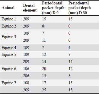
Seven horses, six males and one female, were treated at the veterinary hospital of the Federal University of Pampa-Uruguaiana Campus, Rio Grande do Sul, and at the Uruguaiana Military Circle. They were raised on the same farm and fed a diet based on forage, pelleted feed, and whole grain oats. On the day of the evaluation, the animals underwent a physical examination, followed by intravenous sedation with detomidine hydrochloride at a dose of 20 µg/kg for dental evaluation, blood collection, and radiological examination. In a stock, the intraoral cavity was cleaned for subsequent examination and characterization of the tissues affected by PD using a dental headstall, headlight, and speculum. The presence of gingival recession, absence of purulent and/or bloody secretion in all teeth, periodontal pocket with irregular margin, presence of necrotic tissue, food entrapment, and absence of tooth mobility were identified in the evaluation of PD. The depth of the periodontal pocket was measured with the aid of a millimeter periodontal probe, and the measurements obtained are explained in Table 1.
Table 1. Measurement of the periodontal pocket in the teeth on days 0 and 30 (mm).
The teeth with PD underwent cleaning, debridement, and curettage of the periodontal pocket, followed by the intralesional application of 1 ml of platelet-enriched plasma (Fig. 1). We used a protocol for obtaining PRP as recommended by Carneiro et al. (2017). Peripheral blood was collected from the external jugular vein in a volume equivalent to 10 times the amount of PRP to be applied in each tooth using a 40 12-gauge needle and syringe, homogenized, and stored in tubes containing 0.2 ml of citrate. After the collected blood had rested for 5 minutes, platelet values were quantified using a blood count in an automated machine (Sysmex pocH-100iV Diff™). The samples were then centrifuged (Novatecnica NT 812 800 g) for 10 minutes at a rotation of 120 G, followed by a 10-minute rest. Approximately two-thirds of the plasma near the platelet button was discarded using a pipette, and the remaining material was centrifuged again at 240 G for 10 minutes.
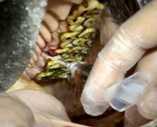
Fig. 1. An intraoral image of a horse. Periodontal disease and intralesional treatment with platelet-enriched plasma.
At the end of centrifugation, the material remained at rest for an additional 10 minutes, when approximately half of the upper plasma was discarded. The final plasma platelet count was performed, and the tripled platelet count was observed in only one animal (two teeth). In the other horses, platelet gel containing values below those recommended by Carneiro et al. (2017) was applied. In all cases, the animals were sedated with detomidine hydrochloride prior to the application of platelet-enriched plasma. The periodontal pocket was curetted using a long Schroeder curette and wiped with gauze. The speculum was then removed, and the animals were allowed to rest for 60 minutes.
Regarding the radiological examination on day zero, only 1 out of the 11 elements evaluated presented radiographic alterations whose images were compatible with loss of alveolar definition and cranial and caudal roots, tooth size smaller than physiological, and area of bone lysis (Fig. 2).
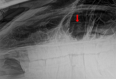
Fig. 2. Laterolateral radiographic image of the adult horse’s middle head. On day zero, loss of alveolar definition of the cranial and caudal roots of the upper left molar (209), inferior tooth size, and bone lysis area (arrow) can be observed.
Thirty days later, all horses were re-evaluated to characterize the periodontal lesions and to measure the depth of the periodontal pocket to verify the treatment effectiveness. Radiographic evaluation was performed only on patients who presented with alterations on day zero. The images remained unchanged, with the same alterations observed previously. In the re-evaluation, 100% of the animals showed clinical improvement in gingival recession, as demonstrated by the healthy appearance of the mucosa with a thin, pink, firm, and odorless margin. Regarding the depth of the periodontal pocket, a reduction in millimeters was observed in nine elements, while two elements remained unchanged (Table 1).
Ethical approval
The Animal Use Ethics Committee of the Federal University of Pampa approved the experimental protocol under protocol number 011/2017.
Discussion
The sedation used on horses ensured accurate detailing of periodontal lesions and measurement of periodontal pocket depth and proved to be efficient for carrying out examinations and treating the animals. Authors such as Valverde (2005) and Velázquez (2009) suggest that, in cases where long and deep sedation is required, other alpha² agonists such as xylazine hydrochloride or romifidine, associated or not with opioids, as well as benzodiazepines. However, in the treated cases, the use of detomidine alone was efficient in performing the required procedures.
Seven animals with 11 maxillary dental elements that presented with PD were treated; all were horses of advanced age. No connection between the condition and premature tooth loss was observed, considering the age of the horses and dental integrity, consistent with the study carried out by Telhado et al. (2004).
The clinical findings were the presence of gingival recession and absence of secretion in all teeth, a periodontal pocket with irregular margin, necrotic tissue, food entrapment, and tooth mobility. On day 30, there was clinical improvement of gingival recession in 100% of the animals, demonstrated by the healthy appearance of the mucosa with a thin, pink, firm, and odorless margin, characteristics mentioned as normal by Klugh (2010).
Regarding the depth of the periodontal pocket, a reduction in millimeters was observed in nine elements (81%), while two elements (18%) remained unchanged, according to measurements performed on days 0 and 30, as presented in Table 1. A significant improvement in the depth of the periodontal pocket and in the appearance of the mucosa was observed, which suggests that the high concentrations of growth factors probably contributed to this effect, which is consistent with the findings of Chung et al. (2022), who studied the application of platelet-rich plasma in the treatment of periodontal disease in dogs. It is important to note that sulci of up to 5 mm are considered normal for the equine species, and that the return to this depth evidences the improvement of the condition studied in the present work (Klugh, 2010).
Regarding the radiographic changes identified on day 0 in tooth 209, a re-evaluation was performed after 30 days, and the images remained unchanged. Soikkonen et al. (1998) reported that alveolar bone loss is the most common radiographic finding in periodontal disease.
Although two-dimensional images are used, the correlation between radiological studies, which demonstrate decreased alveolar radiopacity, and periodontal pocket depth measurement makes it efficient to monitor the progression of PD. Radiological images showing a decrease in radiopacity of alveolar bone surrounding teeth with periodontal disease are effective in monitoring the progression of this disease (Hausmann, 2000). However, in the present study, radiographic evaluation could have provided additional relevant information if performed for a period longer than 30 days, especially regarding alveolar bone resorption and tissue regeneration. Comparative radiographic documentation over time would have enabled a more objective assessment of periodontal lesions, contributing to a more robust analysis of the therapeutic response.
Regarding PRP preparation, a protocol according to Carneiro et al. (2017) was used. The ideal platelet quantity for application must be at least three times greater than the initial count. However, obtaining high platelet counts was quite difficult, requiring multiple blood draws and sample processing on several alternate days. Tripled platelet values were achieved in only one animal (two teeth), which demonstrated complete qualitative and quantitative clinical improvement after 30 days. Studies using PRP in human dentistry, as described by Camargo et al. (2012) and Pagni et al. (2022), have shown promising results, from hemostasis to faster healing, with improved tissue regeneration.
In other equines, platelet gel containing values below those recommended by Carneiro et al. (2017) was applied, achieving similar satisfactory results, validating the study by Cáceres et al. (2011), who stated that PRP and PPP have the capacity for tissue remodeling and contraction, in addition to demonstrating a similar biological response as for gingival repair. In agreement, Creeper and Ivanovski (2012) described that when tested in vitro, PPP and PRP promote migration and proliferation of gingival fibroblasts, with no difference between autologous and allogeneic preparations.
Conclusion
These case reports showed that the intralesional application of platelet-rich plasma combined with curettage is an effective and promising therapy for the treatment of periodontal disease in horses. The established plasma volume was sufficient for successful treatment. Further studies with more representative samples, involving different horse breeds and different evaluation periods, are essential for improving the methodology used in equine PD treatment protocols.
Acknowledgments
On behalf of all authors, we thank Flávia Giribone Acosta Duarte for translating this work.
Conflict of interest
The authors declare that there is no conflict of interest.
Funding
None.
Authors’ contributions
All authors contributed equally to this study.
Data availability
All data were provided in the manuscript.
References
Allen, T. 2003. Common dental abnormalities. In Manual of equine dentistry. S.I: Muleicorn Press, pp: 71–107.
Asfaha, S., Cenac, N., Houle, S., Altier, C., Papez, M.D., Nguyen, C., Steinhoff, M., Chapman, K., Zamponi, G.W. and Vergnolle, N. 2007. Protease-activated receptor-4: a novel mechanism of inflammatory pain modulation. Br. J. Pharmacol. 150, 176–185.
Baker, G.J. 1970. Some aspects of equine dental disease. Equine Vet. J. 2, 105–110.
Bellows, J., Berg, M.L., Dennis, S., Harvey, R., Lobprise, H.B. and Synder, C.J., Stone, A.E.S. and Wetering, A.G.V. 2019. AAHA dental care guidelines for dogs and cats. J. Am. Anim. Hosp. Assoc. 55(2), 49–69.
Berghoff, W.J., Pietrzak, W.S. and Rhodes, R.D. 2006. Platelet-rich plasma application during closure following total knee arthroplasty. Orthopedics 29(7), 590–598.
Cáceres, M., Martínez, C., Martínez, J. and Smith, P.C. 2012. Effects of platelet-rich and -poor plasma on the reparative response of gingival fibroblasts. Clin. Oral Implants Res. 23(9), 1104–1111; doi:10.1111/j.1600-0501.2011.02274.x
Camargo, G.A.C.G., Oliveira, R.L.B., Fortes, T.M.V. and Santos, T.S. 2012. Utilização do plasma rico em plaquetas na odontologia. Odontol. Clín. Cient. 11(3), 187–190.
Carneiro, A.L.M., Duarte, C.A., Döwich, G., Góss, G.C., Lagreca, L.F.J. and Bataglin, C.F. 2017. Alternativa de protocolo para obtenção de plasma rico em plaquetas (PRP) em equinos. In Salão Internacional de Ensino Pesquisa e Extensão. Santana do Livramento, 9.
Chung, C.S., Wei, Y.F. and Lin, L.S. 2023. Submucosal injection of activated platelet-rich plasma for treatment of periodontal disease in dogs. J. Vet. Dent. 40(1), 19–27; doi: 10.1177/08987564221124165
Creeper, F. and Ivanovski, S. 2012. Effect of autologous and allogenic platelet-rich plasma on human gingival fibroblast function. Oral Dis. 18, 494–500.
Dusse, L.M.S., Macedo, A.P., Batschauer, A.P. and Carvalho, M.G. 2008. Plasma Rico em Plaquetas (PRP) e sua aplicação em odontologia. Braz. Journ. Clin. Analyses. 40(3), 193–197.
Easley, K.J. How to properly perform and interpret an endoscopic examination of the equine oral cavity. In Proceedings of the Annual Convention of the American Association of Equine Practitioners, AAEP, SanDiego, United States of America, 2008, p 54.
Edwards, S.G. and Calandruccio, J.H. 2003. Autologous blood injections for refractory lateral epicondylitis. J. Hand Surg. Am. 28, 272–278.
Eke, P.I., Dye, B.A., Wei, L., Slade, G. D., Evans, G.O.T., Borgnakke, W.S., Taylor, G.W., Page, R.C., Beck, J.D. and Genco, R.J. 2015. Update on prevalence of periodontitis in adults in the United States: nHANES 2009 to 2012. J. Periodontol. 86, 611–622.
Gieche, J.M. How to assess the equine periodontium. In Proceedings of the Annual Convention of the American Association od Equine Practitioners, Baltimore, United States of America, 2010, 56.
Handin, R.J., Lux, S.E. and Stossel, T.P. 1995. Blood: principles, practice of hematology. Estados Unidos da América, Nova York, NY: J.B. Lippincott, p: 2190.
Harvey, C.E. and Emily, P.P. 1993. Small animal dentistry. Estados Unidos da América, Saint Louis: Mosby, p: 413.
Hausmann, E. 2000. Radiographic and digital imaging in periodontal practice. J. Periodontol. 71(3), 497–503.
Klugh, D.O. 2010. Principles of equine dentistry. 1. Ed. London, Reino Unido: CRC Press, p: 256.
Margolis, D.J., kantor, J., Santanna, J., Strom, B.L. and Berlin, J.A. 2001. Effectiveness of platelet release for the treatment of diabetic neuropathic foot ulcers. Diabetes. Care. 24, 483–488.
McPhee, T. and Cowley, G. 1981. Essentials of periodontology and periodontics, 3ed. Inglaterra: Blackwell Scientific, p: 492.
Miles, A.E.W. and Grigson, C. 1990. Colyer’s variations and diseases of the teeth of animals. Inglaterra, Cabridge: Cambridge University Press, p: 672.
Miron, R.J., Gruber, R., Farshidfar, N., Sculean, A. and Zhang, Y. 2024. Ten years of injectable platelet-rich fibrin. Periodontol 2000. 94, 92–113; doi:10.1111/prd.12538
Napolitano, M., Matera, S., Bossio, M., Crescibene, A., Costabile, E., Almolla, J., Almolla, H., Togo, F., Giannuzzi, C. and Guido, G. 2012. Autologous platelet gel for tissue regeneration in degenerative disorders of the knee. Blood Transfusion 10, 72–77.
Pagni, G., Tavelli, L. and Rasperini, G. 2022. The evolution of surgical techniques and biomaterials for periodontal regeneration. Dent. Clin. North. Am. 66, 75–85.
Sampson, S., Reed, M., Silvers, H., Meng, M. and Mandelbaum, B. 2010. Injection of platelet-rich plasma in patients with primary and secondary knee osteoarthritis: a pilot study. Am. J. Phys. Med. Rehabil. 89, 270961–226969.
Soikkonen, K., Wolf, J., Närhi, T. and Ainamo, A. 1998. Radiographic periodontal findings in an elderly Finnish population. J. Clin. Períodontol. 25(6), 439–445.
Telhado, J., Aganin, J., Diele, C.A. and Marinho, M.S. 2004. Incidência de cálculo dentário e doença periodontal em cães da raça Pastor Alemão. Ciênc. Bras. 5(2), 99–104.
Valverde, A. Sedation for standing procedures. In Proceeding of the North American Veterinary Conference, Orlando, United States of America, 2005, Vol. 19, p 19.
Velázquez, J.L. Equine dentistry. In Proceeding of the International Congress of World Equine Veterinary Association, Guarujá, Brazil, 2009, p 1.