





| Case Report | ||
Open Vet. J.. 2026; 16(6): 3826-3831 Open Veterinary Journal, (2026), Vol. 16(6): 3826-3831 Research Article Suspected cutaneous metastasis of mammary carcinoma in a dog: A case reportDongwook Kim1†, Huisu Lee2†, Yawon Hwang2 and Gonhyung Kim1*1Department of Veterinary Surgery, College of Veterinary Medicine, Chungbuk National University, Cheongju, South Korea 2Bupyeong SKY Animal Medical Center, Incheon, South Korea †These authors contributed equally to this work. *Corresponding Author: Gonhyung Kim. Department of Veterinary Surgery, College of Veterinary Medicine, Chungbuk National University, Cheongju, South Korea. Email: ghkim [at] cbu.ac.kr Submitted: 08/09/2025 Revised: 05/05/2026 Accepted: 19/05/2026 Published: 16/06/2026 © 2025 Open Veterinary Journal
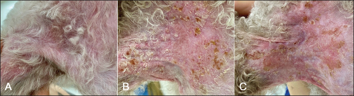
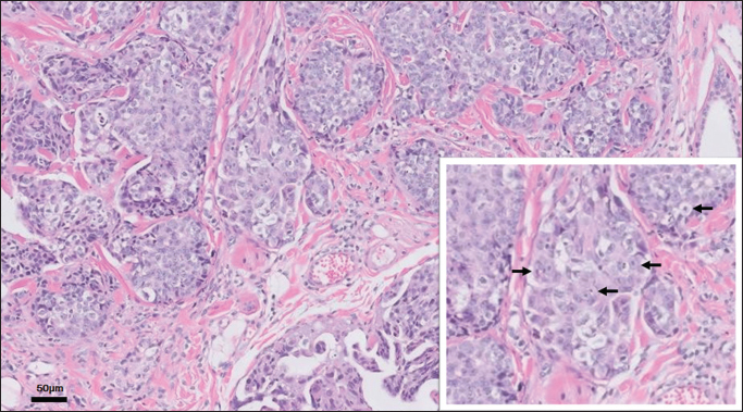
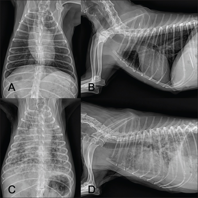
ABSTRACTBackground: Cutaneous metastases of tumors are rare in dogs. In particular, only a few cases of cutaneous metastases from mammary gland tumors have been reported. Case Description: A 13-year-old neutered female Poodle presented with localized pruritus of the right axilla. Physical examination revealed redness and crusting. A skin taping test confirmed Malassezia infection. However, despite antifungal treatment, erythema and pruritus worsened, accompanied by increased crust formation. Immunosuppressive therapy was briefly attempted for a suspected immune-mediated disorder; however, it was ineffective. Subsequent skin biopsy and histopathological evaluation revealed adenocarcinoma with widespread vascular invasion. The tumor was positive for cytokeratin 7 and negative for cytokeratin 20, estrogen receptor, progesterone receptor, and p63. Multiple mammary masses were also palpated. Based on the clinical and histological findings, a diagnosis of metastatic mammary adenocarcinoma was made. Although palliative therapy was initiated, the dog developed acute respiratory distress 2 weeks after the biopsy. Thoracic radiographs showed multiple pulmonary nodules, which were presumed to be metastases. Conclusion: Although metastatic skin tumors are rare in dogs, they can mimic dermatitis and should be included in the differential diagnosis. Keywords: Dog, Malignant, Mammary gland tumor, Metastasis, Skin. IntroductionCanine mammary gland tumors (MGTs) are a prevalent type of tumor, accounting for 14.5% of all tumors in dogs (Dhein et al., 2024). Another study reported that MGTs represented 46.7% of all tumors, of which approximately 51.6% were malignant (Zheng et al., 2022). The average age at which malignant and benign MGTs occur is 9.5 and 8.5 years, respectively (Sorenmo et al., 2009). Malignant tumors are typically larger than benign lesions and have a greater potential for metastasis (Sorenmo et al., 2009). Malignant MGTs are at risk of developing other primary tumors, and approximately 50% of mammary carcinomas metastasize to regional lymph nodes (Sorenmo et al., 2009; Vazquez et al., 2023). Malignant tumors can spread to distant organs in addition to lymph node metastasis (Vazquez et al., 2023). The lungs are the most frequently affected site for metastasis (Sorenmo, 2003), but metastasis to the liver, spleen, pancreas, and bones has also been documented (Simon et al., 2006; Sleeckx et al., 2011; Vazquez et al., 2023). Cutaneous metastases in dogs are rare, occurring primarily with specific tumor types, such as adenocarcinoma, osteosarcoma, and transitional cell carcinoma (Reed et al., 2013; Mazari et al., 2018; Parachini-Winter et al., 2019). Primary tumors associated with cutaneous metastases originate from the bone, urinary bladder, mammary gland, prostate, duodenum, or lung (Juopperi et al., 2003; Reed et al., 2013; Parachini-Winter et al., 2019; Di Maria et al., 2020; Choi et al., 2022; Greyling et al., 2024). To date, cutaneous metastases from mammary carcinomas have been exceedingly rare in dogs (Simon et al., 2006; Mazari et al., 2018; Varallo et al., 2019; Choi et al., 2022). This study presents the case of a dog with suspected cutaneous metastasis of a malignant mammary tumor. Case DetailsA 13-year-old neutered female Poodle weighing 4.3 kg presented with severe pruritus in the right axilla that had persisted for a week. The dog had a history of diabetes management for several years. On physical examination, redness and crust formation were observed in the right axilla (Fig. 1A). A skin taping test confirmed Malassezia infection. The initial treatment involved a week of topical ketoconazole ointment, but no significant improvement was observed. Skin scraping revealed no additional pathogens. Subsequently, systemic antifungal therapy was initiated with fluconazole (5 mg/kg PO q24 h) and terbinafine (25 mg/kg PO q24 h). Prednisolone (0.5 mg/kg PO q12 h) was administered to alleviate pruritus. Despite 5 weeks of antifungal therapy, the lesions progressed, with increased crusting and erythema accompanied by worsening pruritus (Fig. 1B). Following the antifungal treatment, a brief trial of immunosuppressive prednisolone (1 mg/kg PO q12 h) was conducted for 2 weeks to address a suspected immune-mediated dermatosis. However, it was discontinued due to a lack of improvement and glycemic control concerns (Fig. 1C). A skin biopsy was performed to reach a definitive diagnosis. Four samples were obtained from the right axillary region by punch biopsy. The samples were stained with hematoxylin and eosin. Immunohistochemistry was performed using monoclonal antibodies against cytokeratin 7 (CK7), cytokeratin 20 (CK20), estrogen receptor (ER), progesterone receptor (PR), and p63. Histopathological examination revealed a non-encapsulated, poorly demarcated, and highly infiltrative mass with mild to moderate fibrous stroma. The mass exhibited irregular tubular structures and highly cellular solid islands composed of atypical epithelial cells. The pleomorphic epithelial cells were cuboidal to polygonal with indistinct borders and contained small to moderate amounts of eosinophilic cytoplasm. Neoplastic cells exhibited round to oval nuclei with finely stippled chromatin and single to multiple prominent nucleoli. Anisocytosis and anisokaryosis were marked, with 16 mitotic figures observed per 10 high-power fields. Vascular invasion was widespread (Fig. 2). Mild to intense CK7 expression was observed, whereas neoplastic cells were negative for CK20, ER, PR, and p63. Histopathology indicated a differential diagnosis of primary apocrine sweat gland adenocarcinoma and mammary adenocarcinoma. Multiple incidental masses were palpated around the dog’s mammary glands. The largest mass was ill-defined, warm, and measured approximately 2.5 × 2.5 cm. The dog had a history of ovariohysterectomy (OHE) and surgical excision of mammary tumors diagnosed as complex adenomas without vascular invasion 2 years earlier. At that time, preoperative thoracic radiographs showed no evidence of pulmonary metastases (Fig. 3A and B). Palliative treatment was initiated with piroxicam (0.3 mg/kg PO q24 h). The dog’s body weight gradually declined from 4.3 to 3.6 kg over the course of treatment. Two weeks after skin biopsy, the dog presented with acute respiratory distress. Thoracic radiographs revealed pulmonary nodules suggestive of metastasis (Fig. 3C and D). Clinical symptoms progressively worsened, and no further treatments were pursued at the owner’s request.
Fig. 1. The dog’s right axilla showed erythema and crust formation at the time of admission (A), which gradually worsened during the 5th (B) and 7th (C) weeks of treatment.
Fig. 2. Histopathological examination revealed tubular structures composed of highly pleomorphic atypical neoplastic epithelial cells. Inset: The nuclei displayed finely stippled chromatin with single or multiple prominent nucleoli (arrows).
Fig. 3. Thoracic radiographs. Compared with preoperative images obtained 2 years earlier (A, B), multiple pulmonary masses were identified (C, D). DiscussionAlthough skin tumors account for approximately 30% of primary neoplasms, metastatic cutaneous tumors are rare in dogs (Pakhrin et al., 2007; Dhein et al., 2024). To date, only five dogs have been reported to have cutaneous metastases from MGTs (Simon et al., 2006; Mazari et al., 2018; Varallo et al., 2019; Choi et al., 2022). Among these cases, two reports provided details on the observed clinical symptoms. One dog presented with a single solid mass that was not ulcerated, while another exhibited suppurative lesions accompanied by pain (Mazari et al., 2018; Choi et al., 2022). In the current case, the dog showed pruritus, erythema, warmth, and crust formation, which may mimic dermatitis. Although the clinical presentations in previous reports differ from those in this case, the limited number of documented cases complicates the identification of consistent features of cutaneous metastasis in dogs. Histopathological findings in this dog led to the consideration of primary apocrine sweat gland adenocarcinoma and mammary adenocarcinoma as differential diagnoses. In the immunohistochemical evaluation, the tumor in this case was CK7 positive and negative for CK20, ER, PR, and p63. A similar CK7-positive/CK20-negative immunoprofile has been reported in both primary apocrine gland carcinomas and mammary carcinomas (Fernandez-Flores et al., 2007; Llamas-Velasco et al., 2018; Moon et al., 2022). Although most mammary carcinomas are ER- and PR-positive, triple-negative variants have also been described, further complicating the differential diagnosis (Fernandez-Flores, 2012; Kavisha et al., 2023). p63 has been proposed as a useful marker for distinguishing these entities, as metastatic carcinomas are generally negative for this marker (Ivan et al., 2005; Fernandez-Flores, 2012). However, p63 negativity has also been reported in patients with primary apocrine gland carcinomas (Fernandez-Flores, 2012; Khodakaram-Tafti et al., 2012). Overall, immunohistochemistry alone is insufficient for definitive differentiation between these tumor types due to overlapping immunohistochemical features. Careful correlation of immunohistochemical, clinical, and diagnostic findings is required for these tumors. Apocrine sweat gland adenocarcinomas are relatively uncommon, accounting for only 0.7% to 2.2% of all skin tumors in dogs (Simko et al., 2003). These tumors typically exhibit low degrees of vascular invasion and distant metastasis (Simko et al., 2003; Kycko et al., 2016). However, this dog developed multiple thoracic masses, indicating pulmonary metastases, approximately 2 months after treatment began. The presence of widespread vascular invasion, multiple mammary masses, and the rapid onset of pulmonary metastases strongly support the diagnosis of metastatic MGT (Rasotto et al., 2017). According to a previous scoring system for MGTs (Tavasoly et al., 2013), the tumor in this dog was classified as a poorly differentiated mammary adenocarcinoma. One study indicated that surgical resection of benign MGTs combined with OHE can reduce the risk of new tumors by approximately 50% (Kristiansen et al., 2013). Nonetheless, despite undergoing OHE at the time of tumor excision 2 years earlier, this dog still developed new MGTs. During treatment, the affected area exhibited increased warmth and erythema. Inflammatory mammary carcinoma (IMC) is known for its local invasiveness and can sometimes be misdiagnosed as dermatitis (Clemente et al., 2010). IMC is not histologically classified among MGTs. The diagnosis relies on clinical and histopathological features. Clinical signs of IMC include edema, erythema, firmness, and warmth of the mammary glands, and prominent dermal lymphatic invasion is a key histopathological finding (Clemente et al., 2010). IMC patients are likely to exhibit generalized weakness and weight loss. Severe localized pain is commonly observed during physical examination (Pérez Alenza et al., 2001; Peña et al., 2003; Pérez-Alenza et al., 2004). Although the warmth, erythema, and widespread vascular invasion observed in this dog resemble those of IMC, the absence of evident clinical symptoms around the mammary glands makes it challenging to diagnose IMC. In addition, the dog showed no localized pain around either the axillary tumor or the mammary masses. The lack of histopathological confirmation for the mammary and pulmonary masses is a limitation of this report. However, considering the clinical history, the histopathology of the skin lesions, and the rapid progression of the disease, metastatic mammary adenocarcinoma is the most likely diagnosis. ConclusionThis case describes a suspected metastatic skin tumor that mimicked dermatitis. Considering neoplasia in the differential diagnosis is essential in dogs with atypical and treatment-resistant dermatoses. AcknowledgmentsThis work was supported by the Overseas Dispatch Program of Chungbuk National University in 2025. Conflict of interestThe authors declare no conflict of interest. FundingThis study received no specific grant. Authors’ contributionsD.K., H.L., Y.H., and G.K. drafted the manuscript. D.K. and G.K. have revised and edited the manuscript. H.L. and Y.H. prepared and critically reviewed the manuscript. D.K. edited the references. All authors have read and approved the final version of the manuscript. Data availabilityAll data supporting this study’s findings are available within the manuscript. ReferencesChoi, H., Na, H., Lee, S.K., Bae, S., Oh, T. and Lee, K. 2022. Cutaneous metastasis of mammary gland tumor in a dog: a case report. Korean J. Vet. Res. 62, e2. Clemente, M., Pérez-Alenza, M.D. and Peña, L. 2010. Metastasis of canine inflammatory versus non-inflammatory mammary tumours. J. Comp. Pathol. 143, 157–163. Dhein, E.S., Heikkilä, U., Oevermann, A., Blatter, S., Meier, D., Hartnack, S. and Guscetti, F. 2024. Incidence rates of the most common canine tumors based on data from the Swiss Canine Cancer Registry (2008 to 2020). PLoS One 19, e0302231. Di Maria, F.M., Annoni, M., Roccabianca, P., Antoniazzi, E. and Bertazzolo, W. 2020. Cutaneous metastases of prostatic adenocarcinoma in two dogs. Vet. Clin. Pathol. 49, 459–464. Fernandez-Flores, A., Pol, A., Juanes, F. and Crespo, L.G. 2007. Immunohistochemical phenotype of cutaneous cribriform carcinoma with a panel of 15 antibodies. Med. Mol. Morphol. 40, 212–217. Fernandez-Flores, A. 2012. Immunohistochemical and morphologic evaluation of primary cutaneous apocrine carcinomas and cutaneous metastases from ductal breast carcinoma. Rom. J. Morphol. Embryol. 53, 879–892. Greyling, A., Van Der Weyden, L., Lensink, A.V. and O’Dell, N. 2024. Pulmonary adenocarcinoma with cutaneous metastasis in a dog. Vet. Sci. 11, 312. Ivan, D., Diwan, A.H. and Prieto, V.G. 2005. Expression of p63 in primary cutaneous adnexal neoplasms and adenocarcinoma metastatic to the skin. Mod. Pathol. 18, 137–142. Juopperi, T.A., Cesta, M., Tomlinson, L. and Grindem, C.B. 2003. Extensive cutaneous metastases in a dog with duodenal adenocarcinoma. Vet. Clin. Pathol. 32, 88–91. Kavisha, G., Neeraj, K., Avantika, S., Motilal, B.A., Abhay, B.Y., Suresh, P.C., Himani, P., Prabhu, S.N., Renu, S., Soumen, C. and Sanjay, P. 2023. Immunohistochemical detection of prognostic biomarkers in canine mammary tumors and relation with malignancy and prognosis. Clin. Oncol. 8, 1999. Khodakaram-Tafti, A., Khub, A. and Shirian, S. 2012. Pathological and immunohistochemical characteristics of apocrine sweat gland adenocarcinoma in a dog. Comp. Clin. Pathol. 21, 237–239. Kristiansen, V.M., Nødtvedt, A., Breen, A.M., Langeland, M., Teige, J., Goldschmidt, M., Jonasdottir, T.J., Grotmol, T. and Sørenmo, K. 2013. Effect of ovariohysterectomy at the time of tumor removal in dogs with benign mammary tumors and hyperplastic lesions: a randomized controlled clinical trial. J. Vet. Intern. Med. 27, 935–942. Kycko, A., Jasik, A., Bocian, Ł., Otrocka-Domagała, I., Mikiewicz, M., Śmiech, A., Łopuszyński, W., Dolka, I., Nowak, M. and Madej, J.A. 2016. Epidemiological and histopathological analysis of 40 apocrine sweat gland carcinomas in dogs: a retrospective study. J. Vet. Res. 60, 331–337. Llamas-Velasco, M., Pérez-Gónzalez, Y.C., Daudén, E. and Rütten, A. 2018. GATA3 staining in primary cutaneous apocrine cribriform carcinoma: usefulness to differentiate it from breast cancer metastasis. J. Cutan. Pathol. 45, 348–351. Mazari, B., Qoku, A. and Dhaskali, L. 2018. Study of mammary carcinoma in a dog and its relationship with cutaneous metastasis. ANGLISTICUM 7, 46–52. Moon, C.H., Min, H.A., Lee, H.B., Jeong, S.M. and Kim, D.H. 2022. Retroperitoneal metastatic apocrine gland ductal adenocarcinoma in a Beagle dog. Vet. Sci. 9, 234. Pakhrin, B., Kang, M.S., Bae, I.H., Park, M.S., Jee, H., You, M.H., Kim, J.H., Yoon, B.I., Choi, Y.K. and Kim, D.Y. 2007. Retrospective study of canine cutaneous tumors in Korea. J. Vet. Sci. 8, 229–236. Parachini-Winter, C., Curran, K.M., Pellin, M., Laver, T., Hanot, C., Vernier, T.H. and Séguin, B. 2019. Cutaneous and subcutaneous metastasis of appendicular osteosarcoma in dogs: 20 cases. J. Vet. Intern. Med. 33, 2200–2208. Peña, L., Dolores Perez-alenza, M., Rodriguez-Bertos, A. and Nieto, A. 2003. Canine inflammatory mammary carcinoma: histopathology, immunohistochemistry and clinical implications of 21 cases. Breast Cancer Res. Treatment 78, 141–148. Pérez Alenza, M.D., Tabanera, E. and Peña, L. 2001. Inflammatory mammary carcinoma in dogs: 33 cases (1995-1999). J. Am. Vet. Med. Assoc. 219, 1110–1114. Pérez-Alenza, M.D., Jiménez, A., Nieto, A.I. and Peña, L. 2004. First description of feline inflammatory mammary carcinoma: clinicopathological and immunohistochemical characteristics of three cases. Breast Cancer Res. 6, R300–R307. Rasotto, R., Berlato, D., Goldschmidt, M.H. and Zappulli, V. 2017. Prognostic significance of canine mammary tumor histologic subtypes: an observational cohort study of 229 cases. Vet. Pathol. 54, 571–578. Reed, L.T., Knapp, D.W. and Miller, M.A. 2013. Cutaneous metastasis of transitional cell carcinoma in 12 dogs. Vet. Pathol. 50, 676–681. Simko, E., Wilcock, B.P. and Yager, J.A. 2003. A retrospective study of 44 canine apocrine sweat gland adenocarcinomas. Can. Vet. J. 44, 38–42. Simon, D., Schoenrock, D., Baumgärtner, W. and Nolte, I. 2006. Postoperative adjuvant treatment of invasive malignant mammary gland tumors in dogs with doxorubicin and docetaxel. J. Vet. Intern. Med. 20, 1184–1190. Sleeckx, N., De Rooster, H., Veldhuis Kroeze, E., Van Ginneken, C. and Van Brantegem, L. 2011. Canine mammary tumours, an overview. Reprod. Domest. Anim. 46, 1112–1131. Sorenmo, K. 2003. Canine mammary gland tumors. Vet. Clin. North. Am. Small. Anim. Pract. 33, 573–596. Sorenmo, K.U., Kristiansen, V.M., Cofone, M.A., Shofer, F.S., Breen, A.M., Langeland, M., Mongil, C.M., Grondahl, A.M., Teige, J. and Goldschmidt, M.H. 2009. Canine mammary gland tumours; a histological continuum from benign to malignant; clinical and histopathological evidence. Vet. Comp. Oncol. 7, 162–172. Tavasoly, A., Golshahi, H., Rezaie, A. and Farhadi, M. 2013. Classification and grading of canine malignant mammary tumors. Vet. Res. Forum. 4, 25–30. Varallo, G., Gelaleti, G., Maschio-Signorini, L., Moschetta, M., Lopes, J., De Nardi, A., Tinucci-Costa, M., Rocha, R. and De Campos Zuccari, D. 2019. Prognostic phenotypic classification for canine mammary tumors. Oncol. Lett. 18, 6545–6553. Vazquez, E., Lipovka, Y., Cervantes-Arias, A., Garibay-Escobar, A., Haby, M.M., Queiroga, F.L. and Velazquez, C. 2023. Canine mammary cancer: state of the art and future perspectives. Animals (Basel) 13, 3147. Zheng, H.H., Du, C.T., Yu, C., Zhang, Y.Z., Huang, R.L., Tang, X.Y. and Xie, G.H. 2022. Epidemiological investigation of canine mammary tumors in mainland China between 2017 and 2021. Front. Vet. Sci. 9, 843390. | ||
| How to Cite this Article |
| Pubmed Style Kim D, Lee H, Hwang Y, Kim G. Suspected cutaneous metastasis of mammary carcinoma in a dog: A case report. Open Vet. J.. 2026; 16(6): 3826-3831. doi:10.5455/OVJ.2026.v16.i6.53 Web Style Kim D, Lee H, Hwang Y, Kim G. Suspected cutaneous metastasis of mammary carcinoma in a dog: A case report. https://www.openveterinaryjournal.com/?mno=293395 [Access: June 26, 2026]. doi:10.5455/OVJ.2026.v16.i6.53 AMA (American Medical Association) Style Kim D, Lee H, Hwang Y, Kim G. Suspected cutaneous metastasis of mammary carcinoma in a dog: A case report. Open Vet. J.. 2026; 16(6): 3826-3831. doi:10.5455/OVJ.2026.v16.i6.53 Vancouver/ICMJE Style Kim D, Lee H, Hwang Y, Kim G. Suspected cutaneous metastasis of mammary carcinoma in a dog: A case report. Open Vet. J.. (2026), [cited June 26, 2026]; 16(6): 3826-3831. doi:10.5455/OVJ.2026.v16.i6.53 Harvard Style Kim, D., Lee, . H., Hwang, . Y. & Kim, . G. (2026) Suspected cutaneous metastasis of mammary carcinoma in a dog: A case report. Open Vet. J., 16 (6), 3826-3831. doi:10.5455/OVJ.2026.v16.i6.53 Turabian Style Kim, Dongwook, Huisu Lee, Yawon Hwang, and Gonhyung Kim. 2026. Suspected cutaneous metastasis of mammary carcinoma in a dog: A case report. Open Veterinary Journal, 16 (6), 3826-3831. doi:10.5455/OVJ.2026.v16.i6.53 Chicago Style Kim, Dongwook, Huisu Lee, Yawon Hwang, and Gonhyung Kim. "Suspected cutaneous metastasis of mammary carcinoma in a dog: A case report." Open Veterinary Journal 16 (2026), 3826-3831. doi:10.5455/OVJ.2026.v16.i6.53 MLA (The Modern Language Association) Style Kim, Dongwook, Huisu Lee, Yawon Hwang, and Gonhyung Kim. "Suspected cutaneous metastasis of mammary carcinoma in a dog: A case report." Open Veterinary Journal 16.6 (2026), 3826-3831. Print. doi:10.5455/OVJ.2026.v16.i6.53 APA (American Psychological Association) Style Kim, D., Lee, . H., Hwang, . Y. & Kim, . G. (2026) Suspected cutaneous metastasis of mammary carcinoma in a dog: A case report. Open Veterinary Journal, 16 (6), 3826-3831. doi:10.5455/OVJ.2026.v16.i6.53 |


