





| Research Article | ||
Open Vet. J.. 2026; 16(6): 3925-3931 Open Veterinary Journal, (2026), Vol. 16(6): 3925-3931 Research Article The diagnostic challenges of diabetes insipidus in dogs: A narrative review of the water deprivation test and complementary biomarkersCarlos Eduardo Fontoura da Silva and Iago Martins Oliveira*Escola de Ciências Médicas e da Vida, Pontifícia Universidade Católica de Goiás, Goiânia, Brazil *Corresponding Author: Iago Martins Oliveira. Escola de Ciências Médicas e da Vida, Pontifícia Universidade Católica de Goiás, Goiânia, Brazil. Email: iago.vetufg [at] gmail.com Submitted: 11/11/2025 Revised: 22/05/2026 Accepted: 30/05/2026 Published: 20/06/2026 © 2025 Open Veterinary Journal
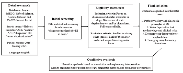
AbstractBackground: Diabetes insipidus (DI) is an uncommon canine endocrinopathy causing polyuria and polydipsia. It may be central, due to antidiuretic hormone (ADH) deficiency, or nephrogenic, resulting from renal insensitivity to ADH. Differentiation between these forms is essential for appropriate treatment. Although the water deprivation test (WDT) is considered the gold standard for confirming the diagnosis, it carries significant risks. Aim: This study aimed to review the pathophysiology and diagnostic methods of DI in dogs, emphasizing the safety and applicability of water deprivation and desmopressin tests, as well as the potential of complementary biomarkers. Methods: The study methodology consisted of a qualitative narrative review using the Scopus, SciELO, Web of Science, and Google Scholar databases. Original articles, literature reviews, and case reports published in English between January 2015 and January 2025 were included. The terms “DI,” “canine,” “diagnosis,” OR “WDT” were established for searching and researching on the platforms. Results: The diagnosis of canine DI begins with the exclusion of other common causes of polyuria and polydipsia, with hyposthenuria as an early indicator. The WDT is considered the gold standard for definitive diagnosis, but it involves considerable risks of dehydration and hypernatremia for the patient. Therapeutic testing with desmopressin is a safe and viable alternative for differentiating between central and nephrogenic forms of the disease. Recent studies highlight copeptin as a promising biomarker for assessing vasopressin activity in the body, while advances in the molecular characterization of AVPR2 and aquaporin-2 suggest a promising future for noninvasive biomarker-based diagnosis in veterinary medicine. Conclusion: The diagnosis of canine DI is complex and requires the combination and interpretation of clinical, laboratory, and therapeutic tests. Although the WDT is considered the gold standard, the desmopressin test offers a safer and more stable alternative. Advances in biomarker research may improve diagnostic accuracy and patient safety in the future. Keywords: Canine, Desmopressin, Endocrinopathy, Polydipsia, Polyuria. IntroductionWater homeostasis is a vital process for maintaining the physiological health of dogs. Diabetes insipidus (DI) is a rare endocrine disorder characterized by a marked impairment in the body’s ability to regulate water properly (Flynn et al., 2025). Clinically, the disease manifests itself through excessive urine production and water intake, accompanied by the production of hypotonic urine with low urinary density and a state of dehydration. The distinction of DI from other common causes of polyuria and polydipsia, such as diabetes mellitus, renal failure, or psychogenic polydipsia, represents a challenge in veterinary diagnosis (Silva and Zanutto, 2021). DI is classified into two main types based on its pathophysiological etiology. Central diabetes insipidus (CDI) results from a deficiency in the synthesis of antidiuretic hormone (ADH) in the hypothalamus or its release from the posterior pituitary into the bloodstream. In contrast, nephrogenic diabetes insipidus (NDI) occurs when the kidneys are unable to respond adequately to ADH, even though the hormone is present and circulating at normal levels. It is essential to correctly differentiate between their forms of manifestation in order to define the appropriate therapeutic protocol (Flynn et al., 2025). Both forms can be classified as primary, when the cause is idiopathic or genetic, or secondary, when they occur as a consequence of other diseases or traumas, such as neoplasms, injuries, or metabolic changes. In dogs, primary CDI is the most common and frequently encountered form, while primary NDI is considered an extremely rare and seldom seen condition. Identifying the type of disease is essential for successful clinical management and treatment, since the wrong therapeutic choice can lead to an unfavorable prognosis and iatrogenic complications for the animal (Ghizzo et al., 2021; Dillberger and Roth-Frank, 2025; Ribeiro et al., 2025). To correctly distinguish between these forms, a crucial factor for clinical management, several diagnostic tests can be used, with the WDT being the most traditional and considered the gold standard for the diagnosis of DI and its differentiation. It assesses the animal’s ability to concentrate urine in response to dehydration. However, this test is complex, requires strict medical monitoring, and poses potential risks to the patient, such as severe dehydration and hypernatremia. The need for an accurate and safe diagnostic method that minimizes the associated risks and allows rapid differentiation of the disease is a factor that drives the search for complementary biomarkers that can assist in clinical practice (Paulin et al., 2025). Despite advances in veterinary endocrinology, the diagnosis of DI in dogs remains challenging due to the limitations and risks associated with traditional diagnostic methods, particularly the WDT (Simões, 2021; Flynn et al., 2025). In addition, most available studies are based on small sample sizes or experimental models, limiting their direct applicability to clinical practice (Paulin et al., 2025). Therefore, there is a growing need to critically evaluate both traditional and emerging diagnostic approaches, including desmopressin testing and novel biomarkers such as copeptin (CoP). This study aimed to review the pathophysiology and diagnostic methods of canine DI, emphasizing the safety and applicability of the water deprivation and desmopressin tests, and the potential of emerging biomarkers. The hypothesis of this study is that the combination of a detailed clinical analysis with the judicious use of the WDT, together with the evaluation of complementary serum biomarkers, can offer greater accuracy and safety in the differential diagnosis of DI in dogs, overcoming the challenges and risks inherent in the isolated test. Materials and MethodsThis study is characterized as a narrative review of the literature, with a qualitative, descriptive, and exploratory approach, addressing complete and original articles, literature reviews, and case reports as documents for the study. The inclusion criteria were primarily based on studies published between January 2015 and January 2025, written in English, and focusing on canine DI. However, seminal studies outside this period, non-canine comparative models, and classical references were also included when considered essential to support pathophysiological concepts and historical diagnostic approaches. The searches were conducted in the Scopus, SciELO, Web of Science, and Google Scholar databases, according to keyword searches, and supplemented by a manual search on the CAPES Journal Portal. The search terms used were as follows: “DI” AND “canine” AND “diagnosis” OR water deprivation test (WDT). The selected documents were subjected to a detailed reading of the abstracts and subsequently analyzed in full to ensure relevance and adherence to the objectives of the research in question. The content analysis of the included articles was performed qualitatively. The information was organized and synthesized based on the thematic axes of the study, such as the pathophysiology and diagnostic mechanisms, the methodology of the WDT, and the clinical applicability of complementary biomarkers. The data were not subjected to statistical analysis, since the objective is the interpretation and critical discussion of the existing literature, and not the generation of new data. The methodological process of article selection, inclusion, and synthesis is summarized in Figure 1.
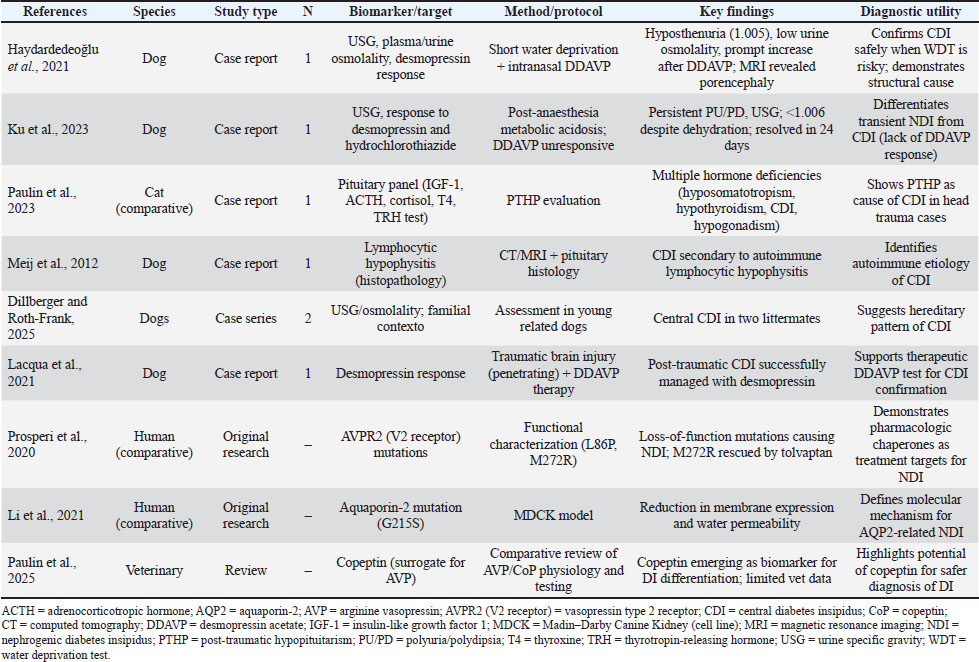
Fig. 1. Flowchart of the methodological process used in the narrative review on diagnostic methods for diabetes insipidus in dogs. The review included database search, initial screening, eligibility assessment, final inclusion, and qualitative synthesis steps, focusing on the analysis of the pathophysiology, diagnostic methods, and emerging biomarkers of the disease. Although this study did not follow a systematic review protocol, efforts were made to ensure methodological rigor through structured database searches, predefined inclusion criteria, and critical qualitative analysis of the selected studies. No formal quality scoring system was applied, as the aim was interpretative synthesis rather than quantitative comparison. Ethical approvalNot needed for this manuscript. ResultsThe diagnosis of DI in dogs requires a systematic, stepwise approach to exclude frequent causes of polyuria and polydipsia, such as chronic kidney disease and diabetes mellitus. Central to this investigation is the identification of hyposthenuria (urine specific gravity below 1.008). In primary or idiopathic forms of DI, this finding often represents the sole laboratory abnormality (Ghizzo et al., 2021; Silva and Zanutto, 2021). The absence of hyposthenuria does not rule out the disease, particularly in partial central DI, where urine density may remain within the isosthenuric range (1.008–1.015). Consequently, a systematic interpretation of routine laboratory tests is fundamental for clinical screening prior to confirmatory testing (Simões, 2021). As established in previous studies, clinical recognition of DI requires the careful correlation of hydration status, urinary concentration, and systemic balance (Schmidt et al., 2009). The WDT remains the traditional gold standard for assessing an animal’s ability to concentrate urine during induced dehydration. This protocol involves total water restriction under continuous monitoring of body weight, hydration status, and urinary concentration; however, it must be immediately interrupted if the animal loses >5% of its body weight (Simões, 2021; Flynn et al., 2025). Despite its accuracy, the risks, including severe dehydration, hypernatremia, and potential mortality, position the WDT as a test of last resort. It is strictly contraindicated for patients with dehydration or concomitant systemic disease (Croton et al., 2019; Haydardedeoğlu et al., 2021). Given these limitations, a therapeutic trial with desmopressin has become the most practical and safe alternative in clinical practice. Studies in dogs and cats demonstrate that conjunctival or intranasal administration of desmopressin for 5 to 7 days, with monitoring of urine specific gravity and water intake, allows for both diagnosis and treatment (Lacqua et al., 2021; Ku et al., 2023). A positive response, defined as a >50% reduction in water consumption and increased urine concentration, confirms central DI, while no response supports nephrogenic DI or psychogenic polydipsia (Croton et al., 2019; Ghizzo et al., 2021). In addition, desmopressin testing provides therapeutic benefit and diagnostic confirmation in patients with traumatic brain injury or post-hypophyseal dysfunction, where central DI is often transient and reversible (Croton et al., 2019; Paulin et al., 2023). In a recent case in cats, acute pituitary dysfunction secondary to hemorrhage and inflammation was associated with transient DI (Hardjo et al., 2025), reinforcing the need to investigate concomitant intracranial conditions before performing confirmatory tests for the disease. In recent years, the search for complementary and less invasive biomarkers has advanced considerably. Although human endocrinology has long explored CoP, a stable substitute peptide derived from the same precursor as vasopressin (AVP), veterinary studies have begun to investigate its potential applicability in routine clinical practice. Comparative reviews emphasize that plasma CoP strongly correlates with AVP release and offers a safer, single-sample alternative to dynamic testing, particularly in differentiating between central and nephrogenic DI (Flynn et al., 2025; Paulin et al., 2025). In parallel with the above, experimental research has elucidated genetic molecular targets such as AVPR2 and aquaporin-2 (AQP2), which regulate renal water permeability. Mutations in these genes lead to nephrogenic DI due to vasopressin resistance or incorrect folding of aquaporin, as demonstrated in cell models using Madin-Darby Canine Kidney (MDCK) lines (Prosperi et al., 2020; Li et al., 2021). These findings pave the way for future molecular and pharmacogenetic testing in veterinary endocrinology. In addition, the quantification of oxytocin and vasopressin in alternative fluids, such as saliva, has been proposed as a noninvasive diagnostic adjunct for specific cases (De Santis et al., 2025). Although still experimental, salivary hormone analysis represents a step toward the integration of biomarker-based clinical diagnosis in veterinary endocrinology. The combined assessment of CoP, AVP, and their receptors may soon increase the accuracy and safety of DI diagnosis in dogs, reducing dependence on dehydration protocols. In summary, the diagnosis of canine DI remains a multifactorial process of exclusion that begins with ruling out the most common causes of polyuria and polydipsia. Its complexity and potential risks justify the preference for therapeutic testing with desmopressin and, in the near future, approaches based on more specific biomarkers. As demonstrated by recent studies (Flynn et al., 2025; Paulin et al., 2025), the integration of CoP measurement, molecular receptor analysis, and noninvasive sampling is likely to redefine diagnostic accuracy, patient safety, and physician decision-making in central and nephrogenic forms of DI in future treatments. A summary of the main studies addressing diagnostic approaches to DI in dogs is presented in Table 1. Table 1. Biomarker and diagnostic studies of diabetes insipidus in dogs organized by author, study design, number of samples and main findings.
Overall, the findings highlight that traditional diagnostic methods, particularly the WDT, remain central but are increasingly complemented by practical approaches such as desmopressin testing and emerging biomarkers. However, variability in study design, sample size, and methodological rigor across the literature limits direct comparison between studies. DiscussionThe clinical interpretation of DI in dogs requires careful differentiation from other causes of polyuria and polydipsia, as these signs are nonspecific and common to several endocrinopathies and renal disorders. In the studies evaluated, hyposthenuria (urine specific gravity < 1.008) has been consistently reported as an early and sometimes unique finding in dogs with central DI (Ghizzo et al., 2021; Silva and Zanutto, 2021). However, its absence does not rule out the disease, particularly in partial or transient cases, in which urinary concentration may remain within the isosthenuric range (Simões, 2021). Therefore, it is essential to systematically exclude the most prevalent causes of polyuria and polydipsia, including chronic kidney disease, diabetes mellitus, hyperadrenocorticism, and liver dysfunction, before proceeding with specific endocrine tests (Flynn et al., 2025). The WDT has historically been considered the gold standard for diagnosing DI and distinguishing its forms. However, studies consistently describe it as a complex and potentially dangerous procedure that requires continuous medical monitoring due to the risk of hypernatremia, hypovolemia, and severe dehydration (Simões, 2021; Ku et al., 2023). Reports such as those by Croton et al. (2019) and Haydardedeoğlu et al. (2021) illustrate how WDT can exacerbate neurological or systemic complications, especially in patients with trauma or previous dehydration, reinforcing that this test should be reserved for strictly stable cases under controlled conditions. Given these limitations, therapeutic testing with desmopressin represents a clinically practical alternative in both clinical and home settings. The diagnostic reliability of the test is reinforced by several studies demonstrating that a reduction in water intake and an increase in urine specific gravity after desmopressin administration confirm central DI, while the absence of response suggests nephrogenic DI or psychogenic polydipsia (Ghizzo et al., 2021; Simões, 2021; Ku et al., 2023). In addition, cases of post-traumatic CDI (Croton et al., 2019) and pituitary dysfunction secondary to lymphocytic hypophysitis or cystic lesions (Meij et al., 2012; Evenhuis et al., 2021; Paulin et al., 2023) demonstrate that the desmopressin test also has therapeutic value, serving to confirm the diagnosis and restore osmotic balance. In recent years, the exploration of complementary biomarkers has opened up new perspectives for the diagnosis of DI with greater safety and accuracy. The peptide CoP, a stable byproduct of preprovasopressin, has gained prominence as a surrogate marker for arginine vasopressin (AVP) secretion, showing a strong correlation with vasopressin activity in human and experimental models (Paulin et al., 2025). Although veterinary studies remain scarce, preliminary data suggest that CoP quantification could allow differentiation between central and nephrogenic DI without subjecting the patient to dehydration protocols, representing a major diagnostic advance (Flynn et al., 2025). In addition, molecular studies have clarified the mechanisms underlying nephrogenic DI. Research on vasopressin V2 receptor (AVPR2) mutations and AQP2 dysfunctions has demonstrated that specific point mutations (M272R and G215S) can impair water channel passage and renal response to vasopressin, leading to hereditary or acquired forms of NDI. These molecular insights not only elucidate pathophysiological mechanisms but also highlight potential therapeutic targets, including pharmacological chaperones such as tolvaptan, capable of restoring receptor functionality in specific genotypes (Prosperi et al., 2020; Li et al., 2021). Recent investigations have also proposed noninvasive alternatives for measuring AVP and related peptides in saliva, expanding the potential for biomarker application in veterinary endocrinology. De Santis et al. (2025) demonstrated the feasibility of quantifying salivary vasopressin and oxytocin in dogs, which could eventually replace plasma-based tests, reducing stress and risk during sample collection. These advances converge toward the development of integrated diagnostic algorithms, combining clinical, hormonal, and molecular data for safer and faster differentiation of DI subtypes. Finally, accurate etiological determination remains critical for treatment and prognosis. Cases of CDI secondary to head trauma (Croton et al., 2019), pituitary inflammation (Meij et al., 2012), or cystic lesions (Paulin et al., 2023) highlight the need to investigate structural and inflammatory causes to avoid misdiagnosis and inappropriate therapies. Current evidence reinforces that the integration of clinical findings, therapeutic tests, and emerging biomarkers represents the most effective strategy for the diagnosis of canine DI (Gooch et al., 2025). In the near future, the incorporation of CoP assays and molecular receptor profiles into veterinary diagnostics could significantly reduce the risks and complexity associated with traditional tests, promoting both diagnostic accuracy and patient safety. An important limitation of the available literature is the predominance of small sample sizes, case reports, and experimental models, which restrict the generalizability of findings to routine clinical practice. In addition, the heterogeneity of study designs and diagnostic criteria limits direct comparison across studies. Potential publication bias should also be considered, as studies with positive findings are more likely to be reported. From a clinical perspective, the integration of traditional and emerging diagnostic tools appears to be the most rational approach. While the WDT remains a reference method, its risks justify prioritizing safer alternatives, such as desmopressin trials in most clinical scenarios. The incorporation of biomarkers such as CoP may, in the future, allow earlier and less invasive diagnosis. However, further validation in canine populations is required before routine clinical implementation. ConclusionThis review shows that the diagnosis of DI in dogs remains challenging due to its nonspecific nature and its potential commonality among other pathologies, as well as its clinical signs and the risks associated with traditional functional tests. Although the WDT remains the diagnostic gold standard, its clinical applicability is limited. The therapeutic desmopressin test currently represents the safest and most viable option for confirming central DI. Emerging evidence on biomarkers, particularly CoP, and advances in the molecular understanding of AVPR2- and AQP2-related genetic mechanisms indicate a shift toward more objective and less invasive diagnostic approaches. However, veterinary validation is still scarce. Future research should focus on the standardization of these biomarkers and the integration of molecular and biochemical data into clinical algorithms to improve diagnostic accuracy and patient safety. AcknowledgmentsThe Pontifical Catholic University of Goiás for its support in conducting this document. FundingThis study did not receive any funding. Authors’ contributionConceptualization, C. E. F. S. and I. M. O.; methodology, C. E. F. S. and I. M. O.; investigation and literature review, C. E. F. S. and I. M. O.; writing – original draft preparation, C. E. F. S.; writing – review and editing, I. M. O.; supervision, I. M. O. Both authors have read and agreed to the published version of the manuscript. Conflicts of interestThe authors declare that there are no conflicts of interest. Data availabilityAll data supporting the findings of this study are available within the manuscript. ReferencesCroton, C., Purcell, S., Schoep, A. and Haworth, M. 2019. Successful treatment of transient central diabetes insipidus following traumatic brain injury in a dog. Case Rep. Vet. Med. 1, 3563675. doi: 10.1155/2019/3563675 De Santis, M., Soncin, M., Bertazzo, V., Martelli, L., Fornasiero, D., Mutinelli, F. and Contalbrigo, L. 2025. A scoping review on salivary oxytocin and vasopressin measurement in the dog. Animals 15, 2421; doi:10.3390/ani15162421 Dillberger, J.E. and Roth-Frank, M. 2025. Central diabetes insipidus in two related juvenile deerhounds. J. Vet. Intern. Med. 39, e1007; doi:10.1002/vrc2.1007 Evenhuis, J., Epstein, S.E., Della-Maggiore, A. and Reagan, K.L. 2021. Congenital pituitary cyst resulting in adipsic central diabetes insipidus and secondary hypernatremia in a cat. J. Vet. Diagn. Invest. 33, 2055116921990294; doi:10.1177/2055116921990294 Flynn, K., Hatfield, J., Brown, K., Vietor, N. and Hoang, T. 2025. Central and nephrogenic diabetes insipidus: updates on diagnosis and management. Front. Endocrinol. 15, 1479764; doi:10.3389/fendo.2024.1479764 Ghizzo, J., Kuser, G. J. and Ghizzo, J. B. 2021. Diabetes insípido central em cão da raça Yorkshire terrie. Relato de caso. Pubvet, 16, 170; doi: 10.31533/pubvet.v16n03a1057.1-3 Gooch, K. L., Granger, K., Yoon, P., Olmo, C., Mui, M. L., Hoppers, S. E. and Hall, K. E. 2025. Acquired central diabetes insipidus secondary to severe, chronic otitis media and interna in an 8-year-old Spanish Mastiff dog. Vet. Rec. Case Rep. 13, e70155; doi: 10.1002/vrc2.70155 Hardjo, S., Giles, A., Seddon, K. and Thomson, C.E. 2025. Pituitary Apoplexy in a Cat With Presumed Acute Diabetes Insipidus. J. Vet. Emerg. Crit. Care. 35, 308–313; doi:10.1111/vec.70004 Haydardedeoğlu, A.E., Çolakoğlu, E. and Alihosseini, H. 2021. A case of porencephaly complicated by central diabetes insipidus in a dog. Med. Weter. 77, 157–159; doi:10.21521/mw.6489 Ku, D., Lee, D., Yun, T., Koo, Y., Chae, Y., Choi, D., Choi, M., Kang, B.T., Yang, M.P. and Kim, H. 2023. Transient distal renal tubular acidosis with nephrogenic diabetes insipidus after general anaesthesia in a dog. Vet. Med. Sci. 9, 1483–1487; doi:10.1002/vms3.1165 Lacqua, A.J., Rozanski, E.A., Fry, D.A., Gregorich, S.L. and Sato, A.F. 2021. Diagnostic findings and successful management of central diabetes insipidus following a self-inflicted penetrating brain injury in a dog. Braz. J. Vet. Med. 43, 1521; doi:10.29374/2527-2179.bjvm001521 Li, Q., Lu, B., Yang, J., Li, C., Li, Y., Chen, H., Li, N., Duan, L., Gu, F., Zhang, J. and Xia, W. 2021. Molecular characterization of an Aquaporin-2 mutation causing nephrogenic diabetes insipidus. Front. Endocrinol. 12, 665145; doi:10.3389/fendo.2021.665145 Meij, B.P., Voorhout, G., Gerritsen, R.J., Grinwis, G.C.M. and Ijzer, J. 2012. Lymphocytic panhypophysitis resulting in central diabetes insipidus in a dog. J. Comp. Pathol. 147, 503–507; doi:10.1016/j.jcpa.2012.04.006 Paulin, M.V., Gleasure, S. and Snead, E.C. 2023. Multiple pituitary hormone deficiencies in a kitten: hyposomatotropism, hypothyroidism, central diabetes insipidus and hypogonadism. Can. Vet. J. 64, 245–251. Paulin, M.V., Schermerhorn, T., Unniappan, S. and Snead, E.C.R. 2025. Arginine vasopressin and copeptin: comparative review and perspective in veterinary medicine. Front. Vet. Sci. 12, 1528008; doi:10.3389/fvets.2025.1528008 Prosperi, F., Suzumoto, Y., Marzuillo, P., Costanzo, V., Jelen, S., Iervolino, A., Guarino, S., La Manna, A., Miraglia Del Giudice, E., Perna, A.F., Zacchia, M., Cordat, E., Capasso, G. and Trepiccione, F. 2020. Characterization of five novel vasopressin V2 receptor mutants causing nephrogenic diabetes insipidus reveals a role of tolvaptan for M272R V2R mutation. Sci. Rep. 10, 22731; doi:10.1038/s41598-020-73089-x Ribeiro, R.R., Trentin, T.C., Camelo, P.M.L. and Oliveira, I.M. 2025. Central diabetes insipidus in a young dog – Case report. Arq. Bras. De. Med. Vet. E. Zoot. 77, e13310; doi:10.1590/1678-4162-13310 Schmidt, C., Emanuelli, M.P., Cargnelutti, J.F., Wolkmer, P., Salbego, F.Z. and Lopes, S.T.D.A. 2009. Diabete insípido central em um cão. Ciênc. Rural 39, 922–925; doi:10.1590/S0103-84782009000300046 Silva, D.D.D. and Zanutto, M.D.S. 2021. Diabetes insipidus central em dois cães. Med. Vet. (UFRPE). 15, 357–362; doi:10.26605/medvet-v15n4-3542 Simões, A. I. A. 2021. Abordagem clínica à diabetes insipidus em cães e gatos. Master’s Dissertation, Escola Universitária Vasco da Gama, Coimbra. | ||
| How to Cite this Article |
| Pubmed Style Silva CEFD, Oliveira IM. The diagnostic challenges of diabetes insipidus in dogs: A narrative review of the water deprivation test and complementary biomarkers. Open Vet. J.. 2026; 16(6): 3925-3931. doi:10.5455/OVJ.2026.v16.i6.61 Web Style Silva CEFD, Oliveira IM. The diagnostic challenges of diabetes insipidus in dogs: A narrative review of the water deprivation test and complementary biomarkers. https://www.openveterinaryjournal.com/?mno=295851 [Access: June 26, 2026]. doi:10.5455/OVJ.2026.v16.i6.61 AMA (American Medical Association) Style Silva CEFD, Oliveira IM. The diagnostic challenges of diabetes insipidus in dogs: A narrative review of the water deprivation test and complementary biomarkers. Open Vet. J.. 2026; 16(6): 3925-3931. doi:10.5455/OVJ.2026.v16.i6.61 Vancouver/ICMJE Style Silva CEFD, Oliveira IM. The diagnostic challenges of diabetes insipidus in dogs: A narrative review of the water deprivation test and complementary biomarkers. Open Vet. J.. (2026), [cited June 26, 2026]; 16(6): 3925-3931. doi:10.5455/OVJ.2026.v16.i6.61 Harvard Style Silva, C. E. F. D. & Oliveira, . I. M. (2026) The diagnostic challenges of diabetes insipidus in dogs: A narrative review of the water deprivation test and complementary biomarkers. Open Vet. J., 16 (6), 3925-3931. doi:10.5455/OVJ.2026.v16.i6.61 Turabian Style Silva, Carlos Eduardo Fontoura Da, and Iago Martins Oliveira. 2026. The diagnostic challenges of diabetes insipidus in dogs: A narrative review of the water deprivation test and complementary biomarkers. Open Veterinary Journal, 16 (6), 3925-3931. doi:10.5455/OVJ.2026.v16.i6.61 Chicago Style Silva, Carlos Eduardo Fontoura Da, and Iago Martins Oliveira. "The diagnostic challenges of diabetes insipidus in dogs: A narrative review of the water deprivation test and complementary biomarkers." Open Veterinary Journal 16 (2026), 3925-3931. doi:10.5455/OVJ.2026.v16.i6.61 MLA (The Modern Language Association) Style Silva, Carlos Eduardo Fontoura Da, and Iago Martins Oliveira. "The diagnostic challenges of diabetes insipidus in dogs: A narrative review of the water deprivation test and complementary biomarkers." Open Veterinary Journal 16.6 (2026), 3925-3931. Print. doi:10.5455/OVJ.2026.v16.i6.61 APA (American Psychological Association) Style Silva, C. E. F. D. & Oliveira, . I. M. (2026) The diagnostic challenges of diabetes insipidus in dogs: A narrative review of the water deprivation test and complementary biomarkers. Open Veterinary Journal, 16 (6), 3925-3931. doi:10.5455/OVJ.2026.v16.i6.61 |


