





| Research Article | ||
Open Vet. J.. 2026; 16(6): 3320-3333 Open Veterinary Journal, (2026), Vol. 16(6): 3320-3333 Research Article Remodeling of intercalated disc proteins in dogs with myxomatous mitral valve disease and dilated cardiomyopathy dogs with supraventricular arrhythmiasDmitrij Oleynikov*Veterinary Clinic “Belij Klyk”, Moscow, Russia *Corresponding Author: Dmitrij Oleynikov. Veterinary Clinic “Belij Klyk,” Moscow, Russia. Submitted: 12/12/2025 Revised: 24/04/2026 Accepted: 04/05/2026 Published: 05/06/2026 © 2025 Open Veterinary Journal
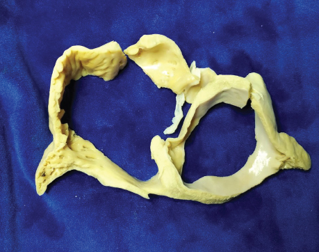
ABSTRACTBackground: Atrial remodeling is a complex process involving tissue, cellular mechanical, and electrical functions, and coherence. There is a lack of information about atrial cell remodeling in two most represented diseases in canines: myxomatous mitral valve disease (MMVD) and dilated cardiomyopathy (DCM). Moreover, there is little data about the expression of intercalated disk (ICD) proteins in diseases associated with atrial arrhythmias, such as atrial extrasystoles (focal atrial tachycardia) and atrial fibrillation (AFib). Aim: To estimate the remodeling of several proteins associated with ICD: connexin 40 (Cx40) and 43 (Cx43), desmin (Des), plakoglobin (PG), and N-cadherin (N-cad). Methods: Fourteen dog specimens were used for a histomorphological and immunohistochemical study of the atrium myocardium at six zones: right anterior (A) pulmonary vein (PV), right (RA) and left atrium at A and posterior portions, and interatrial septum (IAS). Results: DCMAFib (n=3) showed decreased expression of all proteins; DCMFAT (n=5) showed decreased PG in both atria and PV; N-cad—in atria and IAS; Cx43 and Cx40—in atria; and Des showed maladaptive accumulation in PV and left atrium. MMVDAFib (n=3) had decreased PG in the PV and atria; decreased N-cad—in PV, IAS, and atria; Cx43 and Cx40—decreased in PV and RA, with dispersion in other regions; and maladaptive Des accumulation in the PV and atria. MMVDFAT (n=2) showed decreased atria N-cad and adaptive Des accumulation. Conclusion: Differences between atrial remodeling in dogs with MMVD and DCM were described, presenting complicated interactions between ICD proteins within arrhythmia. The AFib paradigm can be reconsidered as a simple consequence of atrial dilatation due to DCM. In these cases, we found more profound markers of alterations in ICD proteins associated with some genetic diseases. In MMVD, we suspect that AFib is a logical consequence of structural atrial remodeling. In addition, we found signs of cardiomyocyte disruption and loss of cell-to-cell contacts. Keywords: Atrial fibrillation, Connexin, Dilated cardiomyopathy, Myxomatous mitral valve disease, Plakoglobin. IntroductionAtrial myocardial remodeling is an umbrella term describing the structural and functional changes that atrial tissue undergoes in response to various pathological factors. This remodeling may be helpful in the first steps to compensate for alterations caused by genetic or secondary factors. However, it leads to a decompensated state characterized by alterations in tissue architecture, cellular function, metabolism, and electrophysiology over time. Fibrosis is the most mentioned parameter of atrial myocardium remodeling because it is strongly associated with atrial electric and mechanical dysfunction. This condition typically leads to atrial arrhythmia and atrial fibrillation (AFib) as the most dangerous consequences (Nattel et al., 2020). Atrial remodeling occurs in both most commonly presented diseases in dogs: myxomatous mitral valve disease (MMVD) and dilated cardiomyopathy (DCM). MMVD is a disease of older, small-breed dogs with breed predisposition: cavalier king Charles spaniel, chihuahua, yorkshire terrier, dachshund, toy poodle, and toy terrier. The pathogenesis of MMVD involves mitral valve insufficiency and volume overload, left chamber dilatation, and retrograde heart failure, which manifests as pulmonary edema (Keene et al., 2019; Crosara et al., 2010; Guglielmini et al., 2020; Baron Toaldo et al., 2020; Santos et al., 2024; Spalla et al., 2025). DCM is a disease of big and giant breeds that can affect middle-aged dogs. It usually manifests as left heart systolic dysfunction and dilatation, often including the right heart, predisposing to pulmonary edema and ascites. Additionally, DCM is followed by ventricular and atrial arrhythmia, with AFib, sometimes as an early manifestation (Wess et al., 2010; Vollmar et al., 2019; Eberhard and Wess, 2020; Friederich et al., 2020; Wess, 2022). Both of these diseases are associated with significant atrial enlargement, but atrial arrhythmia is relatively rare in cases of MMVD, and AFib is extremely uncommon. However, DCM often coexists with ventricular and atrial rhythm alteration, including AFib (Wess et al., 2010; Wess, 2022). Atrial remodeling is associated with these diseases and is described in the context of fibrosis. Unfortunately, information on the remodeling of structural proteins that regulate electrical properties [such as connexin 40 (Cx40) and 43 (Cx43)], cell-to-cell adhesion [plakoglobin (PG), N-cadherin (N-cad)], and internal structure stabilization [desmin (Des)] is limited (Severs et al., 2008). These proteins represent different parts of the intercalated disk (ICD), which consists of three parts: gap junction, fascia adherence, and desmosome. In this study, we aimed to describe changes associated with MMVD and DCM in intercalated disc compartments: gap junction by Cx40&Cx43, desmosome by intermediate filament Des, and fascia adherence by PG/gamma catenin and N-cad. Materials and MethodsFourteen dogs were included in this study: eight with DCM (either with atrial premature complexes—the DCMFAT group,—or with focal atrial tachycardia and AFib—the DCMAFib group; n=5 and n=3, respectively), five with MMVD (either with atrial premature complexes or focal atrial tachycardia—the MMVDFAT group—and with AFib—the MMVDAFib group; n=3 and n=2, respectively), and one healthy dog. Based on echocardiographic and electrocardiographic findings, we compared animals with a DCM phenotype and arrhythmia with those with MMVD to delineate differences in myocardial remodeling. Instrumental diagnostics. All dogs included in the study underwent a complete clinical examination and standard blood analysis. Additionally, echocardiographic and electrocardiographic studies were performed by an experienced operator following a standard protocol. After euthanasia, gross pathological examination of the heart was performed. Tissue samples were collected following the macroscopic examination. The heart sections were fixed in 10% buffered formalin. The medial portions of both atria were excised as a single block, and the ostium of the right anterior (A) pulmonary vein (PV) was used to obtain an additional sample. The atrial block was subsequently divided into A and posterior (P) portions, including the interatrial septum (IAS) (Fig. 1).
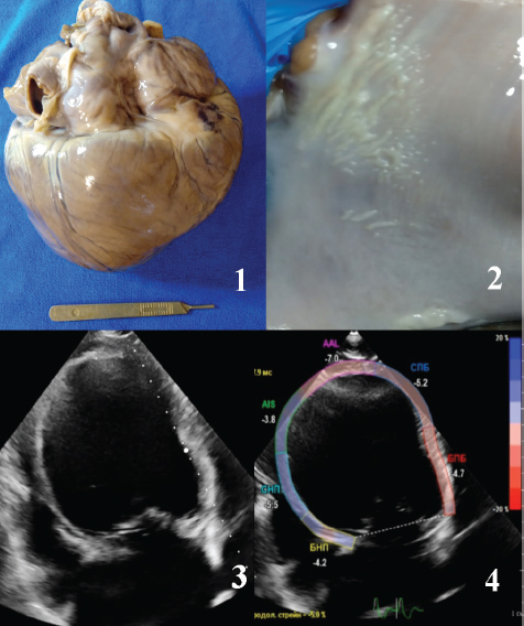
Fig. 1. Cross section of both atria. The study material from the A and P walls of both atria and IAS was taken. Subsequently, the tissue samples were sent to the pathomorphological laboratory for histological and immunohistochemical analyses. Samples were routinely processed, sectioned, and stained with hematoxylin and eosin and Masson’s trichrome (MT). Immunohistochemical labeling was performed on heart sections from all cats. In the laboratory adequate portions of heart samples were incubated with anti-Cx43 antibody (rabbit polyclonal antibody to Cx43/GJA1, Affinity Biosciences AF5339, Uniprot P19022, RRID:AB_2837824), anti- Cx40 antibody (rabbit polyclonal antibody to Cx40/GJA5, Affinity Biosciences DF13633, Uniprot P36382, RRID:AB_2846652), anti-N-cad antibody (rabbit polyclonal antibody to N-cad, RRID:AB_2837725, Uniprot P19022, Affinity Biosciences AF5239), anti-Gamma-Catenin/PG antibody (rabbit polyclonal antibody to Catenin gamma, RRID:AB_2833308, Uniprot P14923, Affinity Biosciences AF0124) and anti-Des antibody (rabbit polyclonal antibody to Des, Affinity Biosciences DF6138, Uniprot P17661, RRID:AB_2838105). The process was performed using Leica bond system and buffered saline for negative control. Des expression was estimated according to the classification for DCM human patients: I—normal type of dispersion, located predominantly near Z-discs; IIa—elevated expression and accumulation, but organized near Z-discs; IIb—elevated expression, but with aggregate formation across cytoplasm and near Z-discs; III—significantly decreased expression or absence of Des (Pawlak et al., 2019). PG/gamma catenin expression was assessed based on its localization in the cell’s longitudinal tips (typical intercalated discs location) or in the cytoplasm. Cx43 and Cx40 presentation was estimated as a typical location in the cell tips, lateralization, and subjective level of cytoplasmic expression. N-cad expression was analyzed according to its intensity and location in the intercalated disc zone at the cell tip. Ethical approvalApproved by the clinical ethics committee. ResultsThe dogs from the DCM group (n=8) had a typical DCM phenotype on echocardiography, including dilation of all four cardiac chambers, decreased systolic function, reduced left ventricle global strain, and signs of anterograde and retrograde heart failure (Fig. 2). Electrocardiographic study in the DCMFAT group (n=5) showed a high burden of premature atrial complexes and paroxysmal focal atrial tachycardia. The DCMAFib group (n=3) showed typical AFib with a tachycardic pattern.
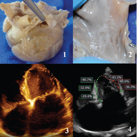
Fig. 2. Dog from the DCM group. 1 – Heart macromorphology, all chambers are dilated. 2 – Fibrous ridges on the left artium endocardium, due to regurgitation jet. apical 4-chamber view echocardiography, dilatation of all chambers. 4–global longitudinal strain of the left ventricle – 5%, ejection fraction 11.5%. All dogs in the DCMFAT group received the following standard medications for heart failure therapy: pimobendan, furosemide, spironolactone, and benazepril/ramipril. Antiarrhythmic therapy included sotalol/amiodarone for DCMFAT and digoxin combined with diltiazem for DCMAFib. One dog from the DCMAFib group was also medicated with amiodarone because of very aggressive AFib and the absence of typical therapy. The dogs from the MMVD group showed typical echocardiographic findings: mitral valve deformation with degenerative leaflet margins, signs of prolapse and chordae tendinea rupture, extreme left atrial dilatation, PV dilatation, left ventricle dilatation with normal systolic function, and signs of retrograde heart failure (American College of Veterinary Internal Medicine stage system grade C) (Fig. 3). Electrocardiographic study in the MMVDFAT group (n=3) showed a repeating pattern of premature atrial complexes (bi- or trigeminy) and rare cases of atrial couplets. One dog exhibited a 2-minute paroxysmal focal atrial tachycardia episode. The MMVDAFib group (n=2) showed a typical tachycardic pattern. One dog, monitored over a long period, showed a tendency for interatrial blockade before AFib started.
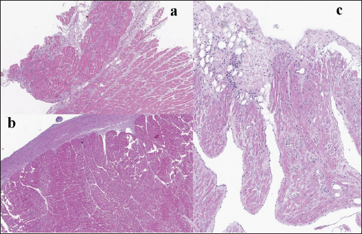
Fig. 3. Dogs from MMVD group. 1- marcomorphology of the heart, severe dilatation of the left atrium, wall is extremely thinned with transparent zones. 2 – endocardial scars and jet lesions in the left atrium. 3 – left apical 4-chamber view echocardiography, dilatation of left chambers. 4 – global longitudinal strain of the left ventricle – 35%, ejection fraction 58%. All dogs in the MMVDFAT group were on the following standard medications for heart failure therapy: pimobendan, furosemide, spironolactone, and benazepril/ramipril. Antiarrhythmic therapy included amiodarone for MMVDFAT and digoxin plus diltiazem for MMVDAFib. One dog experienced spontaneous conversion to the sinus rhythm, but returned to AFib with aggressive tachycardic form without any effects from the therapy after 2 days. Gross pathology. Dogs with DCM showed severe biventricular and biatrial dilatation. The endocardium of the ventricles was thickened and pale, and fibrous ridges in the left atrium (LA) could be visualized, possibly due to jet lesions (Fig. 2). Dogs in the MMVD group showed extremal LA dilatation with a thinned and opalescent external wall. Severe mitral and tricuspid valve deformation with a large coaptation defect was evident. The left atrial endocardium was thickened, stiff, and pale, with numerous jet lesions (Fig. 3). Atrial structural remodeling at the tissue level was relatively uniform in the DCM group. Significant interstitial fibrosis and cardiomyocyte dystrophy were observed in the left atrium, with signs of partial necrosis/apoptosis and intermediate endomyocardial fibrosis (Fig. 4a). In the right atrium (RA), we observed less intensity of interstitial fibrosis and more prominent endomyocardial fibrosis (Fig. 4b). In one DCMAFib case, we found significant inflammatory mononucleated cell infiltration (Fig. 4c).
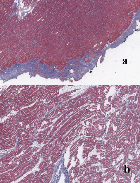
Fig. 4. DCM dogs histomorphology. a – left atrium with significant interstitial fibrosis, lipid cells proliferation, cardiomyocytes dystrophy, partial apoptosis/necrosis. H&E stain. b – RA significant endomyocardial fibrosis, with mild interstitial fibrosis. H&E stain. c – atrial tissue mononuclear inflammatory cells (lymphocytes) infiltration. H&E stain. In the MMVD group, we observe predominantly endocardial fibrosis and weak interstitial fibrosis in LA, but RA had less prominent endocardial fibrosis than in LA and mild interstitial fibrosis (Fig. 5).
Fig. 5. MMVD dogs histomorphology. a – left atrium significant endomyocardial proliferation and fibrosis, with moderate interstitial fibrosis. MT stain. b – RA moderate interstitial fibrosis, with fatty tissue proliferation. MT stain. Immunohistochemical studyWe analyzed protein expression at four anatomical points: PV, IAS, LA, and RA, with subdivision to A and P zones. Therefore, we describe our findings according to the anatomical zones. Pulmonary veinsThe PV of healthy subjects had a strong expression of Des with accumulation in the Z-disc zone and cytoplasmic presence. In the MMVDFAT group, Des was presented as type IIa—with high cytoplasmic presence and strong expression in Z-discs. In dogs with DCMFAT, we observed type IIb accumulation with predominant Des aggregates found in the cytoplasm but not clearly identified in Z-discs. Similar findings were observed in the MMVDAFib group, whereas the DCMAFib group showed type III Des expression. PG was typically found in Z-discs in healthy PV and was similarly located in the MMVDFAT group. In the MMVDAFib and DCMFAT groups, we found protein aggregates across the cytoplasm, with or without location in Z-discs. However, in the DCMAFib group, almost absolute absence of PG was observed in cardiomyocytes (Fig. 6).
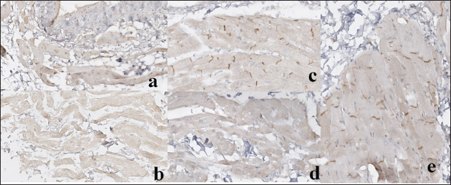
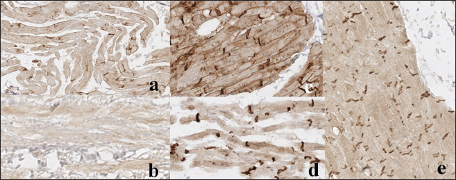
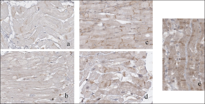
Fig. 6. Immunohistochemistry for PG in PV. a – DCMFAT dog, cytoplasmatic accumulation; b -DCMAFib dog, protein expression is absent; c – MMVDFAT dog, normal representation; d – MMVDAFib dog, cytoplasmatic accumulation; e – healthy dog. See description in the text. N-cad was located in cell-to-cell adhesion sites in normal atrial myocardium, as it was admitted in the MMVDFAT and DCMFAT groups. In the MMVDAFib group, non-uniform expression with an intermitted pattern was observed in some connection zones. The DCMAFib group showed almost complete protein absence in its typical location. Cx43 in a healthy dog was located in the cell adhesion zones, a pattern also observed in the MMVDFAT group. Certain structural variations were found in the DCMAT group, but the Cx43 location was typical. In the MMVDAFib group, we observed consolidated aggregates of Cx43, but still in the Z-disc zone; in the DCMAFib group—almost complete absence of protein expression in the adherence zone, but a weak reaction in the cytoplasm (Fig. 7).
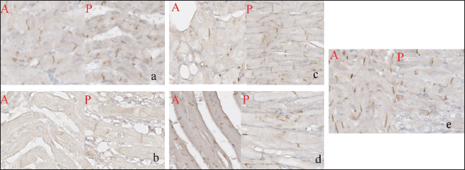
Fig. 7. Immunohistochemistry for Cx43 in PV. a – DCMFAT dog, expression decreased with cytoplasmatic accumulation; b – DCMAFib dog, polymorphic expression; c – MMVDFAT dog, normal representation; d – MMVDAFib dog, polymorphic expression; e – healthy dog. See description in the text. Cx40 was present as cytoplasmic near Z-disc aggregates in the normal dog. This form was also present in the MMVDFAT group. In contrast, Cx40 was present only as cytoplasmic aggregates in the DCMFAT group. The MMVDAFib and DCMAFib groups showed only weak cytoplasmic expression. Left atriumThe left atrium was subdivided into A and P portions. In the healthy subjects, we found normal and uniform Des expression. In the MMVDFAT group, we discovered higher Des expression (type IIa), equal in both portions. The MMVDAFib and DCMFAT groups exhibited cytoplasmic aggregates and high-intensity expression of type IIb, both in the A and P zones. The DCMAFIB group tended toward type III Des expression, with a more severe case in the P portion. PG was typically located in the Z-disc zone in the normal atrial myocardium without regional differences. A similar pattern was observed in the MMVDFAT group. Polymorphism of PG aggregates in the cytoplasm, especially in the P portion, was observed in the MMVDAFib group. In the DCMFAT group, only an intermittent signal was detected in the typical zone. The DCMAFib group showed a weak, intermittent signal in the P wall and an almost absent signal in the A portion (Fig. 8).
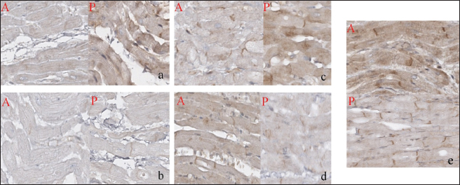
Fig. 8. Immunohistochemistry for PG in the left atrium (A/P). a – DCMFAT dog, cytoplasmatic accumulation; b -DCMAFib dog, protein expression is absent; c – MMVDFAT dog, normal representation; d – MMVDAFib dog, cytoplasmatic accumulation; e – healthy dog. See description in the text. N-cad in the healthy dog showed strong expression in the adhesion zones. In the MMVDFAT group, N-cad expression was less active, and cytoplasmic aggregates and alterations were present mostly in the A wall. The MMVDAFib group showed similar changes, but the severity was accentuated in the P portion. The DCMFAT group showed decreased protein expression, especially in the A portion. The DCMAFib group showed similar findings but more severe findings, with the greatest severity remaining in the A wall (Fig. 9).
Fig. 9. Immunohistochemistry for N-cad in the left atrium (A/P). a – DCMFAT dog, protein expression is absent; b -DCMAFib dog, protein expression is absent; c – MMVDFAT dog, expression decreased; d – MMVDAFib dog, expression decreased; e – healthy dog. See description in the text. Cx43 in the healthy dog had a typical location with a strong reaction in the Z-discs. The MMVDFAT group continued to show a strong reaction in the typical area, but with cytoplasmic aggregate formation, mostly in the P wall. The MMVDAFib group exhibited strong but polymorphic Cx43 expression, with cytoplasmic aggregates and lateralization. The DCMFAT group exhibited weaker expression and cytoplasmic aggregates with higher levels of P portion alterations. The same dispersion pattern was found in the DCMAFib group, but the alterations were much more prominent, especially lateralization and loss of Cx43 proteins in the P wall. Cx40 in the normal myocardium presented as accumulations in the cytoplasm and at Z-discs, with high expression levels. In the MMVDFAT group, significant cytoplasmic aggregates were found. The MMVDAFib group showed a similar tendency to aggregate formation and lateralization, but Cx40 expression was significantly decreased in the P wall. The DCMFAT group presented with decreased Cx40 expression and protein lateralization, with the P portion more affected. The DCMAFib group showed an almost total loss of Cx40 expression, with minimal residual lateralization. Right atriumDes expression was typically presented in the healthy subject, without differentiation between A and P zones. In the MMVDFAT and DCMFAT groups, the distribution was similarly type IIa, with more changes found in the A wall. In the MMVDAFib and DCMAFib groups, Des expression tended to be type III of expression in the A wall and preserved as type IIb in the P wall. PG expression in the healthy dog had a typical form associated with Z-discs. This pattern was unchanged in the MMVDFAT group, whereas the MMVDAF group showed decreased polymorphic expression, with an accent on the A wall. The DCMFAT group exhibited polymorphism and decreased expression. PG was almost absent in the P portion and entirely absent in the A wall in the DCMAFib group. N-cad was normally distributed in the healthy dog’s myocardium. In the MMVDFAT group, expression was decreased and had a polymorphic structure. In the MMVDAFib group, expression was severely depressed. In the DCMFAT group, myocardium N-cad was less present and had significant polymorphism in the P wall. The DCMAFib group exhibited no expression. Cx43 expression in normal cardiomyocytes was strongly present in typical zones. In the MMVDFAT group, the proteins were strongly expressed but had a significant lateralization pattern. The MMVDAFib group exhibited significantly decreased expression, polymorphism, and lateralization. The DCMFAT group presented with a normal distribution but with signs of lateralization. In the DCMAfib group, CX43 was severely underexpressed, polymorphic, and tended to be lateralized in the A portion (Fig. 10).
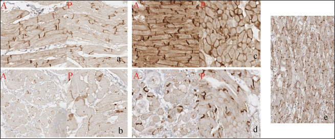
Fig. 10. Immunohistochemistry for Cx43 in the RA (A/P). a – DCMFAT dog, normal representation with lateralization; b -DCMAFib dog, expression decreased with lateralization; c – MMVDFAT dog, normal representation with lateralization; d – MMVDAFib dog, expression decreased with lateralization; e – healthy dog. See description in the text. In healthy RA, Cx40 had a typical presentation. In the MMVDFAT group, CX40 was expressed mostly as cytoplasmic aggregates and was decreased in the P wall. In the MMVDAFib group, the protein was significantly underexpressed with sporadic cytoplasmic aggregates. Only cytoplasmic aggregates were found in the DCMFAT group. CX40 was almost entirely absent in dogs from the DCMAFib group (Fig. 11).
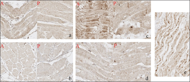
Fig. 11. Immunohistochemistry for Cx40 in the RA (A/P). a – DCMFAT dog, expression decreased; b -DCMAFib dog, protein expression is absent; c – MMVDFAT dog, normal representation; d – MMVDAFib dog, expression decreased; e – healthy dog. See description in the text. Interatrial septumDes was normally expressed in the healthy dog. A similar type IIa distribution was observed across all disease groups. PG was present in the Z-disk area in the healthy dog. A similar pattern was found in the MMVDFAT, DCMFAT, and MMVDAFib groups, but with different polymorphism levels. In the DCMAFib group, expression was severely decreased. N-Cad was normally located in the adhesion zones of healthy dogs. In the myocardium of the MMVDFAT group, there were no changes in distribution, whereas the MMVDAFib group showed only decreased expression and polymorphism. The dogs from the DCMFAT group showed severely decreased N-cad presentation, and it was almost absent in the DCMAFib group (Fig. 12).
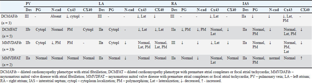
Fig. 12. Immunohistochemistry for N-cad in the IAS. a – DCMFAT dog, expression decreased; b -DCMAFib dog, expression decreased; c – MMVDFAT dog, normal representation; d – MMVDAFib dog, expression decreased; e – healthy dog. See description in the text. Cx43 was typically presented in a normal dog. The same pattern was observed in the MMVDFAT group. The MMVDAFib group exhibited polymorphism and lateralization. The DCMFAT group showed strong expression with significant polymorphism, lateralization, and aggregates. The DCMAFib group showed significantly decreased and intermittent expression, with zones of signal lateralization. IAS Cx40 expression had a normal distribution. The MMVDFAT group showed a high expression level with aggregates in the cytoplasm. In the MMVDAFib group, the signal was significantly decreased, but cytoplasmic and Z-disc aggregates were found. In the DCMFAT group, expression was significantly decreased with only protein aggregates. The DCMAFib myocardium showed almost no protein expression with sporadic cytoplasmic aggregates. A summary of the protein expression distribution is presented in Table 1.
Table 1. Summary of protein expression changes in PV, IAS, left and RA. DiscussionThe key finding of this study is that atrial cardiomyocytes in the DCM groups presented with more severe alterations in the observed ICD proteins. Despite the fact that some findings are similar in cases of DCMAFib and MMVDAFib, some changes in proteins are presented earlier in the disease course of DCM than those of MMVD. Connexins are proteins that form connexons in gap junctions. Because of their low resistance, these proteins play a crucial role in electric stimuli transduction. In the normal dog, we found typical Cx43 expression, similar to that of the working atrial myocardium, along with less intense but naturally occurring Cx40 expression (Verheule, 2002). In this study, we observed loss of Cx40 in PV in cases of MMVDAFib and DCMAFib, which could be a marker of the “common terminal path” in myocardium remodeling, predisposing to full uncoupling between muscle stripes in veins, to spontaneous depolarization, and to prolonged conduction (Chaldoupi et al., 2009). Simultaneously, Cx43 was significantly decreased only in the DCMAFib group, whereas it was mostly unchanged in the MMVDFAT group. Moderate decreases and polymorphic aggregates of Cx43 in the DCMFAT and MMVDAFib groups suggest that PV remodeling occurs earlier in DCM. This could be interpreted as DCM cardiomyocytes being more affected and prone to conductance decrease, potentially due to genetic factors. Data from the left atrium demonstrate a significant decrease in connexin levels in both the DCM groups. The literature contains conflicting reports regarding Cx43 expression in AFib. Some studies have shown that Cx43 may be increased in acute or paroxysmal AFib, whereas others have reported a significant decrease in chronic cases (Nattel et al., 2020; Yeh et al., 2006; Elvan et al., 1997; Maesen et al., 2022). Similar discrepancies were observed for Cx40 expression. However, a genetic study suggests that patients carrying a GAJ5 mutation accompanied by decreased Cx40 expression are more prone to sustained AFib (Wirka et al., 2011; Gollob et al., 2006). Our findings indicate that the left atrial myocardium is more susceptible to AFib initiation, even at the stage characterized only by premature atrial complexes, likely due to disrupted cell-to-cell electrical impulse conduction. In contrast, the MMVD groups, which generally preserve connexin expression, exhibited rare AFib episodes. In these dogs, the underlying mechanism of AFib likely involves factors other than connexin pathology in the left atrium. Another point that we should highlight is the changes in the RA. In the DCMAFib dogs, Cx43 was similarly depleted as in the left atrium. Moreover, similar findings were observed in the MMVDAFib group, which could explain the development of AFib. In some way, the RA myocardium seems more vulnerable. One possible explanation is an association with atrial distension. In typical MMVD, gradual left atrial enlargement occurs with time-dependent adaptation, whereas the RA generally remains unaffected, except in cases of chordal rupture with severe regurgitation or in the presence of pulmonary hypertension. Studies have shown that, at terminal stages, there may be a transition from postcapillary pulmonary hypertension to a combined or even precapillary pattern, and that pulmonary vasculature remodeling consistent with pulmonary hypertension can occur even at earlier stages (Sakarin et al., 2021; Woo and Kim, 2024; Sakarin et al., 2024). This hypothesis is supported by an experimental rat model of monocrotaline-induced pulmonary hypertension, in which decreased Cx43 expression and lateralization were demonstrated in right ventricular cardiomyocytes (Uzzaman et al., 2000). In our study, Cx43 lateralization and the presence of protein aggregates in the DCMFAT group may represent a marker of altered intracellular trafficking and intercalated disc disruption. Cx40 expression was reduced in both the DCM and MMVDAFib groups, indicating more profound alterations in DCM cardiomyocytes that are observed in MMVD dogs only at terminal stages. Interestingly, the IAS appeared less affected. Both Cx43 and Cx40 were severely depleted in cases of DCMAFib. However, Cx43 was mostly preserved in DCMFAT, MMVDFAT, and MMVDAFib. This finding could be explained by the higher stability of the IAS in cases of both diseases, as the highest wall tension and wall stress usually affect the outer walls and regurgitant jets, often vectored to the PV ostia or lateral wall. Nonetheless, undergoing remodeling was evidenced by decreased Cx40 expression in the DCMFAT and MMVDAFib groups, whereas this protein remained stable in MMVDFAT dogs. N-cad and PG play an important role in cell adhesion by anchoring myofibers at adhesion sites and transducing mechanical forces. Disruption of these proteins leads to cellular “sliding,” loss of coupling, and arrhythmia. PG is a member of the catenin family that functions as a transmembrane-associated protein that links N-cad to cytoskeletal proteins (Vite and Radice, 2014). PG itself may contribute to arrhythmogenesis, and its dysfunction has been associated with arrhythmogenic cardiomyopathy (Kim et al., 2013). Its depletion leads to reduced conduction, which has been linked to decreased Cx43 expression (Noorman et al., 2013; Jin et al., 2024). In our study, PG was absent in the PV of the DCMAFib group and significantly decreased in the DCMFAT and MMVDAFib groups. This pattern indicates structural remodeling with intercellular uncoupling that is present in dogs with DCM even at the early stages. A similar picture was observed in the RA and LA, with PG absent in the DCMAFib and markedly reduced in the DCMFAT and MMVDAFib. Although no data are available on the specific role of PG in AFib, in the context of our observation of a single case with myocardial inflammation and adipose tissue proliferation, we may suspect that dogs with DCM have a polygenic genetic cardiomyopathy associated with local inflammation or that PG alteration represents part of a “common terminal pathway” (Asimaki et al., 2011). IAS remained largely unaffected, except in the DCMAFib group. N-cad alterations are more complicated, as its loss leads to myofiber disarray, intercalated disc disruption, and connexon alterations. In some studies, AFib was associated with an increase in N-cad (Adam et al., 2010), but most studies showed decreased N-cad in patients with wild-type DCM or tachycardia. Additionally, one morphological study indicated that intermitted N-cad expression in the intercalated discs zones could be a valuable marker of the DCM (Akar et al., 2004; Ito et al., 2021). This assumption was further strengthened by a transgenic mousetransgenic mice study, where deleted N-cad expression in adult animals led to systolic dysfunction, arrhythmias, and sudden cardiac death (Kostetskii et al., 2005). In our study, PV showed N-cad absence or depletion in patients with DCMAFib and MMVDAFib, respectively, and disconnection on the fibrillatory stage. In the context of PG, we should assume that the parallel absence of catenin and N-cad shows more complex changes in the DCM group and possible genetic predisposition beyond typical factors. In DCMFAT, N-cad remained relatively unchanged, which could indicate that PV N-cad changes present as an isolated finding or serve as a marker of severe remodeling. This should be addressed in a more specific study. The absence of detectable N-cad in the left atrium in both the DCMAFib and DCMFAT groups is another possible marker of genetic predisposition in the DCM groups. In this context, N-cad appears to be a more relevant indicator of myocardial disruption than PG. A plausible explanation lies in the concept of chamber dilatation: in DCM, the LA is exposed to increased pressure earlier than the PV, which may accelerate the degradation of cardiomyocyte junctions, either in the presence of genetic factors or as part of the remodeling process. Conceptually, the remodeling hypothesis is supported by N-cad depletion in the MMVD groups. Within a genetic framework, the presence of relatively preserved PG–N-cad complexes argues against these alterations being solely a consequence of remodeling. The findings from the RA and IAS further support the concept of distinct remodeling pathways in patients with DCM and MMVD. Des has been intensively studied in patients with heart failure and animal models. A key publication relevant to this study demonstrated a connection between Des and AFib, describing the role of Des in Purkinje fibers and the association between high expression of Des in some arrhythmogenic zones in the atrial myocardium (Kugler et al., 2024). Similarly, in our study, we found high Des expression in PV of the healthy dog. In human medicine, there is a classification of Des cardiomyocytes expression, which was used as the DCM severity scale. Overall, it includes several grades: I—normal presence; IIa—increased expression, but with typical localization; IIb—increased expression but losing anatomical localization and connection; III—loss of expression (Pawlak et al., 2012; Pawlak et al., 2013; Pawlak et al., 2019). In our study, we found a loss of Des expression in the DCMAFib group, whereas the DDCMFAT and MMVDAFib groups showed type IIb accumulation. These findings suggest a cytoskeletal adaptation process to increased pressure in PV by increased Des production. This assumption is based on the type IIa accumulation in MMVDFAT, which is the stage prior to the MMVDAFib. The distribution observed in the left and right atrial walls showed type III expression in DCMAFib, which aligns with human and dog studies (Pawlak et al., 2013; Janus et al., 2016). In the DCMFAT group, RA was less affected than LA, with type IIa versus type IIb Des expression, respectively. This could be explained by the less aggressive pressure effect in the RA compared with that in the left atrium. Expression was more dispersed in the MMVD group. MMVDFAT showed type IIa in both atria, which is likely explained by the long-term period of adaptation to the increased atrial pressure. MMVDAFib showed type IIb in the left atrium and type III in the RA. We suspect that this rapid loss of Des in RA is associated with the maladaptive response of RA to high-speed contractions and rapid pressure rise, which limits effective remodeling. In all cases, IAS showed only adaptive type IIa Des accumulation. This study has several limitations. The retrospective design introduces substantial information gaps regarding the progression of heart failure, electrocardiography changes, and the time course of remodeling. Another limitation is the absence of data on troponin and other biomarkers. Given the observed foci of myocardial inflammation, underlying myocarditis, AFib-associated inflammation, or arrhythmogenic cardiomyopathy cannot be excluded. A further limitation is the lack of detailed population data, including age, body weight, and breed. These issues could be addressed in a prospective study with a stricter design. In conclusion, several differences in atrial remodeling were identified between dogs with MMVD and those with DCM, suggesting more complex interactions among intercalated disc proteins during heart failure. These data highlight the arrhythmogenic factors that may differ among the disease entities. Our findings may contribute to a shift in the paradigm of AFib as merely a consequence of DCM, as we identified PG and N-cad alterations that have not been described in typical wild-type canine DCM. Additionally, myocardial inflammation documented in one dog with AFib may help explain the reduced effectiveness of standard therapy. In MMVD, AFib appears to represent a logical consequence of structural atrial remodeling, although there is evidence of cardiomyocyte disruption and loss of cell-to-cell contacts during the progression of heart failure. AcknowledgmentsNone. Conflict of interestThe author declares that there is no conflict of interest. FundingNone. Data availabilityAll data were provided in the manuscript. ReferencesAdam, O., Lavall, D., Theobald, K., Hohl, M., Grube, M., Ameling, S., Sommer, M., Rosenkranz, S., Kroemer, H., Schäfers, H., Böhm, M. and Laufs, U. 2010. Rac1-induced connective tissue growth factor regulates connexin 43 and N-cadherin expression in atrial fibrillation. J. Am. Coll. Cardiol. 55(5), 469–480. Akar, F.G., Spragg, D.D., Tunin, R.S., Kass, D.A. and Tomaselli, G.F. 2004. Mechanisms underlying conduction slowing and arrhythmogenesis in nonischemic dilated cardiomyopathy. Circ. Res. 95(7), 717–725. Asimaki, A., Tandri, H., Duffy, E.R., Winterfield, J.R., Mackey-Bojack, S., Picken, M.M., Cooper, L.T., Wilber, D.J., Marcus, F.I., Basso, C., Thiene, G., Tsatsopoulou, A., Protonotarios, N., Stevenson, W.G., Mckenna, W.J., Gautam, S., Remick, D.G., Calkins, H. and Saffitz, J.E. 2011. Altered desmosomal proteins in granulomatous myocarditis and potential pathogenic links to arrhythmogenic right ventricular cardiomyopathy. Circ. Arrhythm. Electrophysiol. 4(5), 743–752. Baron Toaldo, M., Mazzoldi, C., Romito, G., Poser, H., Contiero, B., Cipone, M. and Guglielmini, C. 2020. Echocardiographic predictors of first onset of atrial fibrillation in dogs with myxomatous mitral valve disease. J. Vet. Intern. Med. 34(5), 1787–1793. Chaldoupi, S.M., Loh, P., Hauer, R.N.W., De Bakker, J.M.T. and Van Rijen, H.V.M. 2009. The role of connexin40 in atrial fibrillation. Cardiovasc. Res. 84(1), 15–23. Crosara, S., Borgarelli, M., Perego, M., Häggström, J., La Rosa, G., Tarducci, A. and Santilli, R. 2010. Holter monitoring in 36 dogs with myxomatous mitral valve disease. Aust. Vet. J. 88(10), 386–392. Eberhard, J. and Wess, G. 2020. The prevalence of atrial premature complexes in healthy Doberman Pinschers and their role in the diagnosis of occult dilated cardiomyopathy. Vet. J. 259-260, 259. Elvan, A., Huang, X.D., Pressler, M.L. and Zipes, D.P. 1997. Radiofrequency catheter ablation of the atria eliminates pacing-induced sustained atrial fibrillation and reduces connexin 43 in dogs. Circulation 96(5), 1675–1685. Friederich, J., Seuß, A. and Wess, G. 2020. The role of atrial fibrillation as a prognostic factor in doberman pinschers with dilated cardiomyopathy and congestive heart failure. Vet. J. 264, 105535. Gollob, M.H., Jones, D.L., Krahn, A.D., Danis, L., Gong, X.Q., Shao, Q., Liu, X., Veinot, J.P., Tang, A.S.L., Stewart, A.F.R., Tesson, F., Klein, G.J., Yee, R., Skanes, A.C., Guiraudon, G.M., Ebihara, L. and Bai, D. 2006. Somatic mutations in the connexin 40 gene (GJA5) in atrial fibrillation. N. Engl. J. Med. 354(25), 2677–2688. Guglielmini, C., Goncalves Sousa, M., Baron Toaldo, M., Valente, C., Bentivoglio, V., Mazzoldi, C., Bergamin, I., Drigo, M. and Poser, H. 2020. Prevalence and risk factors for atrial fibrillation in dogs with myxomatous mitral valve disease. J. Vet. Intern. Med. 34(6), 2223–2231. Ito, Y., Yoshida, M., Masuda, H., Maeda, D., Kudo-Asabe, Y., Umakoshi, M., Nanjo, H. and Goto, A. 2021. Disorganization of intercalated discs in dilated cardiomyopathy. Sci. Rep. 11(1), 11852. Janus, I., Kandefer-Gola, M., Ciaputa, R., Noszczyk-Nowak, A., Pasławska, U., Tursi, M. and Nowak, M. 2016. The immunohistochemical evaluation of selected markers in the left atrium of dogs with end-stage dilated cardiomyopathy and myxomatous mitral valve disease - a preliminary study. Ir. Vet. J. 69, 18. Jin, Q., Lee, K., Selimi, Z., Shimura, D., Wang, E., Zimmerman, J., Shaw, R., Kucera, J., Parker, K., Saffitz, J. and Kleber, A. 2024. Determinants of electrical propagation and propagation block in arrhythmogenic cardiomyopathy. J. Mol. Cell. Cardiol. 186, 71–80. Keene, B.W., Atkins, C.E., Bonagura, J.D., Fox, P.R., Häggström, J., Fuentes, V.L., Oyama, M.A., Rush, J.E., Stepien, R. and Uechi, M. 2019. ACVIM consensus guidelines for the diagnosis and treatment of myxomatous mitral valve disease in dogs. J. Vet. Intern. Med. 33(3), 1127–1140. Kim, C., Wong, J., Wen, J., Wang, S., Wang, C., Spiering, S., Kan, N.G., Forcales, S., Puri, P.L., Leone, T.C., Marine, J.E., Calkins, H., Kelly, D.P., Judge, D.P. and Chen, H.S.V. 2013. Studying arrhythmogenic right ventricular dysplasia with patient-specific iPSCs. Nature 494(7435), 105–110. Kostetskii, I., Li, J., Xiong, Y., Zhou, R., Ferrari, V.A., Patel, V.V., Molkentin, J.D. and Radice, G.L. 2005. Induced deletion of the N-cadherin gene in the heart leads to dissolution of the intercalated disc structure. Circ. Res. 96(3), 346–354. Kugler, S., Tőkés, A., Nagy, N., Fintha, A., Danics, K., Sághi, M., Törő, K., Rácz, G. and Nemeskéri. 2024. Strong desmin immunoreactivity in the myocardial sleeves around pulmonary veins, superior caval vein and coronary sinus supports the presumed arrhythmogenicity of these regions. J. Anat. 244(1), 120–132. Maesen, B., Verheule, S., Zeemering, S., La Meir, M., Nijs, J., Lumeij, S., Lau, D.H., Granier, M., Crijns, H.J., Maessen, J.G., Dhein, S. and Schotten, U. 2022. Endomysial fibrosis, rather than overall connective tissue content, is the main determinant of conduction disturbances in human atrial fibrillation. Europace 24(6), 1015–1024. Nattel, S., Heijman, J., Zhou, L. and Dobrev, D. 2020. Molecular basis of atrial fibrillation pathophysiology and therapy: a translational perspective. Circ. Res. 127(1), 51–72. Noorman, M., Hakim, S., Kessler, E., Groeneweg, J.A., Cox, M.G.P.J., Asimaki, A., Van Rijen, H.V.M., Van Stuijvenberg, L., Chkourko, H., Van Der Heyden, M.A.G., Vos, M.A., De Jonge, N., Van Der Smagt, J.J., Dooijes, D., Vink, A., De Weger, R.A., Varro, A., De Bakker, J.M.T., Saffitz, J.E., Hund, T.J., Mohler, P.J., Delmar, M., Hauer, R.N.W. and Van Veen, T.A.B. 2013. Remodeling of the cardiac sodium channel, connexin43, and plakoglobin at the intercalated disk in patients with arrhythmogenic cardiomyopathy. Heart Rhythm 10(3), 412–419. Pawlak, A., Gil, R., Grajkowska, W., Nasierowska-Guttmejer, A., Rzezak, J. and Kulawik, T. 2013. Significance of low desmin expression in cardiomyocytes in patients with idiopathic dilated cardiomyopathy. Am. J. Cardiol. 111(3), 393–399. Pawlak, A., Gil, R.J., Kulawik, T., Pronicki, M., Karkucińska-Więckowska, A., Szymańska-Dębińska, T., Gil, K., Lagwinski, N. and Czarnowska, E. 2012. Type of desmin expression in cardiomyocytes - a good marker of heart failure development in idiopathic dilated cardiomyopathy. J. Intern. Med. 272(3), 287–297. Pawlak, A., Rejmak-Kozicka, E., Gil, K.E., Ziemba, A., Kaczmarek, L. and Gil, R.J. 2019. Patterns of desmin expression in idiopathic dilated cardiomyopathy are related to the desmin mRNA and ubiquitin expression. J. Investig. Med. 67(1), 11–19. Sakarin, S., Rungsipipat, A. and Surachetpong, S.D. 2021. Histopathological changes of pulmonary vascular remodeling in dogs affected with pulmonary hypertension secondary to degenerative mitral valve disease. J. Vet. Cardiol. 36, 141–152. Sakarin, S., Rungsipipat, A., Roytrakul, S., Jaresitthikunchai, J., Phaonakrop, N., Charoenlappanit, S., Thaisakun, S. and Surachetpong, S.D. 2024. Proteomic analysis of pulmonary arteries and lung tissues from dogs affected with pulmonary hypertension secondary to degenerative mitral valve disease. PLos One 19(1), 296068. Santos, B.A., Corrêa, J.V., Latini, C.D., Tsunemi, M.H., Alfonso, A., Machado, L.H.D.A. and Lourenço, M.L.G. 2024. Study of the arrhythmogenic profile of dogs with myxomatous mitral valve disease in stages B1 and B2. Vet. Sci. 11(10), 467. Severs, N.J., Bruce, A.F., Dupont, E. and Rothery, S. 2008. Remodelling of gap junctions and connexin expression in diseased myocardium. Cardiovasc. Res. 80(1), 9–19. Spalla, I., Porteiro Vazquez, D.M., Partington, C., Bertuccini, T., Arnaboldi, C., Caccia, M., Caroli, V., Galizzi, A. and Toschi Corneliani, R. 2025. Transient atrial fibrillation in dogs with degenerative mitral valve disease: eight cases (2020-2024). J. Small. Anim. Pract. 66(8), 523–531. Uzzaman, M., Honjo, H., Takagishi, Y., Emdad, L., Magee, A.I., Severs, N.J. and Kodama, I. 2000. Remodeling of gap junctional coupling in hypertrophied right ventricles of rats with monocrotaline-induced pulmonary hypertension. Circ. Res. 86(8), 871–878. Verheule, S. 2002. Tissue structure and connexin expression of canine pulmonary veins. Cardiovasc. Res. 55(4), 727–738. Vite, A. and Radice, G. 2014. N-cadherin/catenin complex as a master regulator of intercalated disc function. Cell. Commun. Adhes. 21(3), 169–179. Vollmar, C., Vollmar, A., Keene, B., Fox, P.R., Reese, S. and Kohn, B. 2019. Irish wolfhounds with subclinical atrial fibrillation: progression of disease and causes of death. J. Vet. Cardiol. 24, 48–57. Wess, G. 2022. Screening for dilated cardiomyopathy in dogs. J. Vet. Cardiol. 40, 51–68. Wess, G., Schulze, A., Butz, V., Simak, J., Killich, M., Keller, L.J.M., Maeurer, J. and Hartmann, K. 2010. Prevalence of dilated cardiomyopathy in Doberman Pinschers in various age groups. J. Vet. Intern. Med. 24(3), 533–538. Wirka, R., Gore, S., Van Wagoner, D., Arking, D., Lubitz, S., Lunetta, K., Benjamin, E., Alonso, A., Ellinor, P., Barnard, J., Chung, M. and Smith, J. 2011. A common connexin-40 gene promoter variant affects connexx-40 expression in human atria and is associated with atrial fibrillation. Circ. Arrhythm. Electrophysiol. 4(1), 87–93. Woo, I.S. and Kim, J.H. 2024. Predictive value of left atrial volumes assessed using real-time three-dimensional echocardiography for pulmonary hypertension in dogs with myxomatous mitral valve disease. Front. Vet. Sci. 11, 1441839. Yeh, H.I., Lai, Y.J., Lee, S.H., Chen, S.T., Ko, Y.S., Chen, S.A., Severs, N.J. and Tsai, C.H. 2006. Remodeling of myocardial sleeve and gap junctions in canine superior vena cava after rapid pacing. Basic Res. Cardiol. 101(4), 269–280. | ||
| How to Cite this Article |
| Pubmed Style Dmitrij Oleynikov. Remodeling of intercalated disc proteins in dogs with myxomatous mitral valve disease and dilated cardiomyopathy dogs with supraventricular arrhythmias. Open Vet. J.. 2026; 16(6): 3320-3333. doi:10.5455/OVJ.2026.v16.i6.4 Web Style Dmitrij Oleynikov. Remodeling of intercalated disc proteins in dogs with myxomatous mitral valve disease and dilated cardiomyopathy dogs with supraventricular arrhythmias. https://www.openveterinaryjournal.com/?mno=303987 [Access: July 23, 2026]. doi:10.5455/OVJ.2026.v16.i6.4 AMA (American Medical Association) Style Dmitrij Oleynikov. Remodeling of intercalated disc proteins in dogs with myxomatous mitral valve disease and dilated cardiomyopathy dogs with supraventricular arrhythmias. Open Vet. J.. 2026; 16(6): 3320-3333. doi:10.5455/OVJ.2026.v16.i6.4 Vancouver/ICMJE Style Dmitrij Oleynikov. Remodeling of intercalated disc proteins in dogs with myxomatous mitral valve disease and dilated cardiomyopathy dogs with supraventricular arrhythmias. Open Vet. J.. (2026), [cited July 23, 2026]; 16(6): 3320-3333. doi:10.5455/OVJ.2026.v16.i6.4 Harvard Style Dmitrij Oleynikov (2026) Remodeling of intercalated disc proteins in dogs with myxomatous mitral valve disease and dilated cardiomyopathy dogs with supraventricular arrhythmias. Open Vet. J., 16 (6), 3320-3333. doi:10.5455/OVJ.2026.v16.i6.4 Turabian Style Dmitrij Oleynikov. 2026. Remodeling of intercalated disc proteins in dogs with myxomatous mitral valve disease and dilated cardiomyopathy dogs with supraventricular arrhythmias. Open Veterinary Journal, 16 (6), 3320-3333. doi:10.5455/OVJ.2026.v16.i6.4 Chicago Style Dmitrij Oleynikov. "Remodeling of intercalated disc proteins in dogs with myxomatous mitral valve disease and dilated cardiomyopathy dogs with supraventricular arrhythmias." Open Veterinary Journal 16 (2026), 3320-3333. doi:10.5455/OVJ.2026.v16.i6.4 MLA (The Modern Language Association) Style Dmitrij Oleynikov. "Remodeling of intercalated disc proteins in dogs with myxomatous mitral valve disease and dilated cardiomyopathy dogs with supraventricular arrhythmias." Open Veterinary Journal 16.6 (2026), 3320-3333. Print. doi:10.5455/OVJ.2026.v16.i6.4 APA (American Psychological Association) Style Dmitrij Oleynikov (2026) Remodeling of intercalated disc proteins in dogs with myxomatous mitral valve disease and dilated cardiomyopathy dogs with supraventricular arrhythmias. Open Veterinary Journal, 16 (6), 3320-3333. doi:10.5455/OVJ.2026.v16.i6.4 |


