





| Case Report | ||
Open Vet. J.. 2026; 16(6): 3584-3589 Open Veterinary Journal, (2026), Vol. 16(6): 3584-3589 Case Report Head-and-neck pruritus pattern of feline atopic skin syndrome: Successful resolution via protective bandaging in a catYoichiro Kasuga1* and Nobuo Murayama21Animal Medical Course, Department of Medical Sports, Faculty of Health Care and Medical Sports Department, Teikyo Heisei University, Ichihara-shi Chiba Prefecture, Japan 2Dermatology Services for Dogs and Cats, Tokyo, Japan *Corresponding Author: Yoichiro Kasuga. Animal Medical Course, Department of Medical Sports, Faculty of Health Care and Medical Sports Department, Teikyo Heisei University, Ichihara-shi Chiba Prefecture, Japan. Email: y.kasuga [at] thu.ac.jp Submitted: 02/01/2026 Revised: 24/04/2026 Accepted: 05/05/2026 Published: 05/06/2026 © 2025 Open Veterinary Journal
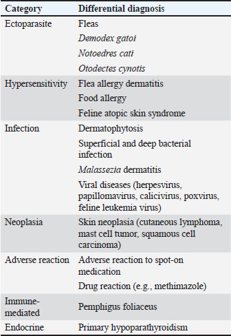
ABSTRACTBackground: Head-and-neck pruritus (HNP) is one of the four reaction patterns associated with feline atopic skin syndrome (FASS). In some cases, the response to glucocorticoid treatment is insufficient, and the optimal therapeutic strategies for refractory HNP remain unclear. Case Description: A 6-year-old spayed female domestic shorthair cat presented with severe pruritus and a characteristic cross-shaped ulcer on its dorsal cervical region. Based on the clinical presentation and routine dermatological examination, infectious and parasitic diseases were considered unlikely. Prednisolone was initiated at 2.73 mg/kg once daily for suspected HNP-type FASS. Despite treatment, the ulcer gradually enlarged. On day 36, a multilayer protective bandage was applied to prevent self-trauma and maintain a moist wound environment. The ulcer then began to contract and completely resolved approximately 35 days after bandaging. No recurrence was observed after bandage removal, and hair regrowth was noted at the ulcer site. Conclusion: These clinical findings suggest that protective bandaging contributed to lesion improvement. One possible explanation is that wound-induced pruritus may have sustained self-trauma, and physical protection may have interrupted this cycle and thereby facilitated healing. Furthermore, although FASS is fundamentally caused by hypersensitivity, the current exclusion-based diagnostic framework may encompass a variety of distinct conditions, including feline idiopathic ulcerative dermatitis and nonhypersensitivity conditions such as behavioral or neurological factors, which may partly explain why glucocorticoid treatment is insufficient in some cases. Although conclusions cannot be generalized from a single case, protective bandaging may be considered a potential adjunctive option in selected refractory HNP cases after other diseases have been reasonably ruled out. Keywords: Feline atopic skin syndrome, Head-and-neck pruritus, Protective bandage, Ulcerative dermatitis. IntroductionIn 2021, a subgroup of the International Committee on Allergic Diseases of Animals (ICADA) published detailed guidelines on the clinical signs and diagnosis of feline atopic syndrome (FAS; Santoro et al., 2021), referred to simply as “the diagnostic guideline,” in which the subgroup proposed replacing the term “non-flea, non-food hypersensitivity dermatitis”—previously used to describe cutaneous manifestations of feline hypersensitivity disorders—with the new designation “feline atopic skin syndrome (FASS)” as part of the broader concept of “FAS.” FASS is characterized by four major reaction patterns: self-induced alopecia, head-and-neck pruritus (HNP), eosinophilic granuloma complex, and miliary dermatitis (Miller, 2012; Jackson and Marsella, 2021; Santoro et al., 2021). According to a retrospective study of 263 cats with a diagnosis of FASS, self-induced alopecia was observed in 31.2%, HNP in 43.0%, eosinophilic granuloma complex in 25.9%, and miliary dermatitis in 60.1% of cases. A total of 37.7% of the cats exhibited at least two reaction patterns simultaneously (Santoro et al., 2021). According to the diagnostic guideline, the diagnosis of FASS is based on a consistent history, characteristic clinical signs, and the exclusion of other conditions with similar manifestations (Santoro et al., 2021; Miller et al., 2023). After infectious and ectoparasitic diseases are ruled out, flea allergy dermatitis must be distinguished from feline food allergy (Santoro et al., 2021). Treatment options for FASS include glucocorticoids, cyclosporine, essential fatty acid supplements, and antihistamines (Jackson and Marsella, 2021; Mueller et al., 2021; Wyatt and Buckley, 2024). HNP is a reaction pattern characterized by intense pruritus that results in excoriations and ulcerations of the face and cervical region. The underlying causes include ectoparasites, hypersensitivity disorders, infections, and neoplasia (Jackson and Marsella, 2021; Santoro et al., 2021). The differential diagnosis for HNP, as outlined in the diagnostic guideline, is summarized in Table 1. Although glucocorticoid treatment is one of the main therapeutic options for FASS, the response is often insufficient in clinical practice, and managing pruritus can thus be challenging (Jackson and Marsella, 2021; Santoro et al., 2021). Therefore, even when HNP is appropriately investigated and FASS is diagnosed, guidance on how to manage an inadequate glucocorticoid response remains limited, representing a clinical challenge. In particular, practical management strategies for cats with refractory HNP-pattern lesions have been insufficiently described, and the clinical role of protective bandaging in such cases remains unclear. The aim of the present report was to describe the clinical course and outcome of a cat with FASS presenting with severe HNP-pattern lesions that failed to respond adequately to glucocorticoid treatment and was successfully managed with protective bandaging. Table 1. Differential diagnosis for head-and-neck pruritus (HNP) in cats. Modified from the International Committee on Allergic Diseases of Animals (ICADA) diagnostic guideline (Santoro et al., 2021).
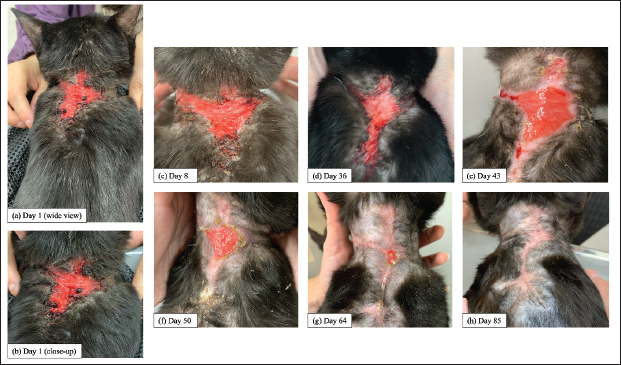
Case DetailsA 6-year-old spayed female domestic shorthair cat weighing 3.66 kg (body condition score: 5/9) was brought to Jiyugaoka Animal Hospital on September 4, 2021, for evaluation of pruritus affecting the dorsal cervical region. According to the patient’s medical history, pruritus had occurred in the same area for approximately 6 months and had been treated at another veterinary clinic with antimicrobial agents and gentamicin ointment; however, the condition gradually worsened despite treatment. The cat was strictly housed indoors, and the owner reported no changes in the living environment or triggering events, such as the application of flea control products to the dorsal cervical area. In addition, no dietary changes were reported during the study period. The initial examination revealed a cross-shaped ulcer extending from the dorsal cervical region to the interscapular area, with crusting along the ulcer margins (Fig. 1a,b). Dermatological evaluations, including impression smears, trichography, tape strip cytological study, and Wood’s lamp examination, revealed no significant abnormalities. Blood testing, skin biopsy, and a dietary elimination trial were recommended to rule out feline food allergy; however, these were not performed because the owner did not consent to these procedures. Therefore, diagnostic certainty was limited, and the diagnosis remained presumptive.
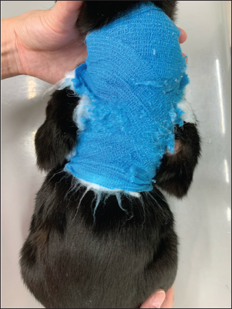
Fig. 1. Clinical photographs of the dorsal cervical lesion during the clinical course. Panel (a) shows a wider view of the lesion at presentation (day 1). Panel (b) shows a close-up view on day 1. Subsequent close-up views were obtained on days 8 (c), 36 (d), 43 (e), 50 (f), 64 (g), and 85 (h). The clinical presentation was consistent with HNP; therefore, the differential diagnosis for this reaction pattern was considered (Table 1). Based on the clinical presentation and routine dermatological examination, ectoparasitic and infectious diseases were deemed unlikely; therefore, FASS, feline food allergy, and cutaneous neoplasia were primarily considered. However, a definitive diagnosis of food allergy or cutaneous neoplasia would have required additional testing, which could not be performed; therefore, treatment was initiated based on a presumptive diagnosis of FASS. Prednisolone (Takeda Pharmaceutical Co., Ltd., Osaka, Japan) was administered at 2.73 mg/kg once daily for 3 days, followed by 2.05 mg/kg once daily for 4 days. On day 8, the owner reported a slight reduction in pruritus; however, the ulcer had enlarged, and prednisolone treatment at 2.73 mg/kg once daily was continued for an additional week (Fig. 1c). On day 15, the owner reported ongoing improvement in pruritus. Prednisolone was prescribed at 2.73 mg/kg once daily for another week, followed by 2.05 mg/kg once daily for the subsequent week. On day 36, the owner stated that the cat continued to receive prednisolone at 2.73 mg/kg once daily because pruritus recurred when the dose was reduced to 2.05 mg/kg once daily. However, the ulcer further enlarged, particularly in the caudal direction (Fig. 1d). The response to prednisolone was insufficient; therefore, physical protection was implemented to prevent self-trauma to the ulcerated area. The wound was first coated with a hydrogel (Intrasite Gel; Smith & Nephew Medical Ltd., Hull, UK) to maintain moisture and promote granulation. Then, it was covered with a nonadherent absorbent dressing (Melolin; Smith & Nephew Medical Ltd.). Next, a layer of padding material (Oltex®; Alcare Co., Ltd., Tokyo, Japan) was applied, and the entire area was secured with a self-adherent elastic bandage (Vetrap®; 3M, St. Paul, MN, USA) (Fig. 2). Prednisolone therapy at 2.73 mg/kg once daily was continued. Protective bandaging was initiated on day 36 and continued until day 71. The bandages were changed at each visit.
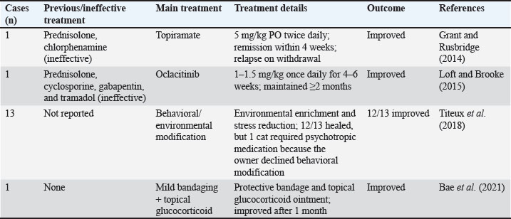
Fig. 2. The appearance of the cat on day 50. This photograph was taken 1 week after the reapplication of the protective bandage on day 43. Wear and tearing of the outer Vetrap® layer are evident in the scapular region, suggesting possible scratching with the hind limbs. On day 43, the ulcerative lesion showed marked improvement, with gradual epithelialization from the wound margins (Fig. 1e). Prednisolone treatment at 2.73 mg/kg once daily was continued. By day 50, the ulcer had further contracted (Fig. 1f). The appearance before rebandaging is shown in Fig. 2, revealing marks suggestive of scratching with the hind limbs through the Vetrap® layer. The prednisolone dosage was reduced to 2.05 mg/kg once daily. By day 64, the ulcer had shrunk to approximately 1 cm2 (Fig. 1g). Prednisolone treatment was maintained at 2.05 mg/kg once daily. By day 71, the ulcerative lesion had completely resolved. Although no findings suggestive of ectoparasitic infestation had been observed during the clinical course, fluralaner (Bravecto® Spot-on for Cats; MSD Animal Health, Summit, NJ, USA) was administered as a precautionary trial treatment. Prednisolone was tapered to 1.37 mg/kg once daily, and because the ulcer had resolved, the protective bandaging was discontinued, and the cat was monitored without bandaging. On day 85, although 2 weeks had passed since the previous visit, no recurrence of ulceration from self-trauma was observed on the dorsal cervical region; instead, hair had begun regrowing from the lesion margins (Fig. 1h). Prednisolone was further tapered to 1.37 mg/kg every other day. A follow-up examination was scheduled for 2 weeks later, but the owner did not keep the appointment. DiscussionIn this case, differential diagnoses were considered based on the clinical presentation, which was consistent with HNP. Based on the clinical presentation and routine dermatological examination, FASS, feline food allergy, and cutaneous neoplasia were considered possible diagnoses. Prednisolone therapy was initiated for suspected FASS, but the ulcer continued to enlarge until day 36, when it was physically covered to prevent self-trauma. The ulcer then began to contract and subsequently healed over the following 35 days. Prednisolone therapy was continued but at reduced dosages, and the lesion did not recur after removal of the protective bandage; moreover, hair regrowth was subsequently observed. Because no dietary changes were made during the entire treatment period and the ulcer resolved completely, feline food allergy and cutaneous neoplasia—initially included in the differential diagnosis—were considered unlikely causes. Based on these findings, a presumptive diagnosis of glucocorticoid-refractory FASS was made. Although FASS is fundamentally a hypersensitivity condition, the exclusion-based diagnostic framework may encompass pathological processes that are not driven by hypersensitivity; this may account for the therapeutic challenges encountered in clinical practice. One representative example is feline idiopathic ulcerative dermatitis (FIUD). Miller (2012) defined FIUD as a rare ulcerative skin disorder of unknown cause in cats, characterized by a solitary ulcer located on the dorsal neck or shoulder region that gradually enlarges over several weeks to months and fails to heal. Thus, FIUD is often diagnosed in cats with ulcerative lesions in the cervical or interscapular regions in clinical settings; however, the diagnostic guideline does not include FIUD in the differential diagnosis for HNP (Table 1). FIUD exhibits a heterogeneous treatment response. Some cases have improved with surgical excision or aggressive glucocorticoid therapy (Spaterna et al., 2003; Miller, 2012). Treatments other than glucocorticoids, as summarized in Table 2, include topiramate (an antiepileptic drug), oclacitinib (a JAK inhibitor), environmental modification and behavior therapy, and physical protection combined with topical corticosteroids (Grant and Rusbridge, 2014; Loft and Brooke, 2015; Titeux et al., 2018; Bae et al., 2021). This diversity in therapeutic response suggests that FIUD is not a single disease entity but a syndrome encompassing multiple underlying pathological conditions. The difference between the HNP reaction pattern of FASS involving the dorsal cervical region and FIUD is not always clear, and the two conditions may overlap. Table 2. Reported treatments for feline idiopathic ulcerative dermatitis (FIUD) other than systemic glucocorticoids.
Thus, the refractoriness of HNP may reflect the possibility that the current FASS diagnostic framework inadvertently encompasses not only hypersensitivity disorders but also nonhypersensitivity conditions, such as behavioral or neurological factors. In fact, reports of FIUD cases improving with environmental modification or behavioral therapy (Titeux et al., 2018), as well as cases responding to antiepileptic medication (Grant and Rusbridge, 2014), suggest that the self-trauma observed in some cats may represent scratching-like behaviors driven by abnormal repetitive behavior or neurologic dysfunction rather than true pruritus. This concept parallels conditions in dogs in which behaviors resembling reactions to pruritus—but not caused by itching—result in skin lesions, such as acral lick dermatitis (a compulsive disorder) and phantom scratching associated with syringomyelia. Taken together, these observations suggest that cases currently diagnosed with FASS are pathophysiologically heterogeneous and that the current exclusion-based diagnostic framework may include nonhypersensitivity conditions, which could partly explain poor responsiveness to glucocorticoids in some cases. Therefore, methods must be devised to distinguish and classify nonhypersensitivity conditions encompassed within FASS. In our patient, marked improvement was observed after the bandage physically protected the ulcer. To explore the possible reasons for this response, we reviewed previous reports describing the use of physical protection in FIUD. These reports suggested that covering the lesion with bandages or fabric materials minimized continuous stimulation and self-trauma, thereby allowing the wound to heal (Bae et al., 2021). Accordingly, we hypothesized that physical protection may be effective because the wound itself may contribute to ongoing pruritic stimulation. Even if the primary disease, such as FASS, had been adequately controlled, persistent wound-induced pruritus could have continued as long as the ulcer remained unhealed. Therefore, although this explanation remains speculative, we consider that physically protecting the ulcer from further self-trauma allowed it to heal, which in turn may have reduced pruritus. Pruritus associated with wounds—particularly chronic wounds—has been well documented in humans. Several studies of patients with chronic wounds have revealed that the prevalence of wound-related pruritus is approximately 28% in cohorts with multiple wound types and up to 68% in populations with chronic leg ulcers (Paul, 2013; Jockenhofer et al., 2019; Papanikolaou et al., 2025). The proposed mechanisms include the release of pruritogenic chemical mediators (e.g., cytokines, growth factors, and histamine) during the wound-healing process. In addition, necrotic tissue, exudate, periwound sclerosis, and edema may impose mechanical stress on the skin and stimulate peripheral nerve endings, thereby inducing itch (Iannone et al., 2019; Papanikolaou et al., 2025). Experimental evidence from a mouse skin-incision model further indicates that interleukin-31 is expressed at the wound site and contributes to itch-related behavior (Xu et al., 2020). Managing wound-associated pruritus in humans is often reported to be challenging (Iannone et al., 2019; Papanikolaou et al., 2025). Studies similar to that of Xu et al. (2020) have not been conducted in cats, and direct experimental evidence demonstrating wound-induced pruritus in this species is lacking. Therefore, this hypothesis remains speculative. Moreover, although the clinical improvement observed after excision of affected tissue (Miller, 2012) may be explained by a reduction in wound-induced pruritus, this interpretation also remains hypothetical at present. The underlying cause of the initial ulcer could not be identified in our patient. However, physical protection of the ulcerated area may have reduced wound-induced pruritus, which significantly contributed to the lesion’s overall resolution. In the HNP pattern of FASS, the reported rate of response to glucocorticoids has been relatively poor (Santoro et al., 2021), and some FIUD cases are similarly unresponsive to glucocorticoid therapy (Grant and Rusbridge, 2014; Loft and Brooke, 2015). At present, no clear guidelines on the management of such refractory cases exist, and such cases are thus considerably challenging for clinicians. When other diseases have been reasonably ruled out, physical protection of the ulcer to allow it to heal, as demonstrated in our patient, may represent a low-risk and low-cost intervention and can be considered a potential therapeutic option. However, tolerability should be considered because bandaging may induce stress and potentially reduce the quality of life in some cats. This study has several limitations. No validated objective scoring system for pruritus or lesion severity was recorded in this case. Nevertheless, serial clinical photographs documented the temporal changes in lesion extent and provided a certain degree of objectivity in the clinical assessment. Given that blood testing, skin biopsy, and a dietary elimination trial were not performed, diagnostic certainty was limited, and the diagnosis of the present case remained presumptive. In addition, this report is limited to the description of a single case; therefore, no definitive conclusions regarding the overall efficacy of physical protection for glucocorticoid-refractory HNP patterns of FASS can be drawn. ConclusionIn a cat with HNP associated with FASS, the condition failed to respond to glucocorticoids but improved after physical protection of the lesion. Glucocorticoid-refractory HNP is a challenging condition for clinicians, and this case suggests that, once other diseases have been reasonably ruled out, wound management through physical protection of the lesion may be considered a potential therapeutic option. We further hypothesize that the mechanisms underlying this improvement involve the prevention of self-trauma and control of wound-induced pruritus generated by the lesion itself. In the future, researchers should investigate other cases of HNP in which glucocorticoids are ineffective despite a diagnosis of FASS, in terms of prioritization of therapeutic options, including physical protection and better characterization of the heterogeneous pathological conditions encompassed within FASS. Ultimately, establishing more precise methods to identify and differentiate the causes of pruritus in cats will lead to more appropriate and targeted treatment strategies. AcknowledgmentsThe authors sincerely thank the staff of Jiyugaoka Animal Hospital for their valuable assistance in the management of this case, which was encountered while the first author was employed at the hospital. Conflict of interestThe authors declare no conflict of interest. FundingThis study received no specific grant. Authors’ contributionsYK and NM wrote the manuscript. YK performed clinical evaluation and clinical care. Both authors contributed to and approved the final version of the manuscript. Data availabilityAll data pertaining to the study findings are available within the manuscript. ReferencesBae, H., Kim, J., Chun, D., Jung, D.I., Park, J., Young Kim, D. and Yu, D. 2021. Idiopathic ulcerative dermatitis in a cat with feline infectious peritonitis. Vet. Med. Sci. 7, 572–576. Grant, D. and Rusbridge, C. 2014. Topiramate in the management of feline idiopathic ulcerative dermatitis in a two-year-old cat. Vet. Dermatol. 25(3):226–e60. Iannone, M., Janowska, A., Dini, V., Tonini, G., Oranges, T. and Romanelli, M. 2019. Itch in chronic wounds: pathophysiology, impact, and management. Medicines 6, 130. Jackson, H. and Marsella, R. 2021. BSAVA Manual of Canine and Feline Dermatology. Gloucester, UK: BSAVA. Jockenhöfer, F., Zaremba, A., Wehrmann, C., Benson, S., Ständer, S. and Dissemond, J. 2019. Pruritus in patients with chronic leg ulcers: a frequent and often neglected problem. Int. Wound. J. 16, 1464–1470. Loft, K.E.S. and Brooke, S. 2015. Feline idiopathic ulcerative dermatitis treated successfully with oclacitinib. Vet. Dermatol. 26, 133–159. Miller, J., Simpson, A., Bloom, P., Diesel, A., Friedeck, A., Paterson, T., Wisecup, M. and Yu, C.M. 2023. 2023 AAHA Management of allergic skin diseases in dogs and cats guidelines. J. Am. Anim. Hosp. Assoc. 59, 255–284. Miller, W.H., Griffin, C.E. and Campbell, K.L. 2012. Muller & Kirk’s small animal dermatology 7th ed. Philadelphia, PA: W. B. Saunders. Mueller, R.S., Nuttall, T., Prost, C., Schulz, B. and Bizikova, P. 2021. Treatment of the feline atopic syndrome - a systematic review. Vet. Dermatol. 32(1), 43–e8. Papanikolaou, M., Paul, J., Nattkemper, L.A., Kirsner, R.S. and Yosipovitch, G. 2025. Prevalence and mechanisms of itch in chronic wounds: a narrative review. J. Clin. Med. 14, 2241. Paul, J. 2013. A cross-sectional study of chronic wound-related pain and itching. Ostomy Wound Manage. 59, 28–34. Santoro, D., Pucheu-Haston, C.M., Prost, C., Mueller, R.S. and Jackson, H. 2021. Clinical signs and diagnosis of feline atopic syndrome: detailed guidelines for a correct diagnosis. Vet. Dermatol. 32(1), 26–e6. Spaterna, A., Mechelli, L., Rueca, F., Cerquetella, M., Brachelente, C., Antognoni, M.T. and Tesei, B. 2003. Feline idiopathic ulcerative dermatosis: three cases. Vet. Res. Commun. 27, 795–798. Titeux, E., Gilbert, C., Briand, A. and Cochet-Faivre, N. 2018. From feline idiopathic ulcerative dermatitis to feline behavioral ulcerative dermatitis: grooming repetitive behaviors as indicators of poor welfare in cats. Front. Vet. Sci. 5, 81. Wyatt, E. and Buckley, L. 2024. Diagnosis and treatment of atopic skin syndrome in cats. Pract 46, 164–179. Xu, J., Zanvit, P., Hu, L., Tseng, P.Y., Liu, N., Wang, F., Liu, O., Zhang, D., Jin, W., Guo, N., Han, Y., Yin, J., Cain, A., Hoon, M.A., Wang, S. and Chen, W. 2020. The cytokine TGF-β induces interleukin-31 expression from dermal dendritic cells to activate sensory neurons and stimulate wound itching. Immunity 53, 371–383. | ||
| How to Cite this Article |
| Pubmed Style Kasuga Y, Murayama N. Head-and-neck pruritus pattern of feline atopic skin syndrome: Successful resolution via protective bandaging in a cat. Open Vet. J.. 2026; 16(6): 3584-3589. doi:10.5455/OVJ.2026.v16.i6.29 Web Style Kasuga Y, Murayama N. Head-and-neck pruritus pattern of feline atopic skin syndrome: Successful resolution via protective bandaging in a cat. https://www.openveterinaryjournal.com/?mno=305257 [Access: June 26, 2026]. doi:10.5455/OVJ.2026.v16.i6.29 AMA (American Medical Association) Style Kasuga Y, Murayama N. Head-and-neck pruritus pattern of feline atopic skin syndrome: Successful resolution via protective bandaging in a cat. Open Vet. J.. 2026; 16(6): 3584-3589. doi:10.5455/OVJ.2026.v16.i6.29 Vancouver/ICMJE Style Kasuga Y, Murayama N. Head-and-neck pruritus pattern of feline atopic skin syndrome: Successful resolution via protective bandaging in a cat. Open Vet. J.. (2026), [cited June 26, 2026]; 16(6): 3584-3589. doi:10.5455/OVJ.2026.v16.i6.29 Harvard Style Kasuga, Y. & Murayama, . N. (2026) Head-and-neck pruritus pattern of feline atopic skin syndrome: Successful resolution via protective bandaging in a cat. Open Vet. J., 16 (6), 3584-3589. doi:10.5455/OVJ.2026.v16.i6.29 Turabian Style Kasuga, Yoichiro, and Nobuo Murayama. 2026. Head-and-neck pruritus pattern of feline atopic skin syndrome: Successful resolution via protective bandaging in a cat. Open Veterinary Journal, 16 (6), 3584-3589. doi:10.5455/OVJ.2026.v16.i6.29 Chicago Style Kasuga, Yoichiro, and Nobuo Murayama. "Head-and-neck pruritus pattern of feline atopic skin syndrome: Successful resolution via protective bandaging in a cat." Open Veterinary Journal 16 (2026), 3584-3589. doi:10.5455/OVJ.2026.v16.i6.29 MLA (The Modern Language Association) Style Kasuga, Yoichiro, and Nobuo Murayama. "Head-and-neck pruritus pattern of feline atopic skin syndrome: Successful resolution via protective bandaging in a cat." Open Veterinary Journal 16.6 (2026), 3584-3589. Print. doi:10.5455/OVJ.2026.v16.i6.29 APA (American Psychological Association) Style Kasuga, Y. & Murayama, . N. (2026) Head-and-neck pruritus pattern of feline atopic skin syndrome: Successful resolution via protective bandaging in a cat. Open Veterinary Journal, 16 (6), 3584-3589. doi:10.5455/OVJ.2026.v16.i6.29 |


