





| Research Article | ||
Open Vet. J.. 2026; 16(6): 4022-4047 Open Veterinary Journal, (2026), Vol. 16(6): 4022-4047 Research Article Protective and restorative role of Myristica fragrans essential oil on testicular and endocrine dysfunction induced by bisphenol AMohamed A. Zarka1, Sajad A. Algazali2, Ali Salah Waday3, and Ghadeer Sabah Bustani3*1Department of Pharmacognosy and Phytochemistry, College of Pharmacy, The Islamic University, Najaf, Iraq 2Department of Anesthesia Techniques, College of Health and Medical Techniques, Al-Mustaqbal University, Babylon, Iraq 3Department of Anesthesia Techniques, College of Medical Techniques, The Islamic University, Najaf, Iraq *Corresponding Author: Ghadeer Sabah Bustani. Department of Anesthesia Techniques, College of Medical Techniques, The Islamic University, Najaf, Iraq. Email: bustani [at] iunajaf.edu.iq Submitted: 11/01/2026 Revised: 20/05/2026 Accepted: 02/06/2026 Published: 30/06/2026 © 2025 Open Veterinary Journal
AbstractBackground: Bisphenol A (BPA) is a widely used endocrine-disrupting chemical that impairs male reproductive health through oxidative stress, hormonal imbalance, and testicular damage. Myristica fragrans (MF), rich in bioactive monoterpenes and phenylpropanoids, has demonstrated antioxidant and hormone-modulating properties, suggesting potential for reproductive protection. Aim: To evaluate the protective and restorative effects of M. fragrans essential oil against BPA-induced reproductive and endocrine toxicity in male rats and to correlate these effects with the oil’s chemical composition as identified by gas chromatography–mass spectrometry (GC–MS) analysis. Methods: Forty adult male rats were divided into four groups (n = 10): control (distilled water), MF-only (200 mg/kg/day), BPA-only (50 mg/kg/day), and BPA + MF (co-treatment). All substances were orally administered for 60 consecutive days. Assessments included sperm motility, viability, morphology, and concentration; chromatin and deoxyribonucleic acid (DNA) integrity (via toluidine blue, aniline blue, and acridine orange stains); serum testosterone and luteinizing hormone (LH) levels; testis weight; and oxidative stress biomarkers total antioxidant capacity (TAC) and malondialdehyde (MDA). The essential oil composition was analyzed via GC–MS. Results: BPA exposure significantly impaired sperm quality, disrupted chromatin condensation and DNA integrity, altered hormone levels (↓testosterone, ↑LH), reduced testicular weight, and elevated oxidative stress markers (↑MDA, ↓TAC) (p < 0.001). Co-treatment with MF significantly restored all evaluated parameters to near-normal levels (p < 0.001 vs. BPA). GC–MS analysis identified sabinene (33.55%), α-pinene (23.08%), β-pinene (14.88%), and myristicin (0.84%) as the major constituents. The MF-only group did not significantly differ from controls in the measured endpoints; however, this finding should not be interpreted as evidence of comprehensive safety. Conclusion: MFE improved the measured sperm quality parameters, endocrine profile, oxidative stress biomarkers, and staining-based cytochemical indicators of sperm nuclear quality in rats exposed to high-dose BPA. However, mating performance, pregnancy rate, litter size, and other direct fertility outcomes were not evaluated; therefore, these findings should not be interpreted as evidence of improved fertility. The observed improvements in sperm quality, chromatin integrity, hormonal profile, and oxidative stress markers suggest that the antioxidant properties of the oil may be associated with its protective effects. However, the precise molecular mechanisms underlying these effects require further investigation. Keywords: Bisphenol A, chromatin integrity, endocrine disruption, Myristica fragrans, oxidative stress. IntroductionMyristica fragrans, commonly known as nutmeg, is a tropical plant traditionally used for culinary and medicinal purposes, with growing scientific interest in its bioactive properties, especially in reproductive health (Sultan et al., 2023). The essential oil of M. fragrans contains several bioactive constituents, including monoterpenes and phenylpropanoids, with compositional variation depending on geographical origin, extraction method, and analytical conditions. Gas chromatography–mass spectrometry (GC–MS) analysis identified sabinene, α-pinene, β-pinene, γ-terpinene, terpinen-4-ol, safrole, and myristicin as representative constituents of the tested oil. Therefore, the interpretation of the biological findings in this manuscript primarily focused on the compounds detected in the current GC–MS profile (Ha et al., 2020). Myristicin is a phenylpropene compound known for its antioxidant, anti-inflammatory, and neuromodulatory properties (Al-Mehana et al., 2021; Seneme et al., 2021). Certain compounds previously reported in M. fragrans, such as eugenol and elemicin, have been described in the literature as having antioxidant properties, antimicrobial, and androgenic activities, potentially contributing to testicular protection and enhancement of spermatogenesis (Zarka and Al Gendy, 2021; Saleh et al., 2024). Safrole and elemicin have also been shown to play promising roles in modulating oxidative stress and supporting endocrine function (Al-Mehana et al., 2012; Götz et al., 2022). Numerous experimental studies have demonstrated that several plant-derived antioxidants, including curcumin, resveratrol, quercetin, Moringa oleifera, and green tea polyphenols, have antioxidant and cytoprotective properties that can attenuate bisphenol A (BPA)-induced testicular toxicity in rodent models [7]. Despite this growing body of evidence, limited information is available regarding the protective effects of M. fragrans essential oil against BPA-induced reproductive damage. Few studies have examined sperm chromatin integrity and deoxyribonucleic acid (DNA) stability as endpoints in this context (Jarwan and Abed, 2024; Dolatabadi et al., 2025). Bisphenol A is commonly found in food can linings, plastic containers, thermal paper receipts, and some dental materials (Manzoor et al., 2022; Al-Furaiji et al., 2025). Exposure occurs mainly through ingestion and skin contact, particularly when plastics are heated or degraded (Kannan and Vimalkumar, 2021). These daily sources contribute to continuous low-level human exposure, raising concerns about endocrine and reproductive health (Balabanic et al., 2011). The antioxidant capacity of the oil is particularly relevant in this context, as BPA exposure is associated with oxidative damage to testicular tissue and hormonal imbalance (Meli et al., 2020; Khodair et al., 2022). Therefore, the phytoconstituents of MFE may help attenuate BPA-induced reproductive toxicity, possibly through antioxidant-related effects that could support redox balance, sperm quality, and hormonal stability (Bustani et al., 2024a,b; Bustani and Kashef Alghetaa, 2024; Katiyar et al., 2024). BPA exposure, even at low doses, compromises key parameters of male fertility, including sperm motility, viability, morphology, and concentration (Ullah et al., 2019; Bustani and Baiee, 2021; Bustani et al., 2025a,b). In addition, BPA can perturb testicular architecture and reduce testicular weight, often along with elevated oxidative stress and lipid peroxidation (Meli et al., 2020; Sabry et al., 2023). Recent research has highlighted the potential of M. fragrans essential oil in protecting against oxidative stress and inflammation, making it a candidate for mitigating reproductive toxicity caused by environmental contaminants (Said et al., 2022). This study aimed to characterize the chemical composition of M. fragrans essential oil using GC–MS and to evaluate its effects on sperm quality, endocrine profile, and testicular status in rats exposed to BPA, a well-known endocrine disruptor. The research further explores the correlation between the identified chemical constituents and the observed biological responses, including sperm DNA and chromatin integrity. Oxidative biomarkers such as increased malondialdehyde (MDA) and decreased total antioxidant capacity (TAC) in the bloodstream are hallmarks of BPA-induced toxicity, which further correlates with DNA fragmentation and chromatin abnormalities, as observed in histochemical stains such as toluidine blue, aniline blue, and acridine orange (Kumar et al., 2018; Al-Amery et al., 2022; Najm et al., 2025). Materials and MethodsAnimalAdult male Wistar rats with an average body weight of approximately 250 g were used in this study. The animals were housed in the Animal House of the College of Science, University of Kufa, under standard laboratory conditions. They were maintained on a 12-hour light / 12-hour dark cycle, with free access to standard laboratory feed and drinking water ad libitum throughout the experimental period. The animals were housed at an optimal room temperature suitable for laboratory use. Study designThe present study was designed to investigate the protective and restorative effects of MFE on male reproductive and endocrine functions in rats exposed to BPA. A total of 40 healthy adult male rats were randomly divided into four experimental groups, with 10 rats in each group: a control group receiving distilled water, a group treated with M. fragrans essential oil (200 mg/kg/day), a group exposed to BPA (50 mg/kg/day), and a co-treatment group receiving both BPA and M. fragrans essential oil at the same respective doses. Animals were randomly allocated to experimental groups using a simple randomization procedure to minimize allocation bias. In addition, to minimize observer bias, sperm analysis and interpretation of chromatin and DNA staining results were performed by an investigator blinded to group allocation. Both BPA and M. fragrans essential oil were administered orally by gavage for 60 consecutive days to ensure controlled daily dosing throughout the experimental period. While the BPA-treated group was expected to exhibit reproductive toxicity and oxidative stress, the potential ameliorative effects of M. fragrans essential oils were assessed in the co-treated group. At the end of the treatment period, reproductive and biochemical parameters, including sperm motility, viability, morphology, and concentration; testicular weight; serum levels of testosterone and luteinizing hormone; blood total antioxidant capacity and MDA levels; and chromatin and DNA integrity, were evaluated using toluidine blue, aniline blue, and acridine orange staining techniques. Plant material and extraction of essential oilsDried seeds of M. fragrans were procured from a certified herbal supplier. To obtain the essential oil, the seeds were coarsely ground and subjected to hydrodistillation using a Clevenger-type apparatus for 3 hours. The resulting oil was dried over anhydrous sodium sulfate and stored in amber vials at 4°C (Zarka and Al Gendy, 2021). GC–MS analysis of Myristica fragrans essential oilThe chemical profile of the essential oil was determined using a Shimadzu GCMS-QP2020 system fitted with an Rtx-5MS capillary column (30 m × 0.25 mm ID × 0.25 µm film thickness). The oven temperature program was set to begin at 60°C (held for 2 minutes), ramped at 4°C/minute to 280°C, and held for 10 minutes. The injector and detector temperatures were maintained at 250°C and 280°C, respectively. Helium was used as the carrier gas at a flow rate of 1.0 ml/minute, and the injection volume was 1µl (split ratio 1:20) (Zarka and Al Gendy, 2021). Mass spectra were acquired in electron ionization mode at 70 eV over a scan range of 40–600 m / z 40–600. Compound identification was based on a comparison with the National Institute of Standards and Technology Mass Spectral Library and verified using published retention index data (Zarka and Al Gendy, 2021). Animal preparationOn the 60th day of the experimental period, the animals were anesthetized via intramuscular injection of ketamine at a dose of 90 mg/kg body weight and xylazine at 40 mg/kg body weight to facilitate the evaluation of the study parameters. Following anesthesia, the bilateral tests, epididymides, and blood samples were carefully collected. After sample collection, the animals were humanely euthanized to complete the experimental procedures (Wellington et al., 2013; Albrecht et al., 2014; Bustani et al., 2022). Epididymal spermatozoaThe left caudal region of the epididymis was washed and placed in physiological saline (2 ml) at 37°C. The spermatozoa were then delicately sectioned into smaller fragments using fine anatomical scissors to release them. This procedure was performed according to the method described by Ngaha Njila et al. (2019) for subsequent sperm analysis (Ngaha Njila et al., 2019). The sperm fixation processA small drop of the sperm suspension was placed on a clean glass slide and left to air dry. The slides were then immersed in a fixative solution consisting of methanol and glacial acetic acid in a 3:1 ratio for 5 minutes. After fixation, the slides were allowed to dry completely at room temperature. As described by Tejada et al. (1984), this careful preparation technique is essential for maintaining the structural integrity of the sperm cells, enabling precise microscopic evaluation. Collection and preparation of testesThe right testes were carefully performed and immediately stored in phosphate-buffered saline at 5°C. After tissue collection, the entire testis was homogenized in 5 mL of PBS to prepare it for analysis. The homogenate was then centrifuged at 10,000 revolutions per minute for 15 minutes, after which the resulting supernatant was collected and stored for subsequent biochemical assessments (Alabedi et al., 2021). Preparation of staining solutions for sperm evaluationEosin-Nigrosin (EN) stainTo prepare the EN stain for assessing rat sperm, a 1% eosin Y solution was initially prepared by dissolving 0.1 g of eosin Y in 10 ml of distilled water with mild heating, then cooled and filtered. Concurrently, we prepared a 10% nigrosin solution by dissolving 1 g of nigrosin in 10 ml of preheated distilled water, then cooled and filtered the solution. We prepared the final working stain by mixing eosin and nigrosin solutions at a 1:4 ratio, which was then used to evaluate sperm viability and morphology (Agarwal et al., 2016; Al-Mousaw et al., 2022). Acridine orange stainThe working solution for ACR staining was prepared by dissolving 50 mg of ACR in 10 ml of distilled water to form a stock solution, which was stored under refrigeration. The working solution was obtained by mixing 1 ml of the stock with 0.5 ml of glacial acetic acid and diluting it to a final volume of 50 ml with distilled water, resulting in a 0.01% concentration of AO suitable for DNA integrity assessment (Tejada et al., 1984; Abbasi et al., 2011; Momeni and Eskandari, 2012; Chandra and Chakraborty, 2017; Bustani et al., 2025a,b). Toluidine blue stainA 5% toluidine blue staining solution was prepared for chromatin structure evaluation using a 50% citrate–phosphate buffer at pH 3.5. We prepared the stain by dissolving the toluidine blue dye directly into the buffer, forming a stable solution for assessing sperm chromatin condensation (Tejada et al., 1984; Pourmasumi et al., 2019). Aniline blue stainThe working solution of aniline blue was made by first dissolving 5 g of aniline blue powder in 100 ml of distilled water to achieve a 5% dye concentration. We prepared a separate 4% acetic acid solution (pH 3.5) by dissolving 4 g of acetic acid in 100 ml of distilled water. Then, we combined the two solutions and precisely adjusted the pH to 3.5 using a pH meter, producing a reliable stain for evaluating histone–protamine transition in sperm chromatin. (Wong et al., 2008; Momeni and Eskandari, 2012; A’laa Hassan Abdul Hussain et al., 2022). Physical assessment of the epididymal spermSperm motilityTo evaluate the percentage of progressive sperm motility, a 10-μl aliquot of the sperm suspension was placed onto a clean, pre-warmed glass slide. The sample was then examined under a light microscope at 400× magnification to determine the proportion of motile sperm, following the procedure described by Al-Amery et al. (2022)Click or tap here to enter text.. Sperm viabilitySperm viability was determined using the EN staining technique, as described in previous studies (Felipe-Pérez et al., 2008; Murcia-Robayo et al., 2018). A 10-μl sample of fresh sperm suspension was combined with 20 μl of EN stain, mixed gently for approximately 10 seconds and then smeared evenly onto a clean, pre-warmed microscope slide. Subsequently, the slide was dried on a slide warmer maintained at 45°C. Once dry, the sample was examined under a light microscope at 400× magnification, with a minimum of 200 sperm cells evaluated. Viable spermatozoa remained unstained, whereas nonviable sperm appeared pink, allowing for accurate differentiation and quantification of sperm viability (Bustani and Baiee, 2021; Bustani et al., 2025a,b). Sperm morphologyTo evaluate sperm morphology, a 10-μl aliquot of the sperm suspension was placed onto a clean, pre-warmed microscope slide. Subsequently, the sample was examined under a light microscope at 400× magnification to identify structural abnormalities and assess overall morphological integrity. This method followed the protocol outlined by Bustani and Baiee (2021), providing insights into the proportion of morphologically normal versus abnormal spermatozoa. Sperm acrosomal integrityAcrosomal integrity was evaluated using sperm smears stained with EN, following the method described by Kaka et al. (2015). The stained slides were examined under a phase-contrast microscope at 1,000 × magnification with oil immersion. A total of 200 spermatozoa were analyzed per sample to determine the percentage of sperm with intact versus detached acrosomes, providing a reliable indicator of acrosomal membrane integrity and fertilization potential (Kaka et al., 2015). Sperm concentrationA 10-μl aliquot of the sperm suspension was accurately diluted with 9,990 μl of a specially prepared counting solution, resulting in a 1:1000 dilution ratio. The diluent consisted of 95% normal saline, 4% formaldehyde, and 1% eosin stain, following the method outlined by Smith and Mayer (1955). This mixture enabled clear visualization and stabilization of sperm cells for counting. Sperm concentration was then determined using a Neubauer hemocytometer chamber, following the standardized techniques reported by Smith and Mayer 1955; Yokoi et al., 2003). Sperm chromatin maturityAir-dried and fixed sperm smears were processed through a standardized staining protocol to assess sperm chromatin maturity. The smears were initially rinsed with distilled water and then hydrolyzed in 0.1-N HCl at 5°C for 5 minutes. After another rinse with distilled water, the samples were stained using 5% toluidine blue dye prepared in a 50% citrate–phosphate buffer at pH 3.5. Each slide was analyzed under a light microscope at 1,000 × magnification, and 200 sperm cells were evaluated. Spermatozoa with unstained or lightly blue-colored nuclei were classified as normal (TB−), indicating mature chromatin, whereas those with dark blue nuclei were classified as abnormal (TB+), indicating chromatin condensation defects. This method provided a detailed assessment of chromatin maturity, contributing to the overall evaluation of sperm quality (Abbasi et al., 2011; Pourmasumi et al., 2019). Sperm nuclear chromatin condensationSperm nuclear chromatin condensation was evaluated using the aniline blue staining method described by Wong et al. (2008). Following fixation, the slides were immersed in a 5% aniline blue solution prepared in 4% acetic acid (pH 3.5) for 5 minutes. After staining, the slides were gently rinsed with distilled water and then counterstained with 0.5% eosin for 1 minute, followed by air drying. The prepared slides were examined under a light microscope at 1,000 × magnification. Sperm cells retaining nuclear histones, indicative of immaturity, appeared dark blue, while mature spermatozoa containing protamines were stained red-pink. At least 100 sperm cells were counted per slide to determine the proportion of chromatin-condensed (mature) versus histone-retaining (immature) sperm, offering insight into sperm nuclear maturity and quality. Sperm DNA fragmentation testTo evaluate DNA fragmentation, fixed sperm smears were immersed in a 0.01% working solution of acridine orange stain for 2 minutes. After staining, the slides were gently rinsed with distilled water and air-dried at room temperature. The stained samples were then examined under a fluorescence microscope, as described by Tejada et al. (1984). Under fluorescent illumination, sperm nuclei emitted green fluorescence when the dye bound to intact, native DNA—indicating well–condensed chromatin. On the contrary, red fluorescence indicated denatured or fragmented DNA, reflecting compromised chromatin integrity. This color-based differentiation served as a qualitative marker for assessing the degree of DNA damage in spermatozoa, which is important for evaluating sperm nuclear integrity and reproductive quality (Tejada et al., 1984; Varghese et al., 2011). Circulatory hormonal evaluationSerum levels of testosterone and luteinizing hormone (LH) in rats were determined using specific enzyme-linked immunosorbent assay kits provided by SunLong Biotech Co., Ltd., China. Blood samples were obtained via cardiac puncture and centrifuged to separate the serum. Testosterone was measured using the cat kit. No: SL1061Ra, while LH was assessed using the cat kit. No: SL1093Ra. Both assays were performed according to the manufacturer’s instructions, with additional extraction steps for each hormone performed following the kit’s detailed protocols. Hormone measurements were performed according to the manufacturer’s instructions. The ELISA kits used in this study were validated for rat serum samples by the manufacturer. To improve measurement reliability, all samples were analyzed in duplicate. The intra- and inter-assay coefficients of variation reported by the manufacturer were <10% and <12%, respectively. Evaluation of the redox systemTo assess oxidative status, 2 primary biochemical markers were evaluated: TAC and MDA levels. TAC was measured using a commercial assay kit (Cat No: BC1315; SunLong Biotech) designed to quantify the overall antioxidant defense in biological samples, including serum and tissue homogenates. The assay involved the preparation of a standard FeSO₄ solution, sequential addition of specific reagents (I, II, and III), and spectrophotometric analysis. Absorbance values were used to construct a standard curve, enabling the calculation of antioxidant capacity in units such as μmol/mg protein, μmol/g tissue, or μmol/ml serum. Lipid peroxidation was determined by quantifying MDA using the MDA content assay kit (cat No: BC0025). The working reagent was prepared by combining liquid reagent I with powder reagent II, which was activated by heating or ultrasonic treatment. After sample extraction and centrifugation, the supernatant was incubated with the MDA reagent mixture at 100°C for 60 minutes. The absorbance was measured at 532 nm and corrected at 600 nm using a spectrophotometer. MDA levels were calculated using a standard formula adjusted to protein content, providing a reliable indicator of oxidative stress. It should also be acknowledged that oxidative stress evaluation in this study was limited to TAC and MDA measurements. We did not analyze key antioxidant enzymes such as superoxide dismutase, catalase, and glutathione peroxidase. Therefore, the involvement of specific antioxidant enzymatic pathways cannot be conclusively determined. Statistical analysisAll statistical analyses were performed using GraphPad Prism version 9, employing a one-way analysis of variance to evaluate differences among the experimental groups. When significant differences were detected, the Holm–Sidak post hoc test was used to perform multiple pairwise comparisons between all experimental groups. Statistical significance was set at a threshold of p < 0.05. Results were expressed using standard significance indicators and presented as the mean ± standard error of the mean (SEM): * p < 0.05, ** p < 0.01, and *** p < 0.001, enabling precise interpretation of treatment effects under varying experimental conditions (Alibraheemi et al., 2021). Before performing analysis of variance, data distribution was assessed using the Shapiro–Wilk test for normality, and Levene’s test was used to evaluate homogeneity of variances. Ethical approvalAll animal procedures in this study were conducted in full compliance with the ethical standards of the animal care and use guidelines and were approved by the Scientific and Ethical Committee of the Faculty of Medicine, The Islamic University. Ethical approval was granted under protocol number [25-13-123]. This study adhered to internationally accepted principles for laboratory animal care, including those outlined in the Guide for the Care and Use of Laboratory Animals (NIH, USA). All efforts were made to minimize animal suffering and reduce the number of animals used. Animals were monitored daily for general health and signs of distress under ARRIVE guidelines. Body weight was recorded periodically, and no mortality occurred during the study. Humane endpoints were predefined, and oral gavage was carefully performed to minimize stress and discomfort. ResultsGC–MS analysis of Myristica fragrans essential oilThe chemical composition of M. fragrans seed essential oil was determined by GC–MS. A total of 16 compounds were identified, representing the major oil constituents based on retention time, Kovats index (KI), and relative concentration. Identification was performed by comparing the mass spectra and KI values with the NIST Mass Spectral Library and supported by published literature data. The essential oil was characterized by a high content of monoterpenes, notably sabinene (33.55%), α-pinene (23.08%), and β-pinene (14.88%), which are known for their antioxidant and anti-inflammatory properties. Other significant constituents included γ-terpinene (5.73%), α-thujene (3.9%), sylvestrene (3.13%), and terpinen-4-ol (3.06%). The oil also contained phenylpropanoids, such as safrole (0.62%) and myristicin (0.84%), which are associated with neuroprotective, antioxidant, and potentially androgenic activities. The complete chemical profile is presented in Table 1. Table 1. Chemical constituents of hydrodistilled M. fragrans seed essential oil.
Evaluation of sperm quality parameters: motility, viability, morphology, and concentrationThe evaluation of sperm motility across the experimental groups revealed significant differences influenced by BPA exposure and the potential restorative role of Myristica fragrans (MF). As illustrated in Fig. 1, the BPA-treated group exhibited a drastic reduction in motility (35.0% ± 2.58%), which was significantly lower than that of the control group (84.5% ± 0.90%), the MF-only group (88.4% ± 1.42%), and the BPA + MF co-treatment group (80.5% ± 1.89%). These results demonstrate that BPA exposure severely impairs sperm motility, confirming its deleterious effect on male reproductive function. On the contrary, rats co-treated with MF and BPA showed a notable improvement in sperm motility compared to the BPA group alone, with a statistically significant difference (p < 0.0001), suggesting that M. fragrans possesses a protective or restorative effect against BPA-induced damage. No significant difference was observed between the MF and control groups (ns), indicating that MF alone does not adversely affect motility and may even support baseline reproductive health. Fig. 2 further supports these findings through Tukey’s 95% confidence interval analysis. The confidence intervals (CIs) for the BPA group comparisons with other groups, particularly BPA versus MF and BPA versus BPA + MF, did not cross the zero line, confirming statistically significant reductions in motility. Conversely, comparisons, such as control versus. MF, fell within the non-significant range, reaffirming the absence of adverse effects from MF alone.
Fig. 1. Percentage of sperm motility. Values are expressed as mean ± SEM. The control group received distilled water, the MF group was administered Myristica fragrans extract at 200 mg/kg/day, the BPA group received bisphenol A at 50 mg/kg/day, and the BPA + MF group was co-treated with both compounds at the same respective doses. All treatments were administered orally for 60 consecutive days. Asterisks indicate statistically significant differences between groups (*p < 0.05, ****p < 0.0001), while "ns" denotes non-significant differences.
Fig. 2. Tukey’s 95% confidence intervals for differences in sperm motility between groups. Tukey’s 95% confidence intervals for differences in sperm motility between groups by Tukey’s multiple comparisons test following one-way analysis of variance. Non-overlap with the 0 line indicates statistically significant differences. Groups: control, MF, BPA, and BPA + MF. Sperm viability analysis revealed significant differences among the experimental groups, indicating the detrimental impact of BPA and the potential protective role of M. fragrans. As shown in Figure 3, the BPA group exhibited a marked reduction in sperm viability (37.4% ± 2.06%) compared with the control (82.0% ± 0.83%) and MF (85.1% ± 0.78%) groups, with highly significant differences (p < 0.0001). This confirms the cytotoxic effect of BPA on sperm cells. However, the co-treatment group (BPA + MF) showed a considerable recovery in viability (75.5% ± 0.72%), which was significantly higher than that of the BPA group alone (p < 0.0001), suggesting that M. fragrans mitigates BPA-induced cytotoxicity. No significant difference was observed between the control and MF groups (ns), indicating that M. fragrans alone does not negatively affect sperm viability and may support or enhance physiological function. These findings are further supported by Figure 4, which presents Tukey’s 95% CIs for pairwise comparisons. Significant differences were observed between BPA and all other groups, particularly BPA versus MF and BPA versus BPA + MF, as indicated by confidence intervals that do not cross the zero line. On the contrary, the interval for control versus MF confirmed non-significance, reinforcing the benign nature of MF treatment alone.
Fig. 3. Percentage of sperm viability. Values are expressed as mean ± SEM. The control group received distilled water, the MF group was administered Myristica fragrans extract at 200 mg/kg/day, the BPA group received bisphenol A at 50 mg/kg/day, and the BPA + MF group was co-treated with both compounds at the same respective doses. All treatments were administered orally for 60 consecutive days. Asterisks indicate statistically significant differences between groups *p < 0.05, ****p < 0.0001, while "ns" denotes non-significant differences.
Fig. 4. Tukey’s 95% confidence intervals for differences in sperm viability between groups. Tukey’s 95% confidence intervals for differences in sperm motility between groups by Tukey’s multiple comparisons test following one-way analysis of variance. Non-overlap with the 0 line indicates statistically significant differences. Groups: control, MF, BPA, and BPA + MF. Sperm morphology analysis revealed a substantial impact of BPA on structural integrity, along with a clear protective effect from MF. As shown in Figure 5, the BPA group showed a marked decline in normal sperm morphology (79.8% ± 0.87%) compared with the control (98.2% ± 0.20%) and MF-treated groups (98.6% ± 0.16%), with statistically significant differences (p < 0.0001). Co-treatment with BPA and MF significantly improved morphology (96.8% ± 0.59%), nearly restoring it to normal levels and showing a highly significant difference when compared to the BPA group (p < 0.0001). No significant difference was observed between the control and MF groups or between the control and BPA+MF (ns) groups, indicating that M. fragrans alone does not adversely affect morphology and successfully mitigates BPA-induced structural abnormalities. These results are further supported by the pairwise comparisons shown in Figure 6, where the Tukey 95% confidence intervals for BPA versus other groups clearly excluded the zero line, confirming significance, whereas the comparisons between the control versus MF and control versus BPA + MF groups remained non-significant.
Fig. 5. Percentage of sperm morphology. Values are expressed as mean ± SEM. The control group received distilled water, the MF group was administered Myristica fragrans extract at 200 mg/kg/day, the BPA group received bisphenol A at 50 mg/kg/day, and the BPA + MF group was co-treated with both compounds at the same respective doses. All treatments were administered orally for 60 consecutive days. Asterisks indicate statistically significant differences between groups ****p < 0.0001, while "ns" denotes non-significant differences.
Fig. 6. Tukey’s 95% confidence intervals for differences in sperm morphology between groups. Tukey’s 95% confidence intervals for differences in sperm motility between groups by Tukey’s multiple comparisons test following one-way analysis of variance. Non-overlap with the 0 line indicates statistically significant differences. Groups: control, MF, BPA, and BPA + MF. Sperm concentration analysis revealed a significant reduction in the BPA-treated group, alongside notable recovery in the co-treatment group with MF. As illustrated in Figure 7, rats exposed to BPA exhibited a markedly decreased sperm concentration (5.16 × 10⁸ ± 1.29 × 10⁷), which was significantly lower than that of the control (8.89 × 10⁸ ± 1.75 × 10⁷), MF (9.98 × 10⁸ ± 1.60 × 10⁷), and BPA+MF (8.81 × 10⁸ ± 1.36 × 10⁷) groups (p < 0.0001). This confirms the damaging effect of BPA on spermatogenesis and male reproductive parameters.
Fig. 7. Percentage of sperm concentration. Values are expressed as mean ± SEM. The control group received distilled water, the MF group was administered Myristica fragrans extract at 200 mg/kg/day, the BPA group received bisphenol A at 50 mg/kg/day, and the BPA + MF group was co-treated with both compounds at the same respective doses. All treatments were administered orally for 60 consecutive days. Asterisks indicate statistically significant differences between groups ****p < 0.0001, while "ns" denotes non-significant differences. Importantly, co-administration of M. fragrans with BPA significantly restored sperm concentration compared with the BPA group (p < 0.0001), nearly matching control levels, a strong protective effect. No statistically significant difference was observed between the control and MF groups (ns), indicating that M. fragrans alone does not impair, and may even support, normal sperm production. These outcomes are reinforced by the pairwise comparison analysis shown in Figure 8, where the 95% confidence intervals from Tukey’s post hoc test confirm significant differences between the BPA and all other groups. Intervals for BPA versus MF and BPA versus BPA + MF notably did not cross the 0 line, while control versus MF remained non-significant.
Fig. 8. Tukey’s 95% confidence intervals for differences in sperm concentration between groups. Tukey’s 95% confidence intervals for differences in sperm motility between groups by Tukey’s multiple comparisons test following one-way analysis of variance. Non-overlap with the 0 line indicates statistically significant differences. Groups: control, MF, BPA, and BPA + MF. Assessment of sperm nuclear integrity and DNA integrityToluidine blue staining (chromatin maturity)The evaluation of sperm chromatin maturity using toluidine blue staining demonstrated significant alterations due to BPA exposure, with notable improvement upon co-treatment with MF. As shown in Figure 9, the BPA-treated group exhibited a substantially higher percentage of spermatozoa with immature or abnormally condensed chromatin (22.28% ± 0.92%) than the control (4.16% ± 0.28%) and MF (4.10% ± 0.17%) groups, indicating significant DNA packaging defects (p < 0.0001). On the contrary, rats in the BPA + MF group displayed a markedly lower percentage of toluidine blue-positive spermatozoa (6.26% ± 0.33%), reflecting a significant reduction in chromatin abnormalities compared with the BPA group (p < 0.0001), approaching normal levels.
Fig. 9. Percentage of sperm chromatin maturity (toluidine blue dye). Values are expressed as mean ± SEM. The control group received distilled water, the MF group was administered Myristica fragrans extract at 200 mg/kg/day, the BPA group received bisphenol A at 50 mg/kg/day, and the BPA + MF group was co-treated with both compounds at the same respective doses. All treatments were administered orally for 60 consecutive days. Asterisks indicate statistically significant differences between groups *p < 0.05, ****p < 0.0001, while "ns" denotes non-significant differences. No significant difference was observed between the control and MF groups (ns), indicating that M. fragrans alone does not negatively impact sperm chromatin quality. This is further supported by Tukey’s 95% confidence intervals in Fig. 10, where all comparisons involving the BPA group (vs. control, MF, and BPA + MF) clearly exclude the zero line, confirming strong statistical significance. Meanwhile, the control versus MF and MF versus BPA + MF comparisons fall within non-significant ranges, reinforcing the protective role of MF.
Fig. 10. Tukey’s 95% confidence intervals for differences in sperm chromatin maturity between groups. Tukey’s 95% confidence intervals for differences in sperm motility between groups by Tukey’s multiple comparisons test following one-way analysis of variance. Non-overlap with the 0 line indicates statistically significant differences. Groups: control, MF, BPA, and BPA + MF. Aniline blue staining (nuclear histone retention)Assessment of nuclear chromatin condensation using aniline blue staining demonstrated significant changes across treatment groups, particularly highlighting the harmful effects of BPA and the corrective influence of MF. As shown in Fig. 11, the BPA-treated group exhibited a markedly elevated percentage of ALB-positive spermatozoa (24.2% ± 0.95%), indicating a high level of retained histones and poor chromatin condensation. This was significantly higher compared with the control (10.15% ± 0.53%), MF (8.3% ± 0.30%), and BPA + MF (11.3% ± 0.30%) groups (p < 0.0001). Co-treatment with MF resulted in a significant reduction in ALB positivity compared with BPA alone (p < 0.0001), restoring chromatin condensation to near-normal levels. No statistically significant difference was observed between the control and MF groups (ns), reinforcing that MF does not adversely affect chromatin packaging and may promote chromatin stability.
Fig. 11. Percentage of sperm nuclear chromatin condensation. Values are expressed as mean ± SEM. The control group received distilled water, the MF group was administered Myristica fragrans extract at 200 mg/kg/day, the BPA group received bisphenol A at 50 mg/kg/day, and the BPA + MF group was co-treated with both compounds at the same respective doses. All treatments were administered orally for 60 consecutive days. Asterisks indicate statistically significant differences between groups **p < 0.01, ****p < 0.0001, while "ns" denotes non-significant differences. These observations are corroborated by Fig. 12, where Tukey’s 95% confidence intervals clearly show significant differences between BPA and the other groups, particularly BPA versus MF and BPA + MF. The confidence intervals for comparisons, such as control versus MF and MF versus BPA + MF, crossed the 0 line, confirming non-significance.
Fig. 12. Tukey’s 95% confidence intervals for differences in sperm nuclear chromatin condensation between groups. Tukey’s 95% confidence intervals for differences in sperm motility between groups by Tukey’s multiple comparisons test following one-way analysis of variance. Non-overlap with the 0 line indicates statistically significant differences. Groups: control, MF, BPA, and BPA + MF. Acridine orange staining (DNA fragmentation)The assessment of sperm DNA fragmentation using acridine orange staining revealed a substantial increase in DNA damage following BPA exposure, while co-treatment with MF markedly reduced this effect. As shown in Fig. 13, the BPA group exhibited significantly higher levels of DNA fragmentation (22.94% ± 0.53%) than the control (6.37% ± 0.14%) and MF (5.09% ± 0.27%) groups (p < 0.0001), indicating the strong genotoxic potential of BPA. Conversely, rats treated with both BPA and MF (7.27% ± 0.57%) showed a notable decrease in DNA fragmentation relative to the BPA group alone (p < 0.0001), reflecting the protective role of MF in maintaining genomic integrity. No significant differences were observed between the control and MF groups (ns), indicating that M. fragrans alone does not impair DNA structure and may support its stability. These observations are confirmed in Fig. 14, where Tukey’s 95% confidence intervals for BPA comparisons (especially versus control, MF, and BPA + MF) exclude the zero line, indicating high statistical significance. On the contrary, the control versus MF and MF versus BPA + MF comparisons crossed the 0 line, confirming the lack of a significant difference between these groups.
Fig. 13. Percentage of sperm DNA fragmentation (acridine orange dye). Values are expressed as mean ± SEM. The control group received distilled water, the MF group was administered Myristica fragrans extract at 200 mg/kg/day, the BPA group received bisphenol A at 50 mg/kg/day, and the BPA + MF group was co-treated with both compounds at the same respective doses. All treatments were administered orally for 60 consecutive days. Asterisks indicate statistically significant differences between groups *p < 0.05, ****p < 0.0001, while "ns" denotes non-significant differences.
Fig. 14. Tukey’s 95% confidence intervals for differences in sperm DNA fragmentation between groups. Tukey’s 95% confidence intervals for differences in sperm motility between groups by Tukey’s multiple comparisons test following one-way analysis of variance. Non-overlap with the 0 line indicates statistically significant differences. Groups: control, MF, BPA, and BPA + MF. Evaluation of testicular function and endocrine hormone profileAnalysis of testicular weight across the experimental groups revealed that BPA exposure significantly reduced testicular mass, whereas MF co-treatment led to partial restoration. As shown in Fig. 15, the BPA group displayed a significantly lower mean testis weight (1.33 ± 0.063 g) than the control (1.61 ± 0.027 g; p < 0.001) and MF groups (1.67 ± 0.056 g; p < 0.0001). However, in the BPA + MF group, the mean testis weight was elevated to 1.47 ± 0.050 g, which, while not significantly different from the control group (ns), was significantly higher than the BPA group alone (p < 0.05), suggesting a restorative effect of MF on BPA-induced testicular atrophy. No significant differences were observed between the control and MF groups (ns) or between the control and BPA+MF groups (ns), indicating that M. fragrans alone does not negatively impact testicular weight and may contribute to its preservation. This result is further confirmed by Fig. 16, which presents Tukey’s 95% confidence intervals for all comparisons. The confidence intervals for control versus BPA and MF versus BPA exclude the 0 line, indicating statistically significant reductions in the BPA group. On the contrary, comparisons such as control versus MF and BPA + MF versus control crossed the 0 line, indicating no significant differences.
Fig. 15. Testis weight. Values are expressed as mean ± SEM. The control group received distilled water, the MF group was administered Myristica fragrans extract at 200 mg/kg/day, the BPA group received bisphenol A at 50 mg/kg/day, and the BPA + MF group was co-treated with both compounds at the same respective doses. All treatments were administered orally for 60 consecutive days. Asterisks indicate statistically significant differences between groups (*p < 0.05, **p < 0.01, ***p < 0.001), while "ns" denotes non-significant differences.
Fig. 16. Tukey’s 95% confidence intervals for differences in testis weight between groups. Tukey’s 95% confidence intervals for differences in sperm motility between groups by Tukey’s multiple comparisons test following one-way analysis of variance. Non-overlap with the 0 line indicates statistically significant differences. Groups: control, MF, BPA, and BPA + MF. Serum testosterone levels revealed a significant decline in rats exposed to BPA, whereas co-treatment with MF restored hormone levels. As shown in Fig. 17, the BPA group exhibited a drastic reduction in testosterone (0.0827 ± 0.0042 ng/ml), significantly lower than all other groups (p < 0.0001). On the contrary, testosterone levels in the BPA + MF group (0.2004 ± 0.0034 ng/ml) were restored to levels comparable to those in the control group (0.1979 ± 0.0053 ng/ml), indicating a strong ameliorative effect of M. fragrans. Rats in the MF-only group demonstrated slightly elevated testosterone levels (0.2214 ± 0.0054 ng/ml) compared to controls, with a statistically significant difference (p < 0.01), suggesting that MF may have a stimulatory effect on endogenous testosterone production under normal conditions. No significant difference was found between the control and BPA+MF groups (ns), further supporting the role of MF in reversing BPA-induced endocrine disruption. These results are further supported by Fig. 18, which displays Tukey’s 95% CIs for group comparisons. The intervals for BPA versus all other groups clearly exclude zero, confirming significant reductions, while control versus MF and control versus BPA + MF comparisons include zero, indicating non-significant differences.
Fig. 17. Testosterone hormone. Values are expressed as mean ± SEM. The control group received distilled water, the MF group was administered Myristica fragrans extract at 200 mg/kg/day, the BPA group received bisphenol A at 50 mg/kg/day, and the BPA + MF group was co-treated with both compounds at the same respective doses. All treatments were administered orally for 60 consecutive days. Asterisks indicate statistically significant differences between groups *p < 0.05, **p < 0.01, ****p < 0.0001, while "ns" denotes non-significant differences.
Fig. 18. Tukey’s 95% confidence intervals for differences in testosterone levels between groups. Tukey’s 95% confidence intervals for differences in sperm motility between groups by Tukey’s multiple comparisons test following one-way analysis of variance. Non-overlap with the 0 line indicates statistically significant differences. Groups: control, MF, BPA, and BPA + MF. Serum LH levels showed that exposure to BPA significantly elevated LH concentration, whereas co-administration with MF normalized it. As shown in Fig. 19, the BPA group had significantly higher LH levels (0.6773 ± 0.0228 ng/ml) than the control (0.3921 ± 0.0034 ng/ml), MF (0.3629 ± 0.0094 ng/ml), and BPA + MF (0.3487 ± 0.0165 ng/ml) groups (p < 0.0001). This elevation is consistent with endocrine dysregulation that may involve disturbance of the hypothalamic–pituitary–gonadal axis. Importantly, LH levels in the BPA + MF group were significantly reduced compared with those in the BPA group (p < 0.0001) and were statistically similar to those in the control and MF groups (ns), indicating that MF co-treatment restored hormonal balance. The MF-only group did not differ significantly from the control group, confirming that MF alone does not affect the regulation of pituitary gonadotropin.
Fig. 19. Luteinizing hormone. Values are expressed as mean ± SEM. The control group received distilled water, the MF group was administered Myristica fragrans extract at 200 mg/kg/day, the BPA group received bisphenol A at 50 mg/kg/day, and the BPA + MF group was co-treated with both compounds at the same respective doses. All treatments were administered orally for 60 consecutive days. Asterisks indicate statistically significant differences between groups ****p < 0.0001, while "ns" denotes non-significant differences. These results are further supported by Tukey’s 95% confidence intervals in Fig. 20, which show significant non-overlapping intervals between the BPA group and all others, confirming the robust elevation of LH by BPA. On the contrary, the intervals for control versus MF and MF versus BPA + MF crossed the 0 line, indicating no significant difference.
Fig. 20. Tukey’s 95% confidence intervals for differences in luteinizing hormone between groups. Tukey’s 95% confidence intervals for differences in sperm motility between groups by Tukey’s multiple comparisons test following one-way analysis of variance. Non-overlap with the 0 line indicates statistically significant differences. Groups: control, MF, BPA, and BPA + MF. Analysis of systemic oxidative stress: total antioxidant capacity and malondialdehyde as a lipid peroxidation biomarkerThe evaluation of blood TAC demonstrated a significant reduction in oxidative defense following BPA exposure, with substantial recovery upon MF co-treatment. As illustrated in Fig. 21, the BPA group exhibited a markedly reduced TAC level (2.34 ± 0.66 µmol/ml) compared with the control (7.82 ± 0.35 µmol/ml) and MF (6.95 ± 0.66 µmol/ml) groups (p < 0.0001). However, the BPA + MF group showed a significantly improved TAC (5.47 ± 0.29 µmol/ml), indicating a protective antioxidant effect from MF (p < 0.001 vs. BPA). No significant difference was found between the control and MF groups (ns), indicating that MF alone does not cause oxidative imbalance and may support normal antioxidant function. Similarly, TAC in the BPA + MF group remained significantly lower than that in the control group (p < 0.05), although considerably higher than that in the BPA group, demonstrating partial restoration of redox status.
Fig. 21. Blood total antioxidant capacity. Values are expressed as mean ± SEM. The control group received distilled water, the MF group was administered Myristica fragrans extract at 200 mg/kg/day, the BPA group received bisphenol A at 50 mg/kg/day, and the BPA + MF group was co-treated with both compounds at the same respective doses. All treatments were administered orally for 60 consecutive days. Asterisks indicate statistically significant differences between groups *p < 0.05, ***p < 0.001, ****p < 0.0001, while "ns" denotes non-significant differences. These findings are validated by Tukey’s 95% confidence intervals in Fig. 22, where comparisons involving the BPA group (versus control, MF, and BPA + MF) reveal confidence intervals well-separated from zero, confirming statistical significance. Comparisons such as control versus MF and MF versus BPA + MF cross the 0 line, indicating no significant difference.
Fig. 22. Tukey’s 95% confidence intervals for differences in total antioxidant capacity between groups. Tukey’s 95% confidence intervals for differences in sperm motility between groups by Tukey’s multiple comparisons test following one-way analysis of variance. Non-overlap with the 0 line indicates statistically significant differences. Groups: control, MF, BPA, and BPA + MF. Malondialdehyde, a key biomarker of lipid peroxidation and oxidative stress, was significantly elevated in rats exposed to BPA, whereas co-treatment with essential oil MF partially ameliorated this effect. As shown in Fig. 23, the BPA group recorded the highest MDA levels (2.76 ± 0.09 µmol/l), which was significantly greater than those in the control (1.91 ± 0.12 µmol/l) and MF (1.61 ± 0.13 µmol/l) groups (p < 0.0001). Rats treated with both BPA and MF (2.53 ± 0.02 µmol/l) displayed moderately reduced MDA levels compared with the BPA-only group, although the difference was not statistically significant (ns), indicating a limited but noticeable protective antioxidant effect.
Fig. 23. Blood malondialdehyde capacity. Values are expressed as mean ± SEM. The control group received distilled water, the MF group was administered Myristica fragrans extract at 200 mg/kg/day, the BPA group received bisphenol A at 50 mg/kg/day, and the BPA + MF group was co-treated with both compounds at the same respective doses. All treatments were administered orally for 60 consecutive days. Asterisks indicate statistically significant differences between groups ***p < 0.001, ****p < 0.0001, while "ns" denotes non-significant differences. The MF group exhibited the lowest MDA levels, slightly below the control group, although the difference between them was not significant (ns), suggesting that MF alone may enhance antioxidant status without promoting oxidative stress. These findings are corroborated by Tukey’s 95% confidence intervals in Fig. 24, where all comparisons involving BPA versus control, MF, and BPA + MF show non-overlapping intervals with the zero line, indicating significant differences. On the contrary, the intervals for control versus MF and BPA versus BPA + MF intersect the 0 line, confirming statistical insignificance in those comparisons.
Fig. 24. Tukey’s 95% confidence intervals for differences in malondialdehyde capacity between groups. Tukey’s 95% confidence intervals for differences in sperm motility between groups by Tukey’s multiple comparisons test following one-way analysis of variance. Non-overlap with the 0 line indicates statistically. DiscussionThis study provides compelling evidence for the protective role of MF (nutmeg) essential oil against BPA-induced reproductive and endocrine dysfunction in male rats. An important limitation of this study is the use of a high BPA dose (50 mg/kg/day). This dose was selected to induce consistent and measurable reproductive, endocrine, and oxidative alterations in a controlled experimental toxicology model. However, it does not reflect the typical low-level environmental exposure encountered by humans. Therefore, the present findings should be interpreted as evidence of the protective effect of MFE under high-dose BPA-induced toxicological stress rather than as a direct simulation of environmentally relevant BPA exposure. The translational relevance of these findings to human environmental exposure remains limited and requires further confirmation using lower, environmentally relevant BPA doses, dose–response designs, and longer term exposure models. Although this dose is suitable for inducing reproducible reproductive and oxidative damage in a controlled toxicological setting, it does not represent typical low-level environmental exposure in humans. Therefore, the present model should be interpreted primarily as a high-dose toxicological model rather than a direct simulation of environmental endocrine disruption. This distinction limits the findings’ translational generalizability to real-world human exposure scenarios (Salian et al., 2009; Liu et al., 2013; Wisniewski et al., 2015). Consistent with these reports, our findings revealed that BPA exposure significantly disrupted sperm motility, viability, morphology, and concentration, along with histopathological changes, reduced testicular weight, and altered key hormonal parameters, such as decreased serum testosterone and elevated LH. These outcomes are consistent with substantial reproductive and endocrine dysfunction following BPA exposure, although specific molecular pathways were not directly assessed in this study (Tohei et al., 2001; Yang et al., 2016). Phytochemical analysis of M. fragrans essential oil using GC–MS revealed a rich composition of bioactive compounds—particularly sabinene (33.55%), α-pinene (23.08%) (Guo et al., 2021), β-pinene (14.88%) (Ha et al., 2020), γ-terpinene, terpinen-4-ol, and phenylpropanoids such as safrole and myristicin (Hussain et al., 2022). These constituents are known for their antioxidant and anti-inflammatory properties, which may contribute to the protective effects observed. Notably, co-administration of M. fragrans oil with BPA led to marked improvements in reproductive parameters. Sperm motility, morphology, viability, and concentration were significantly restored, nearing the values of the control group. This restoration may be related to the antioxidant properties of the oil and its ability to reduce oxidative damage affecting sperm quality (Tohei et al., 2001). The antioxidant properties of sabinene and α-pinene, known for their ROS scavenging activity and membrane-stabilizing effects, may have contributed to the observed protective effects. Sperm chromatin and DNA-related endpoints were assessed using toluidine blue, aniline blue, and acridine orange staining. These techniques provide semi-quantitative cytochemical indicators of chromatin maturity, histone retention, and DNA susceptibility to denaturation. Therefore, the findings should be interpreted as supportive evidence of altered sperm nuclear quality rather than definitive molecular confirmation of genomic integrity or DNA fragmentation. BPA exposure increased the proportion of spermatozoa showing abnormal staining patterns, impaired chromatin packaging, and increased sperm DNA denaturation susceptibility. Co-treatment with M. fragrans essential oil reduced these abnormal cytochemical patterns, indicating a partial improvement in sperm nuclear quality. However, more definitive assays, such as TUNEL, comet assay, sperm chromatin structure assay (SCSA), or molecular markers of DNA damage and repair, are required to confirm these effects at the genomic level. Furthermore, sperm chromatin integrity was assessed via toluidine blue and aniline blue staining, demonstrating that BPA causes chromatin condensation abnormalities and histone retention, which are indicative of defective spermatogenesis and oxidative DNA damage (Liu et al., 2013; Kazemi et al., 2016). Acridine orange staining confirmed DNA fragmentation. Myristica fragrans oil treatment significantly mitigated these abnormalities, indicating an improvement in sperm chromatin status and DNA integrity cytochemical markers, consistent with findings from antioxidant-rich plant extract studies (Rezvanfar et al., 2008). The staining techniques used in this study (toluidine blue, aniline blue, and acridine orange) provide semi-quantitative cytochemical indicators of chromatin status and DNA damage. More definitive molecular assays, such as TUNEL, comet assay, or SCSA, were not performed and should be considered in future studies. The endocrine protective effects of M. fragrans were also remarkable. Testosterone levels in the BPA + MF group returned to near-normal values, whereas LH levels were normalized, indicating partial recovery of endocrine homeostasis. These results suggest that phytochemicals in M. fragrans may contribute to the improvement of hormonal status under BPA exposure; however, the underlying mechanisms, including possible effects on Leydig cell activity or steroidogenic pathways, were not directly investigated in the present study (Wetherill et al., 2007). Myristicin and safrole are believed to modulate the hormonal and neuromodulatory pathways that support steroid biosynthesis. BPA significantly reduced testicular weight, an indirect marker of spermatogenic activity, reflecting germ cell depletion and increased apoptosis (Sabry et al., 2023). Treatment with MFE restored testicular weight to normal levels, improved germ cell survival, and spermatogenic recovery. The protective effects observed in this study may be related to the antioxidant capacity of M. fragrans essential oil. However, the involvement of specific molecular pathways, such as nuclear factor erythroid 2–related factor 2 (Nrf2) signaling or apoptotic regulation, was not investigated in this study and therefore remains to be confirmed in future mechanistic investigations (Dkhil et al., 2019). Oxidative stress is a central mediator of BPA-induced toxicity. BPA exposure led to a significant decrease in TAC and an increase in MDA, a marker of lipid peroxidation. These alterations are consistent with earlier findings (Meli et al., 2020; Sabry et al., 2023). Co-treatment with M. fragrans significantly improved TAC levels and showed a non-significant reduction in MDA compared with the BPA group, indicating a partial but not complete attenuation of oxidative stress. The constituents identified in the present GC–MS analysis—particularly sabinene, α-pinene, β-pinene, and myristicin—may contribute to the observed antioxidant-related effects, inhibition of lipid peroxidation, and improvement in measured oxidative stress biomarkers (Peris et al., 2007; da Silva Maia et al., 2010; dos Santos Hamilton et al., 2016). Although other constituents, such as eugenol and elemicin, have been reported in M. fragrans essential oil in previous studies, they were not detected in the present GC–MS profile and should therefore not be considered direct contributors to the effects observed in this experiment(Jarwan, 2018). The reproductive, hormonal, and oxidative stress parameters of the M. fragrans-only group did not significantly differ from those of the control group. However, this observation should not be interpreted as evidence of comprehensive safety. The present study was not designed as a toxicological safety assessment of M. fragrans essential oil. Important safety-related endpoints, including liver function, kidney function, hematological toxicity, and histopathological examination of major organs, were not evaluated. Myristica fragrans essential oil contains biologically active compounds, including safrole and myristicin, which require careful dose-dependent toxicological evaluation. Therefore, any conclusion regarding the safety profile of this essential oil should be avoided until dedicated acute, sub-chronic, and chronic toxicity studies are conducted. In summary, this study demonstrates that MFE exerts a protective effect against BPA-induced reproductive toxicity. The treatment improved sperm parameters, chromatin integrity, hormonal profile, and oxidative stress markers, playing a beneficial role in preserving testicular function under toxic stress. These protective effects may be associated with M. fragrans essential oil’s antioxidant properties and ability to mitigate oxidative damage in testicular tissue. Although pathways such as Nrf2-mediated antioxidant responses or apoptosis regulation may contribute to these effects, these molecular mechanisms were not investigated in this study. This study also has several limitations. First, the study did not evaluate the systemic safety profile of M. fragrans essential oil. Although the MF-only group showed no significant changes in the measured reproductive and oxidative endpoints, the absence of liver, kidney, hematological, and histopathological assessments prevented any definitive conclusion regarding its safety. Although improvements in sperm quality, hormonal balance, and oxidative stress parameters were observed, direct fertility outcomes, such as mating success, pregnancy rate, and litter size, were not assessed. In addition, we did not measure molecular markers related to apoptosis, steroidogenesis, and antioxidant signaling. Therefore, future studies incorporating molecular analyses and reproductive performance assessments are needed to better clarify the mechanisms underlying the protective effects of MFE. In addition, although the MF-only group did not differ significantly from controls in the measured outcomes, this cannot be considered a full safety evaluation of the essential oil at the administered dose (200 mg/kg/day). A comprehensive toxicological assessment, including liver and kidney function tests and histopathological examination of major organs, was not performed. Note that multiple endpoints were analyzed in this study, which may increase the risk of type I error despite the use of Holm–Sidak correction for multiple comparisons within each analysis. Therefore, the results should be interpreted with appropriate caution, and more stringent control of multiple outcome testing may benefit future studies. ConclusionThis study demonstrates that MF markedly attenuates BPA-induced reproductive and endocrine toxicity in male rats. BPA exposure significantly impaired sperm motility, viability, morphology, and concentration, as well as reduced testicular weight, decreased serum testosterone, elevated luteinizing hormone levels, and increased markers of oxidative stress and DNA/chromatin damage, indicating marked reproductive and endocrine dysfunction. Co-treatment with M. fragrans essential oil ameliorated these pathological changes. The essential oil, characterized by a rich composition of bioactive monoterpenes and phenylpropanoids—including sabinene, α-pinene, β-pinene, γ-terpinene, myristicin, and safrole—restored sperm quality, preserved chromatin and DNA integrity, improved hormonal balance, and improved oxidative stress biomarkers, with significant effects on TAC and partial, non-significant improvement in MDA. These effects were supported by sperm chromatin staining findings (toluidine blue, aniline blue, and acridine orange), indicating protection at the cellular and nuclear integrity levels. The administration of M. fragrans essential oil alone did not significantly alter the reproductive, hormonal, and oxidative stress endpoints. Nevertheless, this finding should not be interpreted as proof of safety because the study did not include a comprehensive toxicological assessment, such as liver and kidney function tests, hematological toxicity evaluation, or histopathological examination of major organs. Therefore, safety-related conclusions regarding M. fragrans essential oil should be avoided until specific toxicological studies are conducted. GC–MS profiling of the oil revealed a chemical composition rich in bioactive constituents that may be related to the observed biological outcomes, particularly in association with antioxidant activity. However, specific molecular pathways were not directly investigated in this study. Collectively, these findings indicate that M. fragrans essential oil can attenuate the reproductive and endocrine alterations induced by high-dose BPA exposure in rats. However, the translational implications of these findings should be interpreted cautiously because the BPA dose used in this study represents a toxicological exposure model rather than typical environmental exposure. Further studies using environmentally relevant BPA doses, multiple dose levels, longer exposure periods, and direct fertility outcomes are required before extrapolating these results to human reproductive health. Before considering translational relevance to human reproductive health, future research should aim to delineate its molecular targets, optimize dosing strategies, and evaluate its effects in additional preclinical models and direct fertility-based assessments. Nevertheless, these findings should be interpreted as evidence of protection under experimental high-dose toxicity rather than typical environmental exposure because BPA was administered at a high toxicological dose. AcknowledgmentsThe authors would like to express their sincere gratitude to the institutions and laboratory staff who provided technical support and facilities during the experimental work. The authors also thank all colleagues who contributed to sample handling, animal care, and laboratory analyses throughout the study. Conflict of interestThe authors declare that they have no conflict of interest. FundingThis research received no specific grant from any funding agency in the public, commercial, or not-for-profit sectors. Authors’ contributionsMohamed A. Zarka contributed to the study conception, plant material preparation, essential oil extraction, and GC–MS analysis. Sajad A. Algazali contributed to the experimental design, animal handling, and laboratory procedures. Ali Salah Waday contributed to sample collection, sperm parameter evaluation, and biochemical analyses. Ghadeer Sabah Bustani contributed to study supervision, methodology development, data interpretation, manuscript writing, and critical revision of the manuscript. All authors read and approved the final version of the manuscript. Data availabilityThe data generated and analyzed during the present study are available from the corresponding author upon reasonable request. ReferencesA’laa Hassan Abdul Hussain, D., Al Haideri, H., AL-Zaidi, A.A.N., Abadi, A.K., Bustani, G.S. and Al-Dhalimy, A.M.B. 2022. Ameliorative Effect of The Moringa pterygosperma extract against cyclophosphamide on the reproductive system. Revista Electronica de Veterinaria 23(3), 77–84. Abbasi, M., Alizadeh, R., Abolhassani, F., Amidi, F., Ragerdi, K.I., Fazelipour, S., Hoshino, Y., Sato, E. and Dehpour, A.R. 2011. Effect of aminoguanidine in sperm DNA fragmentation in varicocelized rats: role of nitric oxide. Reproductive Sci. 18, 545–550. Agarwal, A., Gupta, S. and Sharma, R. 2016. Eosin-nigrosin staining procedure. Andrological Evaluation of Male Infertility: A Laboratory Guide 73–77. Alabedi, T., AL-Baghdady, H.F., Alahmer, M.A., Bustani, G.S. and Al-Dhalimy, A.M. 2021. Effects of Ocimum tenuiflorum on Induced Testicular Degeneration by Filgrastim in Wistar Rats. Arch. Razi. Inst. 76, 1555–1559. Al-amery, A.K.M., Obaid, F.N., Jabbar, M.K. and Bustani, G.S. 2022. Protective Effect of Ocimum Tenuiflorum Against Negative Effect Filgrastim on Sperm Parameters of Male Rats. Revista Electronica de Veterinaria 22–27. Albrecht, M., Henke, J., Tacke, S., Markert, M. and Guth, B. 2014. Influence of repeated anaesthesia on physiological parameters in male Wistar rats: a telemetric study about isoflurane, ketamine-xylazine and a combination of medetomidine, midazolam and fentanyl. BMC. Vet. Res. 10, 1–15. Al-Furaiji, Q.C.A., Hasan, H.A., Al-Mehana, W.N.A., Alraih, A.M., Mohammed, M.A., Abdulkareem-Alsultan, G. and Nassar, M.F. 2025. Introducing 4-(2-ethylhexyl) dithieno 2, 3 pyridine as a novel π-bridge in indolocarbazole-based sensitizers for high-performance single and tandem dye-sensitized solar cells. J. Mol. Struct. 1333, 144747. Alibraheemi, N.A.A., Bustani, G.S. and Al-Dhalimy, A.M.B. 2021. Effect of curcumin on LH and FSH hormones of polycystic syndrome induced by letrozole in female rats. Lat. Am. J. Pharm. 40, 179–183. AL-Mehana, W.N.A., AL-Shibly, M.K. and AL-DAEMI, F.H.F. 2021. Optimal conditions for oxalic acid production by Aspergillus niger and A. flavus. Iran. J. Ichthyol. 8, 138–144. Al-Mehana, W.N.A., Yahya, R., Sonsudin, F., Al-Mehana, I.N.A. and Lo, K.M. 2012. 2, 2′-Diethoxy-4, 4′-(E, E)-hydrazinediylidenebis (methanylylidene) diphenol. Struct. Rep. 68, o2990. Al-Mousaw, M., Bustani, G.S., Barqaawee, M.J.A., A.L. and -Shamma, Y.M. Evaluation of histology and sperm parameters of testes treated by lycopene against cyclophosphamide that induced testicular toxicity in Male rats. In AIP Conference Proceedings, 2022 AIP Publishing LLC, Melville, New York, p 20040. Balabanic, D., Rupnik, M. and Klemencic, A.K. 2011. Negative impact of endocrine-disrupting compounds on human reproductive health. Reprod. Fertil. Dev. 23, 403–416. Bustani, G., Alghetaa, H., Mohammed, A., Nagarkatti, M. and Nagarkatti, P. 2025. The aryl hydrocarbon receptor: a new frontier in male reproductive system. Reproductive Biol. Endocrinol. 23, 1–25. Bustani, G., Al-Humadi, N. and Alghetaa, H. 2024. Aryl Hydrocarbon Receptor Modulation and Resveratrol Influence on Dnah1 Expression: implications for Male Reproductive Health. Iraqi. J. Vet. Med. 48, 15–24. Bustani, G.S. and Baiee, F.H. 2021. Semen extenders: an evaluative overview of preservative mechanisms of semen and semen extenders. Vet. World. 14, 1220. Bustani, G.S. and Kashef Alghetaa, H.F. 2024. Exploring the Impact of Aryl Hydrocarbon Receptor (AhR) Modulation on the Blood-Testis Barrier Integrity via Tight Junction Protein-1 function. Iran. J. Vet. Med. 19(3), 415–426. Bustani, G.S., Jabbar, M.K., AL-Baghdady, H.F. and Al-Dhalimy, A.M.B. Protective effects of curcumin on testicular and sperm parameters abnormalities induced by nicotine in male rats. In AIP Conference Proceedings,2022 AIP Publishing LLC, Melville, New York, p 20042. Chandra, A.K. and Chakraborty, A. 2017. Influence of iodine in excess on seminiferous tubular structure and epididymal sperm character in male rats. Environ. Toxicol. 32(6), 1694–1703; doi: 10.1002/tox.22405 da Silva Maia, M., Bicudo, S.D., Sicherle, C.C., Rodello, L. and Gallego, I.C.S. 2010. Lipid peroxidation and generation of hydrogen peroxide in frozen-thawed ram semen cryopreserved in extenders with antioxidants. Anim. Reprod. Sci. 122(1–2), 118–123; doi: 10.1016/j.anireprosci.2010.08.004 Dkhil, M.A., Abdel Moneim, A.E., Hafez, T.A., Mubaraki, M.A., Mohamed, W.F., Thagfan, F.A. and Al-Quraishy, S. 2019. Myristica fragrans kernels prevent paracetamol-induced hepatotoxicity by inducing anti-apoptotic genes and Nrf2/HO-1 pathway. Int. J. Mol. Sci. 20, 993. Dolatabadi, S., Oskuei, S.R., Mehri, S. and Hosseinzadeh, H. 2025. A comprehensive review of medicinal plants and their beneficial roles in alleviating bisphenol A–induced organ toxicity. Naunyn. Schmiedebergs. Arch. Pharmacol. 1–76. dos Santos Hamilton, T.R., de Castro, L.S., de Carvalho Delgado, J., de Assis, P.M., Siqueira, A.F.P., Mendes, C.M., Goissis, M.D., Muiño-Blanco, T., Cebrián-Pérez, J.A., Nichi, M., Visintin, J.A. and Assumpção, M.E.O.A. 2016. Induced lipid peroxidation in ram sperm: Semen profile, DNA fragmentation and antioxidant status. Reproduction 151(4), 379–390; doi: 10.1530/REP-15-0403 Felipe-Pérez, Y.E., De Lourdes Juárez-mosqueda, M., Hernández-González, E.O. and De Jesús Valencia, J. 2008. Viability of fresh and frozen bull sperm compared by two staining techniques. Acta Veterinaria Brasilica 2, 123–130. Götz, M.E., Sachse, B., Schäfer, B. and Eisenreich, A. 2022. Myristicin and elemicin: potentially toxic alkenylbenzenes in food. Foods 11, 1988. Guo, C., Xue, Y., Sun, D., Yin, Y., Hu, F. and Mao, S. 2021. Transcriptome profiling of hepatic and renal mRNAs and lncRNAs under a nutritional restriction during pregnancy in a sheep model. Genomics 113, 2769–2779; doi:10.1016/j.ygeno.2021.06.023 Ha, M.T., Vu, N.K., Tran, T.H., Kim, J.A., Woo, M.H. and Min, B.S. 2020. Phytochemical and pharmacological properties of Myristica fragrans Houtt.: an updated review. Arch. Pharm. Res. 43, 1067–1092. Hamilton, T.R.D.S., Castro, L.S.D., Delgado, J.D.C., De Assis, P.M., Siqueira, A.F.P., Mendes, C.M., Goissis, M.D., Muiño-Blanco, T., Cebrián-Pérez, J.A., Nichi, M., Visintin, J.A. and D'ávila Assumpção, M.E.O. 2016. Induced lipid peroxidation in ram sperm: semen profile, DNA fragmentation and antioxidant status. Reproduction 151, 379–390. Hussain, A., Kausar, T., Sehar, S., Sarwar, A., Ashraf, A.H., Jamil, M.A., Noreen, S., Rafique, A., Iftikhar, K., Quddoos, M.Y., Aslam, J. and Majeed, M.A. 2022. A comprehensive review of functional ingredients, especially bioactive compounds present in pumpkin peel, flesh and seeds, and their health benefits. Food Chem. Adv. 1, Article No. 100067; doi:10.1016/j.focha.2022.100067 Jarwan, D.A. 2018. Orthogonal polynomials and classical orthogonal polynomials. Int. J. Mech. Eng. Technol. 9, 1613–1630. Jarwan, D.A. and Abed, M.M. 2024. Applications of neutrosophic rings whose elements are regular. J. Discrete. Math. Sci. Cryptography. 27, 1599–1604. Kaka, A., Wahid, H., Rosnina, Y., Yimer, N., Khumran, A.M., Sarsaifi, K., Behan, A.A., Kaka, U. and Ebrahimi, M. 2015. Α-Linolenic acid supplementation in BioXcell® extender can improve the quality of post-cooling and frozen-thawed bovine sperm. Anim. Reprod. Sci. 153, 1–7. Kannan, K. and Vimalkumar, K. 2021. A review of human exposure to microplastics and insights into microplastics as obesogens. Front. Endocrinol. (Lausanne) 12, 724989. Katiyar, D., Saxena, R., Kumar, A., Bansal, P., Prakash, S., Ghosh, D. and Nagarajan, K. 2024. A comprehensive review of the protective effects of herbals against toxicity of Bisphenol-A. Toxin. Rev. 43, 329–357. Kazemi, S., Bahramifar, N., Moghadamnia, A.A. and Jorsaraei, S.G.A. 2016. Detection of bisphenol A and nonylphenol in rat’s blood serum, tissue and impact on reproductive system. Electron. Physician 8(8), 2772–2780; doi:10.19082/2772 Khodair, B.A.W., Mohammed, H.Q., Alabedi, G.S.T., Bustani, G.S. and Al-Dhalimy, A.M.B. 2022. Ameliorative and protective effect of Armoracia Rusticana against filgrastim sperm parameters and serum reproductive hormones of adult male rats. Kumar, A., Prasad, J.K., Mustapha, A.R., Amin, B.Y., Din, O., Katiyar, R., Das, G.K., Srivastava, N., Kumar, A., Verma, M.R. and Ghosh, S.K. 2018. Reduction of dissolved oxygen in semen extender with nitrogen gassing reduces oxidative stress and improves post-thaw semen quality of bulls. Anim. Reprod. Sci. 197, 162–169. Liu, J., Wan, Y., Zhao, Z. and Chen, H. 2013. Determination of the content of rosmarinic acid by HPLC and analytical comparison of volatile constituents by GC-MS in different parts of Perilla frutescens (L.) Britt. Chem. Cent. J. 7, 1–11. Maia, M.D.S., Bicudo, S.D., Sicherle, C.C., Rodello, L. and Gallego, I.C.S. 2010. Lipid peroxidation and generation of hydrogen peroxide in frozen-thawed ram semen cryopreserved in extenders with antioxidants. Anim. Reprod. Sci. 122, 118–123. Manzoor, M.F., Tariq, T., Fatima, B., Sahar, A., Tariq, F., Munir, S., Khan, S., Nawaz Ranjha, M.M.A., Sameen, A. and Zeng, X.A. 2022. An insight into bisphenol A, food exposure and its adverse effects on health: A review. Front. Nutr. 9, 1047827. Meli, R., Monnolo, A., Annunziata, C., Pirozzi, C. and Ferrante, M.C. 2020. Oxidative stress and BPA toxicity: an antioxidant approach for male and female reproductive dysfunction. Antioxidants, 9(5), Article No. 405; doi:10.3390/antiox9050405 Momeni, H.R. and Eskandari, N. 2012. Effect of vitamin E on sperm parameters and DNA integrity in sodium arsenite-treated rats. Iran. J. Reprod. Med. 10, 249. Murcia-Robayo, R.Y., Jouanisson, E., Beauchamp, G. and Diaw, M. 2018. Effects of staining method and clinician experience on the evaluation of stallion sperm morphology. Anim. Reprod. Sci. 188, 165–169. Najm, A.H., Al Khawaja, A. and Bustani, G.S. 2025. Curcumin Modulation of Sperm DNA, Hormonal Parameters, and Dnah Gene Expression in Cadmium Chloride-Exposed Rats. Iran. J. Vet. Med. 20(1), 87–98. Ngaha Njila, M.I., Massoma Lembè, D., Koloko, B.L., Yong Meng, G., Ebrahimi, M., Awad, E.A., Hasan Baiee, F., Kenmogne, H., Hambe, M. and Mandenguè, S.H. 2019. Sperm parameters quality and reproductive effects of methanolic extract of Alchornea cordifolia leaves on senescent male rats. Andrologia 51, e13359. Peris, S.I., Bilodeau, J.F., Dufour, M. and Bailey, J.L. 2007. Impact of cryopreservation and reactive oxygen species on DNA integrity, lipid peroxidation, and functional parameters in ram sperm. Mol. Reprod. Dev. 74, 878–892. Pourmasumi, S., Khoradmehr, A., Rahiminia, T., Sabeti, P., Talebi, A.R. and Ghasemzadeh, J. 2019. Evaluation of sperm chromatin integrity using aniline blue and toluidine blue staining in infertile and normozoospermic men. J. Reprod. Infertil. 20, 95. Rezvanfar, M., Sadrkhanlou, R., Ahmadi, A., Shojaei-Sadee, H., Rezvanfar, M., Mohammadirad, A., Salehnia, A. and Abdollahi, M. 2008. Protection of cyclophosphamide-induced toxicity in reproductive tract histology, sperm characteristics, and DNA damage by an herbal source; evidence for role of free-radical toxic stress. Hum. Exp. Toxicol. 27, 901–910. Sabry, R., Williams, M., Lamarre, J. and Favetta, L.A. 2023. Granulosa cells undergo BPA-induced apoptosis in a miR-21-independent manner. Exp. Cell Res. 427, doi:10.1016/j.yexcr.2023.113574 Said, A.A., Nasr, Y., Galal, A.A.A., Abdelhamid, A.E., Mohamed, H.A., Metwally, M.M.M., Said, M.A., Nassan, M.A., Dahran, N. and Mohamed, A.A.R. 2022. Concerns with male infertility induced by exposure to titanium nanoparticles and the supporting impact of pelargonium graveolens essential oil: morphometric records in male-wistar rats. 12, 639. Saleh, A.S., El-Newary, S.A., Mohamed, W.A., Elgamal, A.M. and Farah, M.A. 2024. Pumpkin seeds (Cucurbita pepo subsp. ovifera) decoction promotes Trichinella spiralis expulsion during intestinal phase via “Weep and Sweep” mechanism. Sci. Rep. 14, Article No. 1548; doi:10.1038/s41598-024-51616-4 Salian, S., Doshi, T. and Vanage, G. 2009. Perinatal exposure of rats to Bisphenol A affects the fertility of male offspring. Life Sci. 85, 742–752. Seneme, E.F., Dos Santos, D.C., Silva, E.M.R., Franco, Y.E.M. and Longato, G.B. 2021. Pharmacological and therapeutic potential of myristicin: a literature review. Molecules 26, 5914. Smith, J.T. and Mayer, D.T. 1955. Evaluation of sperm concentration by the hemacytometer method: comparison of four counting fluids. Fertil. Steril. 6, 271–275. Sultan, M.T., Saeed, F., Raza, H., Ilyas, A., Sadiq, F., Musarrat, A., Afzaal, M., Hussain, M., Raza, M.A. and Al Jbawi, E. 2023. Nutritional and therapeutic potential of nutmeg (Myristica fragrans): a concurrent review. Cogent Food Agric. 9, 2279701. Tejada, R.I., Mitchell, J.C., Norman, A., Marik, J.J. and Friedman, S. 1984. A test for the practical evaluation of male fertility by acridine orange (AO) fluorescence. Fertil. Steril. 42, 87–91. Tohei, A., Suda, S., Taya, K., Hashimoto, T. and Kogo, H. 2001. Bisphenol A inhibits testicular functions and increases luteinizing hormone secretion in adult male rats. Exp. Biol. Med. 226, 216–221. Ullah, A., Pirzada, M., Afsar, T., Razak, S., Almajwal, A. and Jahan, S. 2019. Effect of bisphenol F, an analog of bisphenol A, on the reproductive functions of male rats. Environ. Health. Prev. Med. 24, Article No. 41; doi:10.1186/s12199-019-0797-5 Varghese, A.C., Fischer-Hammadeh, C. and Hammadeh, M.E. 2011. Acridine orange test for assessment of human sperm DNA integrity. Sperm Chromatin: Biological and Clinical Applications in Male Infertility and Assisted Reproduction 189–199. Wellington, D., Mikaelian, I. and Singer, L. 2013. Comparison of ketamine–xylazine and ketamine–dexmedetomidine anesthesia and intraperitoneal tolerance in rats. J. Am. Assoc. For Lab. Anim. Sci. 52, 481–487. Wetherill, Y.B., Akingbemi, B.T., Kanno, J., McLachlan, J.A., Nadal, A., Sonnenschein, C., Watson, C.S., Zoeller, R.T. and Belcher, S.M. 2007. In vitro molecular mechanisms of bisphenol A action. Reproductive. Toxicol. 24, 178–198. Wisniewski, P., Romano, R.M., Kizys, M.M.L., Oliveira, K.C., Kasamatsu, T., Giannocco, G., Chiamolera, M.I., Dias-Da-Silva, M.R. and Romano, M.A. 2015. Adult exposure to bisphenol A (BPA) in Wistar rats reduces sperm quality with disruption of the hypothalamic–pituitary–testicular axis. Toxicology 329, 1–9. Wong, A., Chuan, S.S., Patton, W.C., Jacobson, J.D., Corselli, J. and Chan, P.J. 2008. Addition of eosin to the aniline blue assay to enhance detection of immature sperm histones. Fertil. Steril. 90, 1999–2002. Yang, Y.L., Ren, L.R., Sun, L.F., Huang, C., Xiao, T.X., Wang, B.B., Chen, J., Zabel, B.A., Ren, P. and Zhang, J.V.2016. The role of GPR1 signaling in mice corpus luteum. J. Endocrinol. 230, 55. Yokoi, K., Uthus, E.O. and Nielsen, F.H. 2003. Nickel deficiency diminishes sperm quantity and movement in rats. Biol. Trace. Elem. Res. 93, 141–153. | ||
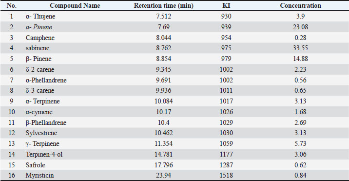
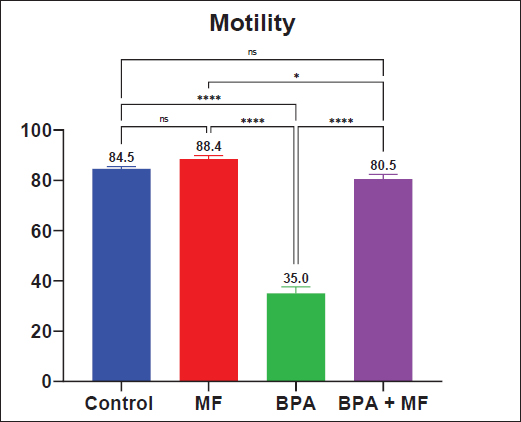
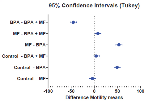
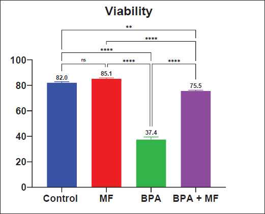
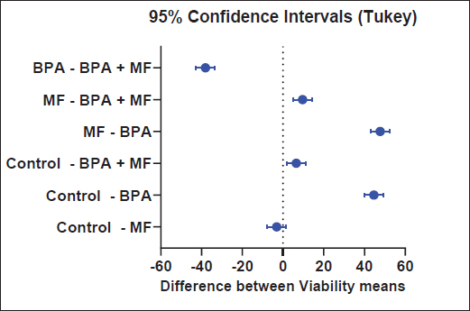
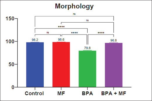
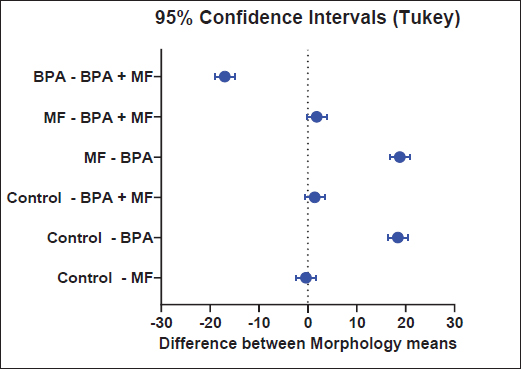
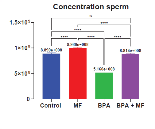
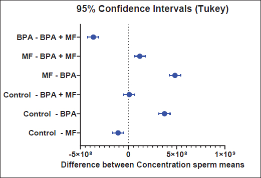
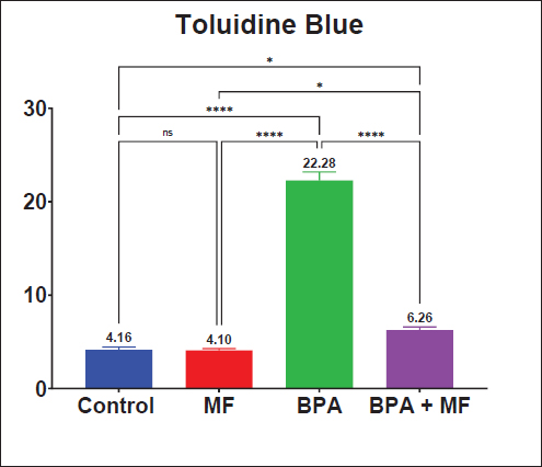
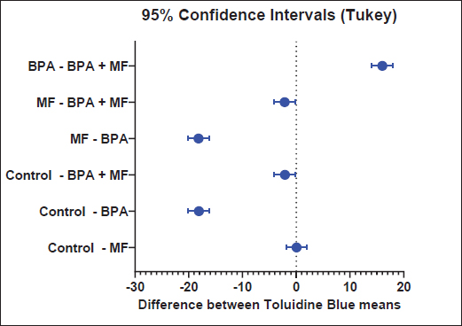
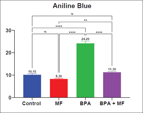
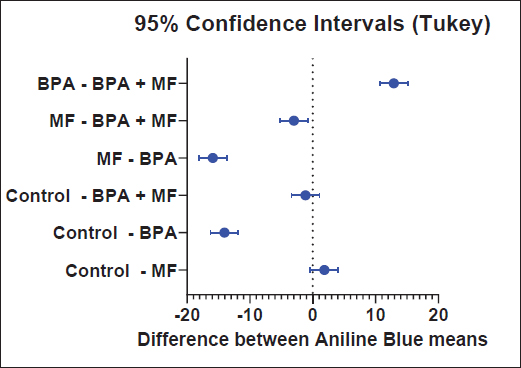
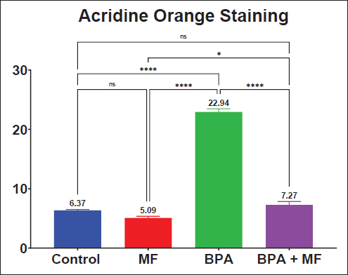
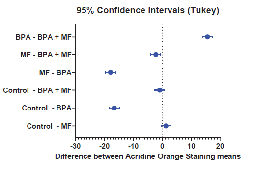
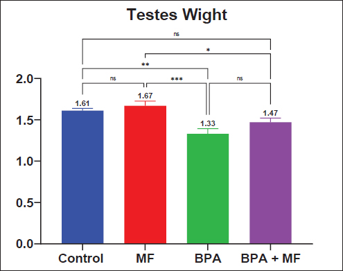
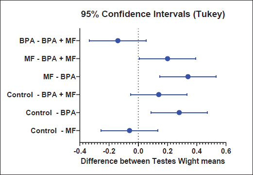
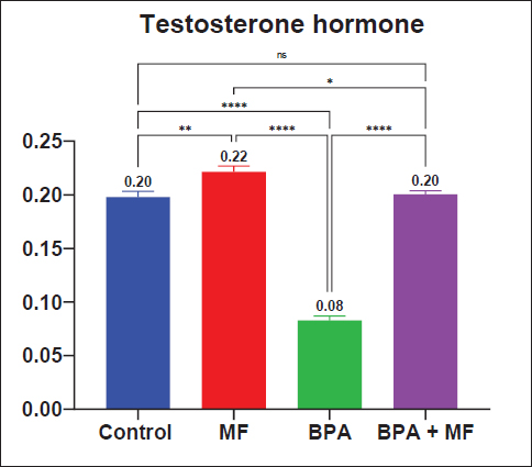
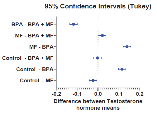
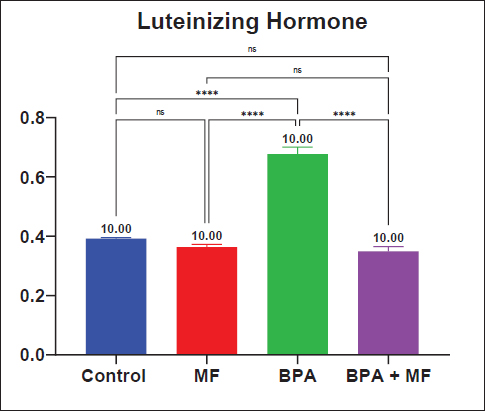
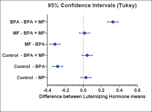
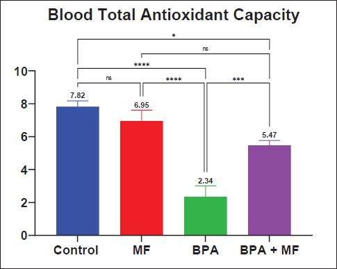
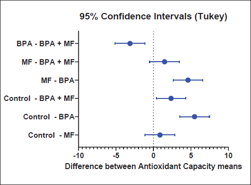
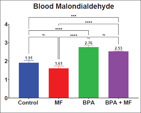
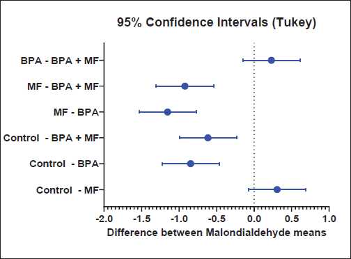
| How to Cite this Article |
| Pubmed Style Zarka MA, Algazali SA, Waday AS, Bustani GS. Protective and restorative role of Myristica fragrans essential oil on testicular and endocrine dysfunction induced by bisphenol A. doi:10.5455/OVJ.2026.v16.i6.69 Web Style Zarka MA, Algazali SA, Waday AS, Bustani GS. Protective and restorative role of Myristica fragrans essential oil on testicular and endocrine dysfunction induced by bisphenol A. https://www.openveterinaryjournal.com/?mno=306420 [Access: June 28, 2026]. doi:10.5455/OVJ.2026.v16.i6.69 AMA (American Medical Association) Style Zarka MA, Algazali SA, Waday AS, Bustani GS. Protective and restorative role of Myristica fragrans essential oil on testicular and endocrine dysfunction induced by bisphenol A. doi:10.5455/OVJ.2026.v16.i6.69 Vancouver/ICMJE Style Zarka MA, Algazali SA, Waday AS, Bustani GS. Protective and restorative role of Myristica fragrans essential oil on testicular and endocrine dysfunction induced by bisphenol A. doi:10.5455/OVJ.2026.v16.i6.69 Harvard Style Zarka, M. A., Algazali, . S. A., Waday, . A. S. & Bustani, . G. S. (2026) Protective and restorative role of Myristica fragrans essential oil on testicular and endocrine dysfunction induced by bisphenol A. doi:10.5455/OVJ.2026.v16.i6.69 Turabian Style Zarka, Mohamed A., Sajad A. Algazali, Ali Salah Waday, and Ghadeer Sabah Bustani. 2026. Protective and restorative role of Myristica fragrans essential oil on testicular and endocrine dysfunction induced by bisphenol A. doi:10.5455/OVJ.2026.v16.i6.69 Chicago Style Zarka, Mohamed A., Sajad A. Algazali, Ali Salah Waday, and Ghadeer Sabah Bustani. "Protective and restorative role of Myristica fragrans essential oil on testicular and endocrine dysfunction induced by bisphenol A." doi:10.5455/OVJ.2026.v16.i6.69 MLA (The Modern Language Association) Style Zarka, Mohamed A., Sajad A. Algazali, Ali Salah Waday, and Ghadeer Sabah Bustani. "Protective and restorative role of Myristica fragrans essential oil on testicular and endocrine dysfunction induced by bisphenol A." doi:10.5455/OVJ.2026.v16.i6.69 APA (American Psychological Association) Style Zarka, M. A., Algazali, . S. A., Waday, . A. S. & Bustani, . G. S. (2026) Protective and restorative role of Myristica fragrans essential oil on testicular and endocrine dysfunction induced by bisphenol A. doi:10.5455/OVJ.2026.v16.i6.69 |


