Open Veterinary Journal, (2026), Vol. 16(6): 3561-3568
Short Communication
10.5455/OVJ.2026.v16.i6.25
Occupational exposure to tick-borne pathogens in veterinary clinic workers from Yucatán, Mexico
Karla Dzul-Rosado1*, Carlos Pérez-Osorio2, Sergio Morales-Garza1, Angel Casanova-Cocom1,
Dayana Lavín-Sánchez1, Henry Noh-Pech1, Juan Arias-León2, Daniela García-Quiroz1,
Fernando Puerto-Manzano1 and Roger I. Rodríguez-Vivas3
1Laboratorio de Enfermedades Emergentes y Reemergentes, Centro de Investigaciones Regionales “Dr. Hideyo Noguchi”, Universidad Autónoma de Yucatán, Mérida, Mexico
2Laboratorio de Enfermedades Infecciosas y Parasitarias I, Facultad de Medicina,
Universidad Autónoma de Yucatán, Mérida, México
3Departamento de Salud Animal y Medicina Preventiva, Facultad de Medicina Veterinaria y Zootecnia,
Campus de Ciencias Biológicas y Agropecuarias, Universidad Autónoma de Yucatán, Mérida, Mexico
*Corresponding Author: Karla Dzul-Rosado. Laboratorio de Enfermedades Emergentes y Reemergentes,
Centro de Investigaciones Regionales “Dr. Hideyo Noguchi”, Universidad Autónoma de Yucatán, Mérida, México.
Email: karla.dzul [at] correo.uady.mx
Submitted: 02/02/2026 Revised: 30/04/2026 Accepted: 11/05/2026 Published: 05/06/2026
© 2025 Open Veterinary Journal
This is an Open Access article distributed under the terms of the Creative Commons Attribution-Non Commercial-No Derivatives License (http://creativecommons.org/licenses/by-nc-nd/4.0/), which permits non-commercial re-use, distribution, and reproduction in any medium, provided the original work is properly cited, and is not altered, transformed, or built upon in any way.
ABSTRACT
Background: Veterinary clinic personnel are exposed to various occupational risks, including the transmission of zoonotic pathogens such as Rickettsia spp. and Ehrlichia spp., which can cause serious diseases in humans. Although exposure to vector-borne agents has been documented among veterinary clinic personnel in Mexico, no previous studies have focused on this population in Yucatán, Mexico.
Aim: To estimate the prevalence of rickettsial pathogens in veterinary clinic personnel in Mérida, Yucatan, Mexico.
Methods: A cross-sectional study was conducted using non-probability convenience sampling in six veterinary clinics. A total of 39 workers over 18 years old who provided informed consent were included. Blood samples were collected by venipuncture and analyzed by indirect immunofluorescence assay to detect antibodies against Rickettsia rickettsii, Rickettsia typhi, and Ehrlichia chaffeensis. Prevalence estimates are presented as percentages with their corresponding 95% confidence intervals, calculated using the Wilson method for binomial proportions.
Results: The prevalence of single-antigen antibody seroreactivity to R. rickettsii, R. typhi, and E. chaffeensis was 41.0% (16/39), 17.9% (7/39), and 0%, respectively. However, the multi-antigen seroreactivity to R. rickettsia and R. typhi was 12.8% (5/39); meanwhile, the multi-antigen seroreactivity to R. rickettsii, R. typhi, and E. chaffeensis was 5.1% (2/39).
Conclusion: This is the first exploratory study in Yucatán and southeastern Mexico to indicate occupational exposure to Rickettsia spp. among veterinary clinic personnel and highlights the need for integrated prevention and surveillance strategies under a One Health approach.
Keywords: Occupational exposure, Personnel in veterinary clinics, Zoonosis.
Introduction
Rickettsial and ehrlichial diseases are emerging zoonoses caused by obligate intracellular Gram-negative bacteria and mainly transmitted by ticks and fleas. Their clinical manifestations range from mild, nonspecific symptoms to severe and potentially fatal outcomes, complicating timely diagnosis (Buckingham et al., 2007; Biggs et al., 2016).
The symptoms of Rickettsia spp. and Ehrlichia spp. infections are often confused with common diseases, leading to underdiagnosis and highlighting the need to strengthen awareness for early detection (Ursery et al., 2025).
Rickettsiosis caused by Rickettsia has several incidence and prevalence rates across Mexico. The prevalence of this genus is higher in the northern and southeastern states of the country (Sánchez-Montes et al., 2021; Torres-Castro et al., 2024). In Mexico, at least 17 species of the genus Rickettsia have been identified, including Rickettsia rickettsii, Rickettsia typhi, Rickettsia parkeri, Rickettsia felis, and Rickettsia akari (Zavala-Castro et al., 2006; Ojeda-Chi et al., 2019d; Sánchez-Montes et al., 2021; Peniche-Lara & Lara-Perera, 2022). The diversity of species identified in the southeastern states of Mexico indicates the persistence of these pathogens in urban and peri-urban environments, as well as the possibility of coinfections, subclinical circulation, and dissemination by synanthropic fauna (Ojeda-Chi et al., 2019c; Sánchez-Montes et al., 2021).
Rickettsia rickettsii infects ticks and causes tick-transmitted rickettsioses in areas of endemicity where ixodid ticks support host transmission during blood feeding. Infected ticks become a primary reservoir of R. rickettsii, providing a lifelong opportunity to transmit and amplify these pathogens in mammalian hosts. Dermacentor variabilis, Dermacentor andersoni, Rhipicephalus sanguineus, and Amblyomma sculptum are confirmed vectors of R. rickettsii in the United States. In Mexico, we found molecular evidence of Rickettsia sp. in Amblyomma mixtum, R. sanguineus sensu lato, and Ixodes affinis collected from dogs in a locality of Yucatan with a history of rickettsiosis outbreaks among its inhabitants (Martínez-Ortiz et al., 2019). Therefore, the authors suggest that these ectoparasites may have played a role in the zoonotic transmission of the bacteria.
In addition, R. typhi, which causes murine or endemic typhus, participates in its classic infection cycle in the presence of mammalian hosts (rodents and humans) and vectors (fleas). Ctenocephalides felis and Ct. canis are some of the main ectoparasites that affect dogs and cats in Yucatán, Mexico (Bolio-González et al., 2012), and R. typhi is transmitted to humans primarily through the feces of infected fleas (Martínez-Ortiz et al., 2019). In Yucatán, significant circulation of Rickettsia spp. has been documented in wild and domestic animals, such as rodents, marsupials, bats, and canines, suggesting the existence of a dynamic zoonotic ecosystem with high transmission potential (Zavala-Castro et al., 2006; Sánchez-Montes et al., 2016; Peniche-Lara and Lara-Perera, 2022).
Human ehrlichiosis is caused by bacteria from the Ehrlichia genus, including Ehrlichia chaffeensis, Ehrlichia ewingii, and Ehrlichia muris subsp. Eauclairensis (Cisneros-Saldaña et al., 2023). The main vectors of this bacterium are Ixodes scapularis (transmitting E. muris subsp. Eauclairensis), Amblyomma americanum, and D. variabilis, which spread E. chaffeensis and E. ewingii. Ticks, such as the white-tailed deer (Odocoileus virginianus) and mazama deer (Mazama temama), become infected after feeding from their primary reservoir host (Cheng et al., 2016; Ojeda-Chi et al., 2019d). Cases of human ehrlichiosis and E. chaffeensis infection in R. sanguineus, A. mixtum, and Amblyomma spp. have been documented in Mexico (Gongóra-Biachi et al., 1999; Ojeda-Chi et al., 2019c; Sosa-Gutierrez et al., 2016). Rickettsiosis and ehrlichiosis pose a significant public health burden because of their high lethality and the costs associated with delayed diagnosis, hospitalization, and intensive care, particularly in regions with limited epidemiological surveillance (Drexler et al., 2015).
Veterinary clinic staff represent a key risk group in this context, given their frequent contact with animals and vectors. This exposure is not limited to veterinarians but also includes assistants, dog groomers, administrative staff, and cleaning staff, who may have less training in biosafety measures. Although occupational exposure to rickettsias in veterinary workers has been documented in different regions of the country (Escárcega-Ávila et al., 2019), studies specifically focused on this sector are lacking in the Yucatán Peninsula. This lack of information limits the design of evidence-based preventive strategies. Furthermore, health authorities have warned about the sustained increase in cases and deaths from rickettsiosis, with case fatality rates close to 50% in some states, prompting national prevention and epidemiological surveillance campaigns (CENAPRECE, 2024).
Therefore, this study aimed to estimate the exposure of veterinary clinic personnel in Mérida, Yucatán, Mexico, to rickettsial and ehrlichial pathogens. It also aims to provide evidence to support early diagnosis and prevention of zoonotic diseases.
Materials and Methods
Study design
Thirty-nine workers from six small-species veterinary clinics participated in a cross-sectional study between December 2022 and January 2023, including veterinarians and staff working in different areas of six small-species veterinary clinics in the city of Mérida, Yucatán, Mexico. Clinic selection was carried out through convenience sampling without probability. Workers over 18 years of age who were actively working in the clinics during the study period and who gave their informed consent after receiving a detailed explanation of the study objectives and procedures were included in the study. We excluded those who refused to participate, had a recent history of hospitalization (last 30 days), or had a previously confirmed diagnosis of Rickettsia spp. or Ehrlichia spp. infection were excluded.
The medical records of the participants were revised to validate their exclusion from the study and to reduce the bias from their self-response.
Blood sampling
Six milliliters of blood were drawn from each participant by venipuncture using anticoagulant-free Vacutainer® tubes (Becton Dickinson, Franklin Lakes, NJ). The samples were individually and properly labeled, followed by transportation under refrigeration (4°–8°C) to the laboratory for analysis. The estimated time from blood collection to centrifugation for serum separation was 1 hour. The Ethylenediaminetetraacetic acid-free tubes were centrifuged at 3,000 g for 30 minutes at 4°C to obtain serum, which was stored in 2 ml vials at 20°C until further processing.
Survey
A structured questionnaire was adapted from previously published instruments addressing occupational exposure to zoonotic and tick-borne pathogens (Escárcega-Ávila et al., 2019), and its content validity was reviewed by subject-matter experts to ensure clarity and relevance. The instrument included the variables previously described and was pilot-tested with a small group of veterinary clinic workers to assess comprehension, response consistency, and overall applicability.
Minor adjustments were incorporated before its final administration to collect information for assessing the risk of occupational exposure to tick-borne pathogens, including: a) sociodemographic variables (age and sex), b) occupational characteristics (clinic role and weekly working hours), c) arthropod exposure, d) number of tick bites, and e) number of animals attended per week.
Indirect immunofluorescence assay (IFAS)
Human sera were examined by IFA using crude antigens from R. rickettsii (representing the SFG Rickettsiae) (Rosado et al., 2013) and R. typhi (representing the TG Rickettsiae) (Zavala-Castro et al., 2014). Slides were prepared in-house for research use only, as reported in previous studies (Dzul-Rosado et al., 2022, 2025; Villarreal-Jimenez et al., 2026) and E. chaffeensis (donated by the University of Texas Medical Branch). The antibody titers were independently obtained for each antigen.
Rickettsia rickettsii and R. typhi were grown in VERO 76 cells (ATCC CRL-1587). Cell cultures were observed for macroscopic changes [R. rickettsii (5-8 días) y R. typhi (12 a 15 días)] as an indication of infection progress (Lugo-Caballero et al., 2018). The cell culture was considered highly compromised when the growth medium acquired a bright yellow color, indicating a low pH, or when the cells detached from the flask surface, indicating a cytopathic effect due to bacterial growth (Ammerman et al., 2008). The infected cells were seeded and suspended in 1X phosphate-buffered saline (PBS) with 10% poly-L-lysine to a final concentration of 5,000–10,000 cells/l (Sigma, Cat. P8920, Alemania). Then, the cell suspension was transferred using 10 l each to 12-well slides (Ted Pella Inc., cat. 260505, California, with a confluence of 80%–100% infected cells (La Scola and Raoult, 1997). Slides were allowed to dry at 37°C for 15 minutes and then fixed (Ammerman et al., 2008). Antigen slides were stored at −20°C until further use.
In-house indirect fluorescent antibody assays were performed to detect reactive antibodies in the samples. For each test batch, a human antigen-specific hyperimmune serum was used as a positive control and PBS containing 1% bovine serum albumin (BSA) as a negative control to validate positive reactions and the absence of unspecific reactions, respectively. Human serum dilutions were prepared in a series of PBS containing 1% BSA. Antigen wells were blocked in PBS containing 1% BSA and 0.01% sodium azide; 10 μl of each serum dilution was added to each well of the antigen sheet and incubated in a humid chamber for 30 minutes at 37°C. The slides were then washed with PBS containing 0.1% Tween 20 for 10 minutes and then washed twice in the same solution for 10 minutes. Fluorescein isothiocyanate–conjugated goat anti-human IgG immune serum (ABCAM, ab6854) diluted 1:100 in PBS containing 1% BSA and 0.01% Tween 20 was added to each well and incubated in a humid chamber for 30 minutes at 37°C. Slides were washed once with 0.1% PBS.
Tween 20 for 10 minutes and once with PBS containing 0.1% Tween 20 and 0.01% Evans blue.
For 10 minutes and then observed under a fluorescence microscope at 400× magnification. Serum
samples yielding distinctly fluorescent Rickettsia sp. at a ≥64 dilution were considered positive (Stewart and Stewart, 2021), and the samples were titrated through serial dilutions to the endpoint.
Statistical analysis
A descriptive analysis was conducted to calculate the prevalence of seroreactivity to R. rickettsii, R. typhi, and E. chaffeensis. The prevalence of multiantigen seroreactivity involving these pathogens was also estimated.
Given the small sample size, prevalence estimates are presented as percentages with their corresponding 95% confidence intervals (IC95%), which were calculated using the Wilson method for binomial proportions.
Ethical approval
The study was conducted in accordance with the Declaration of Helsinki and approved by the Ethics Committee of the O’Horan Hospital (Protocol code CIE-06-2022).
Results and Discussion
A total of 39 veterinary clinic personnel from six veterinary clinics were tested using IFAT. The prevalence of antibody single-antigen seroreactivity of R. rickettsii, R. typhi, and E. chaffeensis was 41.0 % (16/39), 17.9 % (7/39), and 0%, respectively. Multi-antigen seroreactivity to R. rickettsii and R. typhi was 12.8% (5/39); meanwhile, multi-antigen seroreactivity to R. rickettsii, R. typhi, and E. chaffeensis was 5.1% (2/39).
The results obtained show a high seroprevalence of antibodies against R. rickettsii (41%) in veterinary clinic personnel in Yucatán (Table 1). However, further research is needed to associate seroreactivity with exposure to this agent, since other rickettsias, such as R. parkeri or R. felis, are closely related and may cause cross-reactions (Teoh et al., 2017). Complementary tests, including molecular biology and expanded antigen panels for serology tests, are recommended. This finding is consistent with previous studies that identified veterinary workers as a risk group for tick-borne diseases (Nicholson et al., 2010; Alvarez-Hernandez et al., 2015). Other studies have documented a seroprevalence of 21% in veterinary staff exposed to tick-infested dogs (Blanton, 2019). Furthermore, prevalences of only 3%–4% been recorded in the general population in Mexico, which contrasts with the high levels observed in veterinarians (Field-Cortazares et al., 2015). Other studies in Mexico have not found significant correlations between occupation and exposure (Escárcega Ávila et al., 2018).
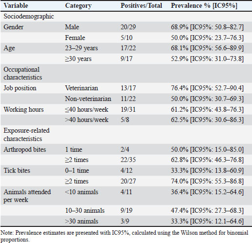
Table 1. Prevalence of single-antigen seroreactivity to R. rickettsii in veterinary clinic personnel (n=39).
The presence of single-antigen seroreactivity against R. typhi (17.9%) in this study suggests additional exposure to flea-associated rickettsiae, reflecting the diversity of vectors in the region. Meta-analyses show that contact with animals significantly increases occupational risk (odds ratio=2.07) (Adebowale et al., 2021). Furthermore, studies in veterinarians have found prevalences for R. typhi (4.6%), although without identifying specific associated factors—likely indicating challenges with statistical power or homogeneous exposure across groups (Teoh et al., 2017). In other studies, seropositivity to R. typhi is associated more with environmental factors (proximity to water, dog ownership) than with bite memory, which coincides with the idea that fleas—imperceptible vectors—also play an important role, especially in urban areas (Devamani et al., 2020).
The observed multi-antigen seroreactivity to R. rickettsii and R. typhi (12.8%) may be explained by antigenic cross-reactivity, a known limitation of IFA-based serology when evaluating closely related Rickettsia spp. This suggests that the dual reactivity may reflect exposure to antigenically related rickettsial species rather than true co-infection (Stewart and Stewart, 2021).
Due to the small number of patients studied in this study, a statistical analysis was not performed to determine the factors associated with the prevalence of single antigen seroreactivity to R. rickettsii in the veterinary clinic staff (Table 1). However, it is important to highlight that the prevalence of single antigen seroreactivity to R. rickettsii was higher in veterinarians than in non-veterinary staff (76.4% vs. 50.0%), suggesting increased exposure among personnel directly involved in clinical animal care. This pattern is consistent with previous studies identifying veterinarians as a high-risk occupational group due to frequent contact with animals and ectoparasites (Nicholson et al., 2010; Escárcega-Ávila et al., 2019). Similar serological evidence of occupational exposure has been reported in veterinarians from other regions, supporting the role of professional activity rather than solely environmental factors (Teoh et al., 2017). Nonetheless, the considerable seroprevalence observed in nonveterinary personnel highlights that occupational risk extends across all job positions within veterinary clinics, emphasizing the need for comprehensive prevention strategies targeting all staff.
A higher frequency of single antigen seroreactivity to R. rickettsii was observed among individuals with a history of ≥ 2 tick bites compared with 0–1 bites (33.5% vs. 74.0%). This finding suggests greater exposure to tick-borne rickettsial agents among participants with repeated contact with the vector, which is consistent with the well-recognized role of ticks in the transmission of spotted fever group rickettsiae (Parola and Raoult, 2001). This finding should be interpreted with caution, as tick bites were not classified by setting and no non-octrolateral comparison group was included; thus, the observed pattern likely reflects general vector exposure rather than clinic-specific occupational risk. Comparable descriptive patterns have been reported in other seroepidemiological studies, where a greater prevalence of rickettsial seropositivity was noted among individuals with a history of tick bites, despite differences in study design and population characteristics (Gual-Gonzalez et al., 2024). Additionally, studies conducted in urban environments have highlighted that frequent interaction with domestic animals and vectors—particularly ticks and fleas—may facilitate unnoticed exposure and transmission, even outside strictly occupational contexts (Anstead, 2025; Eremeeva and Dasch, 2025). In future studies, it is imperative to obtain a larger number of samples to accurately define the factors associated with the prevalence of R. rickettsi and other pathogens in veterinarians and non-veterans working in veterinary clinics to design a vector-borne disease prevention program in the region.
A relevant finding is the absence of single-antigen seroreactivity for E. chaffeensis, with only 5.1% of the samples showing multi-antigen seroreactivity, including E. chaffeensis, which could indicate low circulation of this agent in the area or differences in vector ecology, as has been reported in other regions of Mexico (Alvarez-Hernandez et al., 2015). The absence of single-antigen seroreactivity to E. chaffeensis (0%) is a relevant finding, especially when compared with the current literature on this pathogen in animals within the region. This discrepancy suggests that the risk of human exposure in the working environment in this study could be lower than that in rural populations, where the presence of the pathogen and its vector has been reported (Ojeda-Chi et al., 2019a,b,c,d). These results are consistent with reports of low exposure in other occupational groups, such as agricultural workers in China (0.4%) (Zhang & Diao, 2020). In contrast, studies in the general population of rural areas in South America have reported a higher prevalence (up to 25%), suggesting that the risk depends not only on occupation but also on local ecological and vector factors (Quintero V. et al., 2017). Further studies integrating surveillance in animals, vectors, and humans are needed to determine the actual level of risk in this occupational group more accurately.
Based on these results, targeted measures can be proposed to reduce occupational exposure to rickettsial agents in Yucatán veterinary clinics. Given the central role of tick and flea contact in seropositivity—particularly to R. rickettsii—routine inspection and safe removal of ectoparasites should be implemented after handling animals at risk. Strengthening staff training, improving ectoparasite control within clinics, and encouraging the reporting of bites or high-risk contacts could substantially reduce occupational exposure.
These interventions reduce immediate exposure to vectors and improve personnel protection; however, their impact is greater when integrated into a broader framework that considers the interaction between animals, humans, and the environment. The One Health approach is essential for addressing rickettsiosis and ehrlichiosis, as it enables coordinated surveillance, vector control, improved diagnostics, and targeted risk communication. Together, these strategies support earlier detection and more effective and sustainable prevention of these zoonotic diseases (Meurer, 2025).
Conclusion
Veterinary clinic personnel in Yucatán showed frequent seroreactivity to Rickettsia spp., indicating ongoing occupational exposure. As the first exploratory study conducted in Yucatán and southeastern Mexico, these findings indicate that exposure to rickettsial agents, particularly in relation to contact with ticks, is shared across different job positions within veterinary clinics, including both veterinarians and other staff.
These results underscore the importance of reinforcing preventive practices and surveillance through a One Health approach that integrates occupational training, vector control, and coordinated monitoring to reduce exposure and support early detection.
Acknowledgments
The authors would like to thank the participants and social service students for their enthusiastic participation in this study, as well as the veterinary clinic personnel.
Conflict of interest
The authors have no conflicts of interest to declare.
Funding
This work was supported by Grant CONACYT SALUD 2015-2-261885.
Authors’ contributions
Conceptualization, K.D.R. and R.I.R.V. methodology, K.D.R., R.I.R.V., C.P.O., S.M.G., validation, K.D.R., R.I.R.V. formal analysis, K.D.R., R.I.R.V., C.P.O., D.L.S., D.G.Q., resources, K.D.R.; data curation, K.D.R., R.I.R.V., D.L.S., D.G.Q., A.C.C., J.A.L.; writing—original draft preparation, K.D.R., R.I.R.V., C.P.O., J.A.L., F.P.M. writing—review and editing, K.D.R., C.P.O., H.N.P.M. J.A.L., F.P.M. project administration, K.D.R. Funding acquisition K.D.R.
Data availability
All data supporting the findings of this study are available within the manuscript.
References
Adebowale, O., Fasanmi, O.G., Awosile, B., Afolabi, M. and Fasina, F.O. 2021. Systematic review and meta-analysis of veterinary-related occupational exposures to hazards. Open. Vet. Sci. 2(1), 6–22.
Alvarez-Hernandez, G., Murillo-Benitez, C., Del Carmen Candia-plata, M. and Moro, M. 2015. Clinical profile and predictors of fatal Rocky Mountain spotted fever in children from Sonora, Mexico. Pediatric Infect. Dis. J. 34(2), 125–130.
Ammerman, N.C., Beier-Sexton, M. and Azad, A.F. 2008. Laboratory maintenance of Rickettsia Rickettsii. Curr. Protocols Microbiol. 11(1), 3A.5.1–3A.5.21; doi:10.1002/9780471729259.mc03a05s11
Anstead, G.M. 2025. A One Health perspective on the resurgence of flea-borne typhus in texas in the 21st century part 1 the bacteria, the cat flea, urbanization, and climate change. Pathogens 14(2), 154.
Biggs, H.M., Behravesh, C.B., Bradley, K.K., Dahlgren, F.S., Drexler, N.A., Dumler, J.S., Folk, S.M., Kato, C.Y., Lash, R.R., Levin, M.L., Massung, R.F., Nadelman, R.B., Nicholson, W.L., Paddock, C.D., Pritt, B.S. and Traeger, M.S. 2016. Diagnosis and management of tickborne rickettsial diseases Rocky Mountain spotted fever and other spotted fever group rickettsioses, ehrlichioses, and anaplasmosis — United States. MMWR. Recommendations Rep. 65(2), g1–44.
Blanton, L.S. 2019. The rickettsioses a practical update. Infec. Dis. Clin. North Am. 33(1), 213–229.
Bolio-González, M., Rodríguez-Vivas, R., Sauri-Arceo, C., Gutiérrez-Blanco, E., Morales-Puerto, E., Aranda-Cirerol, F., De Oca-jiménez, R., Manrique-Saide, P., Rosado-Aguilar, J. and Puerto-Nájera, J. 2012. Prevalencia y lesiones cutáneas de Ctenocephalides felis y Ctenocephalides canis en perros del estado de Yucatán. Bioagrociencias 5(1), 15–19.
Buckingham, S.C., Marshall, G.S., Schutze, G.E., Woods, C.R., Jackson, M.A., Patterson, L.E.R. and Jacobs, R.F. 2007. Clinical and laboratory features, hospital course, and outcome of Rocky Mountain spotted fever in children. J. Pediatrics 150(2), 180–184; doi.org10.1016j.jpeds.2006.11.023
CENAPRECE. (2024, January 1). Informes trimestrales SaNAS_2024. HttpsSpps. MxSanasCenaprece2024.
Cheng, C., Fu, W., Ju, W., Yang, L., Xu, N., Wang, Y.M., Li, H., Wang, Y.L., Hu, M.X., Wen, J., Jiao, D., Geng, C. and Sun, Y. 2016. Diversity of spotted fever group Rickettsia infection in hard ticks from Suifenhe, Chinese–Russian border. Ticks Tick-Borne Dis. 7(5), 715–719.
Cisneros-Saldaña, D., Osuna-Álvarez, L.E., Castillo-Bejarano, J.I., Mascareñas-De los Santos, A.H., Vaquera-Aparicio, D.N. and Pérez-Cavazos, S. 2023. First report of pediatric ehrlichiosis in Mexico. Mexico City, Mexico: Boletín Médico Del Hospital Infantil de México, vol. 80(91); doi:10.24875BMHIM.22000056
Devamani, C.S., Schmidt, W.P., Ariyoshi, K., Anitha, A., Kalaimani, S. and Prakash, J.A.J. 2020. Risk factors for scrub typhus, murine typhus, and spotted fever seropositivity in urban areas, rural plains, and peri-forest hill villages in South India a cross-sectional study. Am. J. Trop. Med. Hygiene 103(1), 238–248; doi:10.4269ajtmh.19-0642
Drexler, N.A., Traeger, M.S., Mcquiston, J.H., Williams, V., Hamilton, C. and Regan, J.J. 2015. Medical and indirect costs associated with a Rocky Mountain spotted fever epidemic in Arizona, 2002–2011. Am. Soc. Trop. Med. Hygiene. 93(3), 549–551.
Dzul Rosado, K.R., Peña Bates, C.A., Tello, M.R., Noh-Pech, H.R., Puerto, F.I. and Omodior, O. 2025. SFG and TG seropositivity in humans suspected of TBD in Yucatan, Epidemiol. Infect. 153, e21; doi:10.1017/S0950268824001894
Dzul-Rosado, K., Cámara Herrera, R., Miranda-Schaeubinger, M., Arias-León, J., Peniche-Lara, G., Gilman Robert, H., Mercado-saavedra Brandon, N., Lugo-Caballero, C., López Ávila, K., Tello Martín, R. and Omodior, O. 2022. Socio-ecological determinants of rickettsial seroprevalence in a rural community of Yucatán, Mexico. Infect. Genet. Evol. 102(105291), g1–g8; doi:10.1016j.meegid.2022.105291
Eremeeva, M. and Dasch, G. 2025. Other Rickettsia species (Georgia Southern University, Ed.; 1st ed., Vol. 1). Atlanta: Centers for Disease Control and Prevention.
Escárcega Ávila, A.M., Luna Flores, B.S., De La Mora Covarrubias, A. and Jiménez, F. 2018. Análisis exploratorio de enfermedades Rickettsiales transmitidas por garrapatas en perros de Ciudad Juárez, Chihuahua, México. Acta. Universitaria. 28(3), 72–78.
Escárcega-Ávila, A.M., De La Mora-covarrubias, A., Quezada-Casasola, A. and Jiménez-Vega, F. 2019. Occupational risk for personnel working in veterinary clinics through exposure to vectors of Rickettsial pathogens. Ticks Tick-Borne Dis. 10(2), 299–304; httpsdoi.org10.1016j.ttbdis.2018.10.012
Field-Cortazares, J., Escárcega-Ávila, A.M., López-Valencia, G., Barreras-Serrano, A. and Tinoco-Gracia, L. 2015. Seroprevalence of risk factors associated with Rickettsiosis (Rickettsia rickettsii) in humans in Baja California, Mexico. Gaceta Medica. De Mex. 151(1), 42–46.
Gongóra-Biachi, R.A., Zavala-Velázquez, J., Castro-Sansores, C.J. and González-Martínez, P. 1999. First case of human ehrlichiosis in Mexico. Emerg. Infect. Dis. 5(3), 481.
Gual-Gonzalez, L., Self, S.C.W., Meyer, M., Cantillo-Barraza, O., Torres, M.E. and Nolan, M.S. 2024. Human spotted fever group Rickettsia seroprevalence and associated epidemiologic factors among diverse, marginalized populations in South Carolina. Ticks Tick-Borne Dis. 15(2), 102288.
La Scola, B., and D. Raoult. 1997. Laboratory diagnosis of Rickettsioses: current approaches to diagnosis of old and new Rickettsial diseases. J. Clin. Microbiol. 35(11), 2715–27; doi:10.1128/jcm.35.11.2715-2727.1997
Lugo-Caballero, C., Tello-Martin, R., Dzul-Rosado, K. and Zavala-Castro, J. 2018. Approaches for the successful isolation and cell culture of American Rickettsia species. J. Vector. Borne. Dis. 55(4), 258.
Martínez-Ortiz, D., Torres-Castro, M.A., López-Ávila, K., Koyoc-Cardeña, E. and Manrique-Saide, P. 2019. Rickettsia spp. en garrapatas (Acari Ixodidae) que infestan perros de una comunidad rural con antecedentes de rickettsiosis, Yucatán, México. Rev. Bioméd. 30(2), 43–50; doi:10.32776/revbiomed.v30i2.650
Meurer, I. 2025. The importance of medical knowledge about Q fever in the context of timely diagnosis and treatment and the use of the one health approach in combating this and other neglected zoonotic diseases Letter. Infect. Drug Resist. 18, 5007–5008; doi:10.2147IDR.S567142
Nicholson, W.L., Allen, K.E., Mcquiston, J.H., Breitschwerdt, E.B. and Little, S.E. 2010. The increasing recognition of rickettsial pathogens in dogs and people. Trends. Parasitol. 26(4), 205–212; doi:10.1016j.pt.2010.01.007
Ojeda-Chi, M.M., Rodriguez-Vivas, R.I., Esteve-Gasent, M.D., Pérez De León, A., Modarelli, J.J. and Villegas-Perez, S. 2019c. Molecular detection of rickettsial tick-borne agents in white-tailed deer (Odocoileus virginianus yucatanensis), mazama deer (Mazama temama), and the ticks they host in Yucatan, Mexico. Ticks Tick-Borne Dis. 10(2), 365–370.
Ojeda-Chi, M.M., Rodriguez-Vivas, R.I., Esteve-Gasent, M.D., Pérez De León, A.A., Modarelli, J.J. and Villegas-Perez, S.L. 2019d. Ticks infesting dogs in rural communities of Yucatan, Mexico and molecular diagnosis of rickettsial infection. Transboundary. Emerg. Dis. 66(1), 102–110; httpsdoi.org10.1111tbed.12990
Ojeda-Chi, M.M., Rodriguez-Vivas, R.I., Esteve-Gasent, M.D., Pérez De León, A.A., Modarelli, J.J. and Villegas-Perez, S.L. 2019a. Ehrlichia canis in dogs of Mexico prevalence, incidence, co-infection and factors associated. Comparative Immunol. Microbiol. Infect. Dis. 67, 101351.
Ojeda-Chi, M.M., Rodriguez-Vivas, R.I., Esteve-Gasent, M.D., Pérez De León, A.A., Modarelli, J.J. and Villegas-Perez, S.L. 2019b. Ehrlichia canis in dogs of Mexico prevalence, incidence, co–infection and factors associated. Comparative Immunol. Microbiol. Infect. Dis. 67, 101351.
Parola, P. and Raoult, D. 2001. Ticks and tickborne bacterial diseases in humans an emerging infectious threat. Clin. Infect. Dis. 32(6), 897–928.
Peniche-Lara, G. and Lara-Perera, V. 2022. Rickettsiosis Causada por Rickettsia parkeri, Mexico. 28(2), 478–79; doi:10.3201/eid2802.210454
Quintero V., J.C., Paternina T., L.E., Uribe Y., A., Muskus, C., Hidalgo., M., Gil., J., Cienfuegos G., A.V., Osorio Q., L. and Rojas A., C. 2017. Eco-epidemiological analysis of rickettsial seropositivity in rural areas of Colombia A multilevel approach. PLos Neglected. Trop. Dis. 11(9), e0005892.
Rosado, K., Lara, G., N, R., Zquez, J., Pacheco, R., Labruna, M. and Castro, E. 2013. Rickettsia rickettsii isolation from naturally infected Amblyomma parvum ticks by centrifugation in a 24-well culture plate technique. Open. Vet. J. 3(2), 101–105.
Sánchez-Montes, S., Blum-Domínguez, S., Lozano-Sardaneta, Y.N., Zazueta-Islas, H.M., Solís-Cortés, M., Ovando-Márquez, O., Colunga-Salas, P., Tamay-Segovia, P., Becker, I., Fernández-Figueroa, E. and Rangel-Escareño, C. 2021. Molecular detection of Rickettsia sp. cf. Rickettsia monacensis in Ixodes sp. cf. Ixodes affinis collected from white-tailed deer in Campeche, Mexico. Parasitol. Res. 120(5), 1891–1895.
Sánchez-Montes, S., Guzmán-Cornejo, C., Martínez-Nájera, Y., Becker, I., Venzal, J.M. and Labruna, M.B. 2016. Rickettsia lusitaniae associated with Ornithodoros yumatensis (Acari Argasidae) from two caves in Yucatan, Mexico. Ticks. Tick-Borne. Dis. 7(6), 1097–1101; httpsdoi.org10.1016j.ttbdis.2016.09.003
Sosa-Gutierrez, C.G., Solorzano-Santos, F., Walker, D.H., Torres, J., Serrano, C.A. and Gordillo-Perez, G. 2016. Fatal monocytic ehrlichiosis in woman, Mexico, 2013. Emerg. Infect. Dis. 22(5), 871–874.
Stewart, A.G. and Stewart, A.G.A. 2021. An update on the laboratory diagnosis of Rickettsia spp. infection. Pathogens 10(10), 1319.
Teoh, Y.T., Hii, S.F., Stevenson, M.A., Graves, S., Rees, R., Stenos, J. and Traub, R.J. 2017. Serological evidence of exposure to Rickettsia felis and Rickettsia typhi in Australian veterinarians. Parasites Vectors 10(1), 129.
Torres-Castro, M., Reyes-Novelo, E., Arroyo-Ramírez, A., Lugo-Caballero, C., Panti-May, J.A. and Rodríguez-Vivas, R.I. 2024. Actualización sobre aspectos epidemiológicos de la rickettsiosis en el Trópico De MéXICO. Trop. SubTrop. Agroecosystems. 27(2).
Ursery, L., Mansour, O., Abernathy, H., Wichmann, E., Yackley, A., Siegler, A., Giandomenico, D., Williams, C., Barbarin, A., Reiskind, M.H. and Boyce, R.M. 2025. Enhanced surveillance for tick-borne rickettsiosis and ehrlichiosis in North Carolina Protocol and preliminary results. PLos One 20(5), e0320361.
Villarreal-Jimenez, E., Dzul-Rosado, K., Puerto-Manzano, F., Guillermo-Herrera, J.C., Pech-Noh, H. and Mendez-Dominguez, N. 2026. Ecologic and sociodemographic factors associated with seroprevalence of Rickettsia in Yucatan, Mexico. Epidemiologia 7(2), 30.
Zavala-Castro, J., Zavala-Velázquez, J., Walker, D., Arcila-Ruiz, E., Laviada-Molina, H., Olano, J., Ruiz-Sosa, J., Small, M. and Dzul-Rosado, K. 2006. Fatal human infection with Rickettsia. Emerg. Infect. Dis. 12(4), 672–674.
Zavala-Castro, J.E., Dzul-Rosado, K.R., Peniche-Lara, G., Tello-Martín, R. and Zavala-Velázquez, J.E. 2014. Isolation of Rickettsia typhi from human, Mexico. Emerg. Infect. Dis. 20(8), e30095.
Zhang, Y. and Diao, X. 2020. The changing role of agriculture with economic structural change–The case of China. China Econ. Rev. 62, 101504.