





| Case Report | ||
Open Vet. J.. 2026; 16(6): 3595-3600 Open Veterinary Journal, (2026), Vol. 16(6): 3595-3600 Case Report Emerging human pathogen Sphingomonas paucimobilis associated with tongue lesions in a group of crossbred beef × dairy bulls prior to slaughterPlamen Marutsov and Betina Boneva-Marutsova*Department of Veterinary Microbiology, Infectious and Parasitic Diseases, Faculty of Veterinary Medicine, Trakia University, Stara Zagora, Bulgaria *Corresponding Author: Betina Boneva-Marutsova. Department of Veterinary Microbiology, Infectious and Parasitic Diseases, Faculty of Veterinary Medicine, Trakia University, Stara Zagora, Bulgaria. Email: betina.boneva [at] trakia-uni.bg Submitted: 02/02/2026 Revised: 05/05/2026 Accepted: 15/05/2026 Published: 05/06/2026 © 2025 Open Veterinary Journal
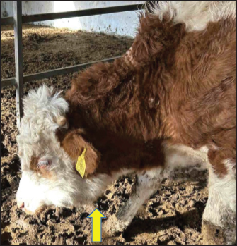
ABSTRACTBackground: Sphingomonas spp. are ubiquitously distributed free-living microorganisms in the natural environment. One of the representatives of the family, called Sphingomonas paucimobilis, is known as an opportunistic pathogen in humans responsible for bone and soft tissue infections. Case Description: In January 2024, signs of drooling, tongue rolling, and local edema in the pharyngeal region were observed in a male beef-dairy cross. Clinical examination revealed several ulcerative lesions on the tongue. The bull was not treated with antibiotics due to the impending slaughter of the entire lot of cattle. During the slaughter of 14 dairy-beef bulls, it was found that 13 of them had necrotic ulcerative lesions on the dorsal and lateral surfaces of their tongues. Tongue samples from only two of the bulls were sent from the slaughterhouse for diagnostic analysis. A pure bacterial culture was isolated from the tongue ulcers following the culturing process. As a result of bacteriological tests on these samples, a strain of S. paucimobilis was isolated, suggesting a possible association with the condition's cause. Over the past two decades, this microorganism has been noted in cases of mastitis in dairy cows in both the USA and the Republic of Türkiye, as well as in instances of polyarthritis in dogs within the USA. Additionally, S. paucimobilis has been reported in cases of septicemia and pneumonia in calves and cows in the Republic of Türkiye. Conclusion: These findings raise concerns about the impact of S. paucimobilis on livestock health and the potential negative effects on farm welfare. Additionally, these bacteria exhibit reduced susceptibility to antibiotics, highlighting the necessity for surveillance to protect both animal and human health. Keywords: Beef-dairy crossbred, Bulls, Sphingomonas paucimobilis, Tongue ulcers. IntroductionSphingomonas paucimobilis is a Gram-negative Bacillus, strictly aerobic, non-fermentative, with catalase and oxidase activity, and ubiquitously distributed that has been reported to be a common inhabitant of water and soil (Ionescu et al., 2022). Virulence factors associated with the bacterium include: presence of pili, phospholipase C, protease, and a flagellum (Laupland et al., 2022). Sphingomonas spp. are bacteria that replace lipopolysaccharide with glycosphingolipids and can grow in a wide range of environments that are hostile to most other bacteria (Kuehn et al., 2013). The lack of endotoxin activity may explain the reduced virulence and the favorable prognosis in infections that do occur (Lin et al., 2010). Sphingomonas paucimobilis is a hetero- and oligotrophic bacterium that can survive in low-nutrient environments and has the ability to form biofilm in water pipes, thus causing bioaccumulation and spreading in drinking water systems (Güneş et al., 2025). The bacterium has also been isolated from hospital settings, such as hospital water systems, dialysis fluid, nebulizers, and equipment for mechanical ventilation (Santarelli et al., 2016). It is believed to be an opportunistic pathogen that has rarely been reported to cause clinical disease in humans and animals (El Beaino et al., 2018). In humans, it has been associated with cases of bacteremia, catheter-related sepsis, diarrhea, peritonitis, meningitis, skin diseases, lung empyema, and pneumonia, oral and visceral infections, endophthalmitis, urinary tract infections, and more. Due to the low virulence of Sphingomonas spp., the mortality rate from this pathogen is very low (Nguyen et al., 2025). Sphingomonas paucimobilis has been identified as a pathogen in animals, causing mastitis in dairy cows, chronic lung infections in goats and cows, joint infections in cows, septicemia in ruminants, and polyarthritis in dogs (Kuehn et al., 2013; Davenport et al., 2013; Cengiz et al., 2015; Kenar et al., 2019). The presented case demonstrates the potential role of S. paucimobilis as a cause of tongue infections in cattle. Case DetailsFarm informationThe feedlot served as a distinct unit where 57 male cattle were raised. All bulls, sourced after weaning from a neighboring farm owned by the same person, were dairy-beef crosses. The animals were housed in six partially covered, single-row boxes featuring dirt floors and open feed alleys. They were separated by age and body weight, with 7 to 10 calves placed in each box. Upon arrival, all calves received a subcutaneous injection of ivermectin for deworming. No vaccinations were administered during their time in the feedlot. Feed and waterAll cattle in the feedlot were fed a balanced total mixed ration that included grain, corn silage, soybean meal, mineral salts, and straw, which was delivered once per day. In addition, large bales of coarse thorny hay [containing yellow bristle grass (YBG) and thistle plants] were available ad libitum. They had free access to water, with each pair of pens sharing a common trough that held approximately 200 l. Case descriptionIn early January 2024, a case of abnormal behavior was identified in a group of seven bulls before slaughter. The entire batch of the oldest fattened calves consisted of 14 animals, divided into two pens. The average age of the group was 17 months, and their body weights varied heterogeneously, ranging from 450 to 530 kg. The smallest bull exhibited symptoms including drooling, tongue rolling, and a noticeable decrease in body weight relative to the rest of the group. However, no abnormalities in food and water intake were observed at this time. Physical examinationThree weeks later, a local edema appeared in the pharyngeal region. The bull's attitude indicated that its head was bent, and the hair coat was coarse and dry, accompanied by a slight increase in the skin tent test (Fig. 1). The affected bull experienced difficulties while swallowing, both when drinking and eating. There were observations of reduced rumination and mild tympani with slight distension of the left paralumbar fossa. The body temperature was measured at a normal 38.2°C, with the pulse recorded at 78 beats per minute and the respiratory rate at 32 breaths per minute. Upon clinical examination of the oral cavity, a 3.5 cm non-bleeding, necrotizing ulcerative lesion was identified on the dorsal surface of the tongue. Furthermore, the regional submandibular lymph nodes were swollen and tender upon palpation. The bull was treated solely with menbuton (Neobuton®, FM Pharm, Subotica, Serbia) at a dosage of 7.5 mg/kg, administered via intramuscular injection, along with a drench of mineral oil. Antibiotics were not utilized due to the imminent slaughter of the entire cattle batch. No disease-suspicious symptoms were observed in the other cattle, so they were not subjected to a physical examination.
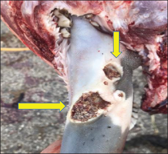
Fig. 1. Depressed bull exhibiting a lowered head posture with pronounced submandibular edema (yellow arrow). Diagnostic methods and laboratory findingsAt the time of slaughter, necrotic ulcers were found on the lateral and dorsal surfaces of the tongue. The bigger, deeper, and pus-containing lesion was located on the lateral surface, while the dorsal lesions were much smaller. The borders of the ulcers were raised and uneven, covered by slough (Fig. 2). During a routine inspection of the carcass, the official veterinarian found swollen submandibular and deep cervical lymph nodes. Out of the 14 bulls in the batch, lesions were identified in 13 (92.8%), while only one bull exhibited no lesions. The other individuals affected exhibited fewer and smaller lesions, primarily located on the lateral side of the tongue. During the inspection of the oral cavity, there was no severe thickening, swelling, or hardening of the tongue, nor was there any formation of abscesses detected. Additionally, no further macro-morphological changes were observed during the post-mortem inspection of the thoracic and abdominal cavity.
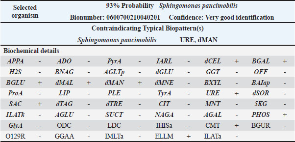
Fig. 2. Scattered ulcers with necrotic bases, confined to the lateral posterior half of the tongue's body (yellow arrows). To determine the possible etiological cause, samples from two bulls (with the most severe changes) were collected, frozen, and sent to the microbiology laboratory of the Faculty of Veterinary Medicine at Trakia University in Stara Zagora, Bulgaria. The tongues with lesions from the remaining animals were subjected to destruction. Microbiological testing: Bacteriological examination of tongue specimens from two bulls was performed. Inoculation material was obtained by searing the surface with a red-hot spatula. A sterile scalpel was then used for cutting, and a sterile swab was employed to collect a sample from within the tongue. Samples were cultured aerobically in liquid broth media (Soyabean Casein Digest Medium, HiMedia™), as well as on solid nutrient media (Blood Agar Base, supplemented with 5% defibrinated ovine blood and MacConkey agar, HiMedia™). Samples were incubated at 37°C for 5 days (Zeynali Kelishomi et al., 2023). The samples were also cultured on Sabouraud dextrose agar (SDA) containing chloramphenicol to isolate pathogenic fungi and incubated at two temperature regimes, 25°C and 37°C, for 2 weeks. The results of the cultivation showed that the bacterium was slow-growing; after 24 hours of aerobic incubation at 37°C on blood agar, small, round, smooth, almost white, non-hemolytic colonies were visible. After 48 hours, these colonies turned yellow. No visible colonies were observed on MacConkey agar after 48 hours of incubation. The organism was catalase-positive and oxidase-positive. It was identified as an aerobe with no fermentative capabilities. It grew slowly in broth media, producing weak turbidity. The cellular morphology observed in the Gram-stained microscopic preparations revealed medium-long Gram-negative rods. The isolate was subjected to phenotypic testing using the automated Vitek 2 system and software (bioMérieux). The identification was done on the colonies obtained from blood agar with 5% sheep blood after 24 hours of incubation. The suspension was prepared in 3 ml of sterile saline (0.45% NaCl). A turbidity of 0.50–0.63 McFarland was measured with a Vitek 2 DensiCHEK, and a GN ID card was used for the automatic identification. The analysis time for the identification results was completed within approximately 6 hours (Table 1). Table 1. VITEK Compaq® 2 system report for the isolated strain.
The results of the culture test were negative for Actinobacillus lignieresii, a common bacteriological agent associated with tongue lesions in cattle. No visible growth of SDA was observed. Susceptibility testing of the isolate to antimicrobials was performed using the Kirby-Bauer disk diffusion technique on Mueller-Hinton medium (HiMedia®, India). Due to the absence of established standards for susceptibility testing of S. paucimobilis, the zone diameter interpretive standards that were used for glucose-nonfermenting bacilli, specifically Acinetobacter spp., were applied in accordance with CLSI criteria (Ionescu et al., 2022). The isolates were found to be sensitive to amoxicillin–clavulanic acid, gentamicin, cefquinome, enrofloxacin, marbofloxacin, tulathromycin, trimethoprim–sulphonamides, and oxytetracycline. The strain demonstrated intermediate results to chloramphenicol and colistin. However, it was resistant to penicillin, cephalexin, and cephapirin. To effectively manage the condition, several control and prevention measures were implemented, including cleaning and disinfecting the water troughs and chlorinating the water source. To minimize the risk of traumatic injuries, sharp edges were removed, and coarse thorny hay was eliminated from the diet. Additionally, mineral lick blocks were provided to support overall health. In this case, it is likely a condition resulting from the combination of a number of risk factors. In their full set, as component causes, they constitute a sufficient cause for the occurrence of the disease. After the introduction of the indicated preventive measures, the owner did not report similar ulcerative-necrotic tongue lesions in subsequent batches of bulls. DiscussionIn animals, S. paucimobilis is reported as a bacterial agent responsible for sporadic infections, including mastitis, arthritis, pneumonia, and septicemia. Due to its opportunistic nature, these conditions are often attributed to factors that can influence overall microbial resistance (Davenport et al., 2013; Cengiz et al., 2015). In this case, the bacterium was isolated from tongue samples of only two slaughtered bulls. While other affected bulls exhibited similar ulcers in the same parts of the tongue, it is speculative to assert that the same agent causes them. The primary viral pathogens that cause tongue lesions are foot-and-mouth disease, vesicular stomatitis, and the bluetongue virus. Official veterinary inspectors conducted epidemiological surveys and sent samples for emerging diseases at both national and international levels, yielding negative results. Antigen tests for bovine pestivirus using (antigen capture ELISA) were also negative. The samples were not tested for bovine papular stomatitis virus; therefore, we could not exclude its potential role. Sphingomonas paucimobilis is associated with infections in bones and soft tissues, often entering the body through traumatic injuries (El Beaino et al., 2018). Possible damage to the surface of the tongue in our case could have resulted from the sharp edges of the water trough, licking sharp objects, or consuming rough, thorny hay. Similar observations were reported in a case from New Zealand, where a 100% prevalence of ulcerative tongue lesions was documented in 25 cull cows at a slaughterhouse. All cows had been fed a diet of baleage hay collected from pastures contaminated with YBG (Setaria pumila) for 3 weeks before their slaughter. The authors assumed that the main cause was the presence of significant amounts of YBG in the diet. The lesions identified were deep, transverse, linear ulcers of the dorsal lingual surface in the lingual fossa immediately rostral to the torus linguae (O’Connell et al., 2024). Previous studies indicated that YBG significantly contributed to lesion formation in the oral cavity and tongue of both cattle (Fava et al., 2000) and horses (Johnson et al., 2012; Kutasi et al., 2018). In this case report, the presence of YBG in the ration was also proven. However, most of the more severe lesions are localized laterally, while dorsal ones were rostral to the fossa linguae. In this instance, histological analyses were not conducted as the samples had been frozen, rendering them unsuitable for examination by the pathologist. The likelihood of coarse, dry hay harboring considerable quantities of YBG and milk thistle was the primary risk factor that likely precipitated superficial mucosal injury, subsequently leading to secondary microbial contamination of the affected lesions. Sphingomonas paucimobilis is ubiquitously distributed and often isolated from soil and water samples. Its nutritional requirements allow it to survive in nutrient-poor substrates. In addition, the bacterium forms a biofilm by depositing strong, adhesive exopolymers that promote bacterial cell attachment, thereby producing and reinforcing the biofilm structure (El Beaino et al., 2018; Tsvetanova et al., 2022). The potential for bacterial growth and accumulation in the feedlot watering troughs represented a significant and constant source contributing to the occurrence of various local infections. However, the possible causes of clinical manifestations are influenced by several factors, including exposure to a high bacterial load, and the combined impact of various risk factors, such as dietary errors, environmental factors, and management practices (Licitra et al., 2021). The probable involvement of S. paucimobilis in a significant number of affected bulls may be due to the common effect of predisposing factors or suggest a potential bioadaptation of the strain to the bovine host. While the latter remains unproven and is purely hypothetical, similar occurrences have been described in other bacterial species (Toft and Andersson, 2010; Barber and Fitzgerald, 2024). In this context, this phenomenon could indicate a potential shift from being predominantly opportunistic to exhibiting more pathogenic behavior. Recent studies have indicated a shift in the perception of the pathogenic potential of S. paucimobilis in humans. This bacterium is now increasingly recognized as an emerging pathogen linked to considerable morbidity (Nguyen et al., 2025). Various studies on the susceptibility of S. paucimobilis and other Gram-negative bacteria have reported an alarming increase in colistin resistance (Ionescu et al., 2022; Aboelnasr et al., 2024). In our case, the isolate's intermediate response to chloramphenicol and colistin indicated reduced susceptibility to these antibiotics. This is of particular concern, as intermediate results are typically the most selective for the development of resistance, warranting continued surveillance given the importance of both drugs in antimicrobial stewardship (Urban-Chmiel et al., 2022; Singh et al., 2025). Resistance to penicillin, cephalexin, and cefapirin is consistent with a widespread decline in the effectiveness of β-lactams, likely due to intrinsic or acquired β-lactamase activity. This pattern of resistance to first-generation β-lactams while maintaining susceptibility to later-generation or combination agents reflects commonly reported resistance trajectories (LaPlante et al., 2022). Earlier research conducted in Bulgaria on antimicrobial resistance of heterotrophic bacteria in drinking water, including S. paucimobilis, has established resistance to beta-lactams and nitrofurantoin (Tsvetanova et al., 2022). Currently, there is no evidence in the literature supporting direct transmission of S. paucimobilis from animals to humans. The route of infection and associated risk factors remain poorly understood, highlighting the need for further research. Within the One Health framework, interactions between animals, humans, and the environment—particularly through common water supplies, soil, or contaminated surfaces—create opportunities for parallel exposure rather than true zoonotic transmission. These shared pathways could play an important role in the transfer of antimicrobial resistance, even in the absence of direct pathogen exchange. Recognizing these connections highlights the broader ecological context in which S. paucimobilis and similar opportunistic pathogens can emerge as threats to human health (Meier et al., 2022). ConclusionIn conclusion, this report suggests the possible role of S. paucimobilis as a pathogen in cattle, responsible for soft-tissue infections. The low detection of this bacterium in veterinary practice is likely due to its low virulence, which often goes unnoticed in most cases. However, cases involving a large number of individuals can have a negative impact on productivity, health, and overall welfare on farms. LimitationA limitation of this case report is that only two tongue samples were submitted for microbiological analysis, although lesions were found in 13 bulls. Furthermore, the freezing of the samples rendered them unsuitable for histological examination. Conflict of interestThe authors declare no conflicts of interest. FundingThis research received no external funding. Authors' contributionsConceptualization: B. B-M., P.M.; methodology: P.M., B.B-M; resources: B.B-M., P.M.; writing—original draft preparation: B.B-M., P.M.; writing—review and editing: B.B-M., P.M. All authors have read and agreed to the published version of the manuscript. Data availabilityData supporting the findings are available from the corresponding author on reasonable request. ReferencesAboelnasr, N.M., Abu-Elghait, M., Gebreel, H. and Youssef, H.I. 2024. Prevalence of colistin resistance among difficult-to-treat Gram-negative nosocomial pathogens: an emerging clinical challenge. Microbial. Biosystems. 9(2), 166–178. Barber, M.F. and Fitzgerald, J.R. 2024. Mechanisms of host adaptation by bacterial pathogens. FEMS. Microbiol. Rev. 48(4), 19. Cengiz, S., Seyitoglu, S., Kiliç Altun, S. and Dinler, U. 2015. Detection of Sphingomonas paucimobilis infections in domestic animals by VITEK® Compaq 2 and polymerase chain reaction. Arch. Med.Vet. 47(1), 117–119. Davenport, A.C., Mascarelli, P.E., Maggi, R.G. and Breitschwerdt, E.B. 2013. Phylogenetic diversity of bacteria isolated from sick dogs using the BAPGM enrichment culture platform. J. Vet. Internal. Med. 27(4), 854–861. El Beaino, M., Fares, J., Malek, A. and Hachem, R. 2018. Sphingomonas paucimobilis-related bone and soft-tissue infections: a systematic review. Int. J. Infect. Dis. 77, 68–73. Fava, E., Rossi, F., Speranzini, G., Nigrelli, A., Rossignoli, G., Gelmetti, D., Mariotti, M.G., Sali, G., Stöber, M., Wolf, P. and Von Boberfeld, O. 2000. Enzootisches Zungenrückengeschwür beim Rind infolge Aufnahme von Borstenhirse-Rispen enthaltendem Heu [Enzootic ulcer in the back of the tongue in cattle after ingestion of hay containing flower clusters of yellow bristle-grass]. Dtsch Tierarztl Wochenschr. 107(9), 351–354. Güneş, Ö., Parlakay, A., Güney, A.Y., Coşkun, Z.N., Üçkardeş, F., Yahşi, A. and Bayhan G, I. 2025. Clinical characteristics, antibiotic susceptibilities, treatment characteristics and outcomes in pediatric patients with Sphingomonas paucimobilis bacteremia. J. Pediatric. Disease/Türkiye. Çocuk. Hastalıkları. Dergisi. 19(4), 191–198. Ionescu, M.I., Neagoe, D.S., Crăciun, A.M. and Moldovan, O.T. 2022. The Gram-negative bacilli isolated from caves—Sphingomonas paucimobilis and Hafnia alvei and a review of their involvement in human infections. Int. J. Environ. Res. Public Health 19(4), 2324. Johnson, P.J., Lacarrubba, A.M., Messer, N.T. and Turnquist, S.E. 2012. Ulcerative glossitis and gingivitis associated with foxtail grass awn irritation in two horses. Equine Vet. Educ. 24(4), 182–186. Kenar, B., Aksoy, A. and Köse, Z. 2019. The new mastitis agents emerged in cattle in Turkey and an investigation of their antimicrobial susceptibility. Kocatepe Vet. J. 12(4), 400–406. Kuehn, J.S., Gorden, P.J., Munro, D., Rong, R., Dong, Q., Plummer, P.J., Wang, C. and Phillips, G.J. 2013. Bacterial community profiling of milk samples as a means to understand culture-negative bovine clinical mastitis. PLos One 8(4), e61959. Kutasi, O., Andrasofszky, E., Szenci, O., Bersenyi, A., Siller, I. and Abonyi, T. 2018. Foxtail grass (Setaria viridis)-induced ulcerative stomatitis-gingivitis resembling viral vesicular stomatitis in horses. Livestock Sci. 215, 41–45. Laplante, K.L., Dhand, A., Wright, K. and Lauterio, M. 2022. Re-establishing the utility of tetracycline-class antibiotics for current challenges with antibiotic resistance. Ann. Med. 54(1), 1686–1700. Laupland, K.B., Paterson, D.L., Stewart, A.G., Edwards, F. and Harris, P.N.A. 2022. Sphingomonas paucimobilis bloodstream infection is a predominantly community-onset disease with significant lethality. Int. J. Infect. Dis. 119, 172–177. Licitra, F., Perillo, L., Antoci, F., Piccione, G., Giannetto, C., Salonia, R., Giudice, E., Monteverde, V. and Cascone, G. 2021. Management factors influence animal welfare and the correlation to infectious diseases in dairy cows. Animals 11(11), 3321; doi:10.3390/ani11113321 Lin, J.N., Lai, C.H., Chen, Y.H., Lin, H.L., Huang, C.K., Chen, W.F., Wang, J.L., Chung, H.C., Liang, S.H. and Lin, H.H. 2010. Sphingomonas paucimobilis bacteremia in humans: 16 case reports and a literature review. J. Microbiol. Immunol. Infection. 43(1), 35–42. Meier, H., Spinner, K., Crump, L., Kuenzli, E., Schuepbach, G. and Zinsstag, J. 2022. State of knowledge on the acquisition, diversity, interspecies attribution and spread of antimicrobial resistance between humans, animals and the environment: a systematic review. Antibiotics 12(1), 73. Nguyen, S., Naushab, M. and Srinivasan, L. 2025. Unveiling a hidden pathogen: the role of Sphingomonas paucimobilis beyond the hospital walls. Cureus 17(5), 17. O’Connell, J., Lawrence, K., Aberdein, D., Gans, C., Schluter, D. and Taylor, H. 2024. Ulcerative stomatitis associated with yellow bristle grass in New Zealand dairy cows. N. Z. Vet. J. 72(4), 236–240. Santarelli, A., Mascitti, M., Galeazzi, R., Marziali, A., Busco, F. and Procaccini, M. 2016. Oral ulcer by Sphingomonas paucimobilis: first report. Int. J. Oral. Maxillofacial. Surg. 45(10), 1280–1282. Singh, S., Sahoo, R.K. and Sahu, M.C. 2025. Understanding recent developments in colistin resistance: mechanisms, clinical implications, and future perspectives. Antibiotics 14(10), 958. Toft, C. and Andersson, S.G.E. 2010. Evolutionary microbial genomics: insights into bacterial host adaptation. Nature Rev. Genet. 11(7), 465–475. Tsvetanova, Z., Tsvetkova, I. and Najdenski, H. 2022. Antimicrobial resistance of heterotrophic bacteria in drinking water-associated biofilms. Water 14(6), 944. Urban-Chmiel, R., Marek, A., Stępień-Pyśniak, D., Wieczorek, K., Dec, M., Nowaczek, A. and Osek, J. 2022. Antibiotic resistance in bacteria—a review. Antibiotics 11(8), 1079. Zeynali Kelishomi, F., Mohammadi, F., Khakpoor, M., Malekmohammadi, R. and Nikkhahi, F. 2023. Isolation of Sphingomonas paucimobilis from an ocular infection and identification using ribosomal RNA gene: first case report from Iran. Clin. Case. Rep. 11(7), 7715. | ||
| How to Cite this Article |
| Pubmed Style Marutsov P, Boneva-marutsova B. Emerging human pathogen Sphingomonas paucimobilis associated with tongue lesions in a group of crossbred beef × dairy bulls prior to slaughter. Open Vet. J.. 2026; 16(6): 3595-3600. doi:10.5455/OVJ.2026.v16.i6.31 Web Style Marutsov P, Boneva-marutsova B. Emerging human pathogen Sphingomonas paucimobilis associated with tongue lesions in a group of crossbred beef × dairy bulls prior to slaughter. https://www.openveterinaryjournal.com/?mno=308957 [Access: June 26, 2026]. doi:10.5455/OVJ.2026.v16.i6.31 AMA (American Medical Association) Style Marutsov P, Boneva-marutsova B. Emerging human pathogen Sphingomonas paucimobilis associated with tongue lesions in a group of crossbred beef × dairy bulls prior to slaughter. Open Vet. J.. 2026; 16(6): 3595-3600. doi:10.5455/OVJ.2026.v16.i6.31 Vancouver/ICMJE Style Marutsov P, Boneva-marutsova B. Emerging human pathogen Sphingomonas paucimobilis associated with tongue lesions in a group of crossbred beef × dairy bulls prior to slaughter. Open Vet. J.. (2026), [cited June 26, 2026]; 16(6): 3595-3600. doi:10.5455/OVJ.2026.v16.i6.31 Harvard Style Marutsov, P. & Boneva-marutsova, . B. (2026) Emerging human pathogen Sphingomonas paucimobilis associated with tongue lesions in a group of crossbred beef × dairy bulls prior to slaughter. Open Vet. J., 16 (6), 3595-3600. doi:10.5455/OVJ.2026.v16.i6.31 Turabian Style Marutsov, Plamen, and Betina Boneva-marutsova. 2026. Emerging human pathogen Sphingomonas paucimobilis associated with tongue lesions in a group of crossbred beef × dairy bulls prior to slaughter. Open Veterinary Journal, 16 (6), 3595-3600. doi:10.5455/OVJ.2026.v16.i6.31 Chicago Style Marutsov, Plamen, and Betina Boneva-marutsova. "Emerging human pathogen Sphingomonas paucimobilis associated with tongue lesions in a group of crossbred beef × dairy bulls prior to slaughter." Open Veterinary Journal 16 (2026), 3595-3600. doi:10.5455/OVJ.2026.v16.i6.31 MLA (The Modern Language Association) Style Marutsov, Plamen, and Betina Boneva-marutsova. "Emerging human pathogen Sphingomonas paucimobilis associated with tongue lesions in a group of crossbred beef × dairy bulls prior to slaughter." Open Veterinary Journal 16.6 (2026), 3595-3600. Print. doi:10.5455/OVJ.2026.v16.i6.31 APA (American Psychological Association) Style Marutsov, P. & Boneva-marutsova, . B. (2026) Emerging human pathogen Sphingomonas paucimobilis associated with tongue lesions in a group of crossbred beef × dairy bulls prior to slaughter. Open Veterinary Journal, 16 (6), 3595-3600. doi:10.5455/OVJ.2026.v16.i6.31 |


