





| Research Article | ||
Open Vet. J.. 2026; 16(6): 3986-4000 Open Veterinary Journal, (2026), Vol. 16(6): 3986-4000 Research Article Alleviation of gentamicin-induced injury in liver and kidney by impact of red grape seed and chia seed extractionRebin Kanabi Majeed1, Nadia Abdulkarim Salih1, and Snur Mohammad Amin Hassan2*1Department of Basic Sciences, College of Veterinary Medicine, University of Sulaimani, Sulaimani, Iraq 2Department of Anatomy and Histopathology, College of Veterinary Medicine, University of Sulaimani, Sulaimani, Iraq *Corresponding Author: Snur Mohammad Amin Hassan. Department of Anatomy and Histopathology, College of Veterinary Medicine, University of Sulaimani, Sulaimani, Iraq. Email: snur.amin [at] univsul.edu.iq; hassan_snur [at] yahoo.com Submitted: 05/02/2026 Revised: 20/04/2026 Accepted: 29/04/2026 Published: 30/06/2026 © 2025 Open Veterinary Journal
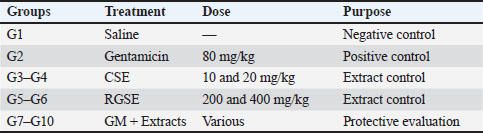
ABSTRACTBackground: Gentamicin is a widely used aminoglycoside antibiotic; however, its clinical application is limited by dose-dependent hepatotoxicity and nephrotoxicity, mainly mediated through oxidative stress, inflammation, and apoptosis. Plant-derived natural antioxidants may provide protection against drug-induced organ injury. Aim: This study aimed to evaluate and compare the protective effects of chia seed extract (CSE) and red grape seed extract (RGSE) against gentamicin-induced hepatorenal toxicity in rats. Previous studies have largely investigated these extracts separately, with limited direct comparative evidence under the same experimental conditions. In addition, the roles of oxidative stress, inflammatory mediators, and apoptotic pathways have not been fully clarified using integrated biochemical, histopathological, and immunohistochemical approaches. Therefore, this study was designed to compare the prophylactic efficacy of CSE and RGSE and to explore the mechanisms underlying their protective effects. Methods: Sixty adult male Sprague–Dawley rats were randomly assigned to 10 experimental groups (n=6/group). Hepatorenal toxicity was induced by gentamicin administration (80 mg/kg body weight). Rats received CSE (10 and 20 mg/kg) or RGSE (200 and 400 mg/kg), either alone or in combination with gentamicin, for 21 days. Liver and kidney function indices, oxidative stress biomarkers (Malondialdehyde (MDA) , Glutathione, glutathione S-transferase, superoxide dismutase, glutathione peroxidase, glutathione reductase, and vitamins C and E), histopathological changes, and immunohistochemical expression of TNF-α and Caspase-3 were assessed. Results: Gentamicin significantly increased oxidative stress, elevated serum liver enzymes (alanine aminotransferase and aspartate aminotransferase), and induced marked hepatic and renal histopathological alterations (p < 0.05). Treatment with both extracts significantly reduced MDA levels, restored antioxidant enzyme activities, improved biochemical indices, and attenuated tissue injury. RGSE exhibited greater protective efficacy than CSE, with more pronounced suppression of TNF-α and Caspase-3 expression. Conclusion: Both chia seed and RGSEs exerted significant protective effects against gentamicin-induced hepatorenal toxicity through antioxidant, anti-inflammatory, and anti-apoptotic mechanisms, with RGSE showing comparatively greater efficacy. Further translational studies are warranted to confirm their clinical applicability. Keywords: Caspase-3, Chia seed, Grape seed, Oxidative stress, TNF-α. IntroductionGentamicin is one of the broad-spectrum aminoglycoside antibiotics used in treating severe bacterial infections, especially from gram-negative organisms. Due to its wide-spectrum anti-microbial activity, it is often used in the management of infections due to sepsis, pneumonia, and urinary tract infections, as well as skin and soft-tissue infections, bone- and joint-related infections (Rajput et al., 2021; Rahimi Monfared et al., 2024; Tunç et al., 2020). Although gentamicin is an effective therapeutic agent, its clinical utility is limited because of its well-established toxic effects, such as nephrotoxicity and hepatotoxicity, associated with high doses and prolonged administration (Thy et al., 2023; Nor et al., 2025). There are multiple intertwined mechanisms by which gentamicin-induced organ injury occurs. Excessive production of reactive oxygen species (ROS), which is now regarded as a significant cause of lipid peroxidation and cellular protein and DNA injury. Moreover, gentamicin triggers inflammatory and apoptotic pathways, thereby worsening structural and functional changes in hepatic and renal tissues. Like all antibiotics, gentamicin undergoes liver metabolism and renal clearance (Kaur, 2021; Zhang et al., 2024; Huang et al., 2025). Therefore, organs related to drug metabolism and excretion, including the liver and kidneys, are also susceptible to GM-induced toxicity. Although gentamicin has a well-known nephrotoxic effect, few protective strategies against gentamicin-induced hepato-renal injury have been reported to be efficient and safe (Moafa et al., 2023; Thakur et al., 2023). Owing to their antioxidant, anti-inflammatory, and antiapoptotc properties, natural products have gained much interest as possible protective agents against drug-induced organ toxicity. Chia seeds (Salvia hispanica L.) and grape seeds (Vitis vinifera) are potent sources of food waste because of the rich biosynthesis of bioactive substances. Several phytochemical constituents, such as omega-3 fatty acids, flavonoids, and other phenolic compounds, which may alleviate oxidative stress and inflammatory responses, were also observed in Chia (Salvia hispanica) seeds. Proanthocyanidins are one of the major classes of polyphenols found in grape seeds, which is also a class of polyphenols that is highly protective to tissues against free radicals in experimental models (Cani et al., 2007; Chen et al., 2024). However, the protective effects of chia seed extract (CSE) and grape seed extract individually have been studied under different experimental conditions; hence, research regarding their comparative efficacy against gentamicin-induced hepatic and renal toxicity is still lacking. Based on data until October 2023, the majority of previous studies have focused particularly on their antioxidant actions; however, the influence of polyphenolic compounds in terms of oxidative stress, inflammation, and apoptosis within an experimental model has been less studied. Moreover, studies directly comparing histopathological, biochemical, and molecular assessments are limited. This is a known limitation due to these gaps, making it difficult to identify which natural adjunct therapy may be more effective in protecting against gentamicin-induced organ damage (Abd Elwahab et al., 2021; Layas et al., 2023; Miao et al., 2023; Mukaromah et al., 2024; Pam et al., 2024). On the basis of this background, the study was performed on the postulate that both CSE and red grape seed extract (RGSE) would protect against gentamicin-induced hepato-renal toxicity by virtue of their antioxidant, anti-inflammatory, and antiapoptotic effects but RGSE might exert a better protection as it has higher polyphenolic content. Thus, this study aimed to investigate whether the protective effects of CSE and RGSE against gentamicin-induced liver and kidney injury can be determined using histopathological, biochemical, and molecular approaches. Moreover, the extract is compared to determine which one offers the best protective effect, possibly as a natural add-on therapy against gentamicin-induced side effects (Pan et al., 2018; Thakur et al., 2023; El Dabee, Y.A. et al., 2024). Materials and MethodsCollection of plantsThe plants were collected from different areas of Sulaimani (Iraq). Chia seeds were acquired from the Khaje Xanm local market in the Sulaimani province, and grapes were collected from Surdash in the Kurdistan region in the middle of August 2024. The plant materials were authenticated by Assistant Professor Dr Ahmed Ali at the Department of Botany, College of Agriculture, University of Sulaimani, Iraq. Voucher specimens of Chia (Salvia hispanica) and grape (Vitis vinifera) seeds were prepared and deposited in the departmental herbarium under assigned reference numbers. Copies of the herbarium sheets have been included in the Supplementary Material. Grape seed extractionAfter removing the skin and pulp, the grape (Vitis vinifera) seeds were manually separated from the fruit, washed thoroughly two or three times with distilled water, and shade-dried at room temperature (25°C–30°C) until a constant weight was obtained. After drying, an electric grinding machine was used to grind them into powder. Extraction was carried out using solvent extraction methods according to the protocol described in Downey et al. (2007) with slight modifications. Briefly, 0.7 g of powdered seed material was macerated in 7 ml of 70% ethanol (1:10 w/v) at room temperature for 48–72 hours with a few shakes. The extract was filtered through Whatman No.1 filter paper and then concentrated under reduced pressure using a rotary evaporator to obtain a semi-solid crude extract. StandardizationThe extract was not a standardized single purified compound but was characterized based on its total polyphenolic content, which has been found to represent >90% of the weight (dry extract) (Downey et al., 2007; Farid et al., 2023). Gas chromatography–mass spectrometry (GC–MS) analysis was conducted on the purified extract for phytochemical profiling to identify the major bioactive constituents. Chia seed extractionChia seeds (Salvia hispanica) were washed, air-dried at ambient temperature, and ground into a fine powder. For the soil lipid fraction, extraction was first achieved using n-hexane by Soxhlet for 6–8 hours. The solvent was evaporated under reduced pressure to yield crude oil extract. GC–MS for grape seed extract (GSE) and Chia seedGas chromatography–mass spectrometry analysis was performed to identify the chemical constituents of GSE and CSE. The GC–MS system was coupled with a capillary column (e.g., HP-5MS). The operating conditions were as follows: Helium gas at a flow rate of 1.0 ml/minute for 1 µl (split mode) at 250°C and electron impact of 70 eV. For chia seed extraction, we would outline the conditions such as the ion source temperature (e.g., 230°C), the transfer line temperature (e.g., 250°C), and the mass scan range (e.g., 50–550 m/z). These settings were chosen to optimize the ionization and detection of the compounds expected from chia seeds. Similar parameters would be detailed for grape seed extraction, potentially with adjustments. For instance, the ion source temperature might be slightly different (e.g., 200°C), the transfer line temperature could be similar or adjusted (e.g., 240°C), and the mass scan range might also be tailored (e.g., 40–600 m/z) based on the anticipated compounds. The data used for the identification of compounds by GC–MS, including detailed chromatograms and spectra, were omitted from the original submission. Consistent with previous studies (Zager, 2007; Senila et al., 2020; Zhang et al., 2024), chia seeds are a source of polyunsaturated fatty acids (>80%) and antioxidant compounds, including omega-3 fatty acids. Experimental animals and designSixty adult male Albino Sprague–Dawley rats (250–300 g, 6–8 weeks old) were used in this study. Animals were randomly allocated into 10 experimental groups (n=6 per group). The sample size (n=6 per group) was determined based on previously published studies and supported by a power-based estimation (α=0.05, power=80%), indicating that this number is sufficient to detect significant biological differences while adhering to ethical principles of animal use. All animals were maintained under standard laboratory conditions (12-hour light/dark cycle, 23°C–25°C) with free access to food and water. The experimental animals were divided into 10 groups,each comprising 6 rats designated as follows (Table 1): Table 1. The experimental design and treatment policy in the studied groups.
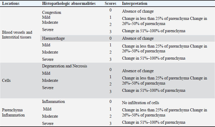
1. Group 1 (G1): Control-negative Rats in this group were administered an equivalent volume of saline (0.9%) for 21 days. 2. Group 2 (G2): Control-positive The rats in this group were administered 80 mg/kg gentamicin for 21 days. 3. Group 3 (G3): Chia seed (Low dose) The rats in this group were administered chia seed at a dose of 10 mg/kg body weight for 21 days. 4. Group 4 (G4): Chia seed (High dose) The rats in this group were administered chia seed at a dose of 20 mg/kg body weight for 21 days. 5. Group 5 (G5): Low-dose grape seeds The rats in this group were administered 200mg/kg of grape seed for 21 days. 6. Group 6 (G6): Grape seed (High dose) The rats in this group were administered 400 mg/kg of grape seed for 21 days. Chia and grape seed extract dosing variation reflects differences in phytochemical composition and bioavailability. Each extract was administered at doses previously reported to be biologically effective and safe. The study was designed to independently evaluate their protective effects rather than to compare them on a dose-equivalent basis. 7. Group 7 (G7): Gentamicin + Chia seed (low dose) The rats in this group were administered gentamicin and chia seed at a dose of 10 mg/kg body weight for 21 days. 8. Group 8 (G8): Gentamicin + Chia seed (high dose) The rats in this group were administered gentamicin and chia seed at a dose of 20 mg/kg body weight for 21 days. 9. Group 9 (G9): Gentamicin + Grape seed (Low) Gentamicin and grape seed were administered to rats in this group at a dose of 200 mg/kg body weight for 21 days. 10. Group 10 (G10): Gentamicin + Grape seed (High dose) The rats in this group were administered gentamicin and grape seed at a dose of 400 mg/kg body weight for 21 days. A 21-day experimental period was selected to model subacute exposure, allowing sufficient time for the development of cumulative organ toxicity, and to evaluate the sustained protective effects of the tested extracts. Gentamicin was administered intraperitoneally, while both grape and chia seeds were dissolved in normal saline and administered by oral gavage. The dose ranges for CSE (10 and 20 mg/kg) and RGSE (200 and 400 mg/kg) were selected from previously reported biologically effective and safe ranges for each extract. These extracts were not administered on a dose-equivalent basis because they differ substantially in phytochemical composition, extract yield, potency, and bioavailability. Therefore, the present study was designed to compare their protective profiles within their respective active dose ranges, rather than to establish the absolute superiority of one extract over the other. Serum biochemical assaysAnesthesia was induced with ketamine hydrochloride (Sigma-Aldrich, St. Louis, MO, USA) and xylazine hydrochloride (Sigma-Aldrich). On day 21 post-experiment, blood samples (approximately ~5 ml) were obtained via cardiac puncture and centrifuged at 3,000 rpm for 15 minutes to obtain serum. Blood tests were performed from a complete blood count using an automated hematology analyzer (Sysmex Corporation, Kobe, Japan). The obtained serum was then used to estimate alanine aminotransferase (ALT), alkaline phosphatase (ALP), aspartate aminotransferase (AST), total protein, albumin, bilirubin, triglycerides, total cholesterol, low-density lipoprotein, and high-density lipoprotein using commercially available assay kits (Bio-Rad Laboratories, Hercules, CA, USA). All biochemical analyses were performed according to the manufacturer’s instructions using an automated enzyme immunoassay analyzer (Bio-Rad Laboratories, Hercules, CA, USA). Assessment of enzymatic and nonenzymatic markers of oxidative stressAt the end of the experimental period, the rats were employed to induce anesthesia by using Ketamine and Xylazine; their livers and kidneys were promptly removed, immersed in cold phosphate buffer solution (pH 7.4) at 4°C, blotted with filter paper, and weighed. One gram of liver was used to generate a 10% tissue homogenate using the identical buffer solution and the Omnitissue homogenizer (10 mm). The homogenate was then incubated for 10 minutes at 4°C; the homogenate level of lipid peroxide was as follows: Malondialdehyde (MDA), glutathione (GSH), glutathione S-transferase (GST), superoxide dismutase (SOD), CAT, GGT, glutathione reductase, and Vit E ascorbic acid and glutathione peroxidase (GPx) contents were measured using standard kits for each test. Histologic examinationLiver and kidney tissue samples were collected from each rat in each group. The specimens were fixed in 10% neutral buffered formalin for a minimum of 48 hours, subsequently dehydrated through a graded series of ethanol, embedded in paraffin, and deparaffinized in xylene. They were then consecutively hydrated in 100%, 95%, 70%, and 60% ethanol, followed by two washes in phosphate-buffered saline (PBS) at the Anwar Shexa Medical City/Sulaimani Governorate Histopathology Lab. Four thin sections (4 μm) from each tissue were mounted on standard and positively charged glass slides for hematoxylin and eosin staining and immunohistochemical staining for TNF-alpha and Caspase-3. Histopathologic assessmentA histopathologist examined the slides in a descriptive-analytical study using an Olympus light microscope (Japan) at 100X and 400X magnification to assess the severity of the liver, kidney, and parenchyma. Histological slices were examined and recorded using an AmscopeTM microscope (Japan) connected to a computer via a webcam. The level of damage to the liver and kidney was evaluated semi-quantitatively by computing the percentage of the damaged region, as shown in Table 2, according to the methodology of Hassan et al. (2022). Subsequent parameters or lesions were evaluated to score and grade the pathological changes in the kidney: Grade 0 denotes unaltered renal tubules and glomeruli. Grades 1–4 are characterized by congestion accompanied by interstitial bleeding and degeneration of renal glomeruli and tubular epithelial cells without significant necrosis (Zhang et al., 2025) with modification. Grade 1: Minor alteration affecting up to 25%; change commencement. Table 2. Score interpretation for different parameters of liver and kidney lesions (Hassan et al., 2022).
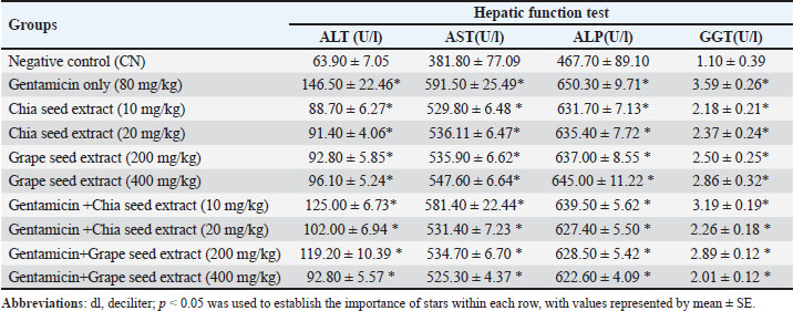
Grade 2: Moderate alteration encompassing 26%–50%. Grade 3: Moderate to severe alteration affecting 51%–75% of the patients. Grade 4: Significant alteration encompassing ≥75%; extensive alterations detected. Immunohistochemical analysisTissue sections were treated with 10 mM citrate buffer (pH 6.0) at 95°C–100°C for 20 minutes for antigen retrieval in a microwave oven, followed by a 15-minute peroxidase blocking step. Nonspecific bindings were inhibited using 5% bovine serum and 0.1% Triton X-100 in PBS prior to a 1-hour incubation with polyclonal primary TNF-alpha (1:600, Biorybt, UK) and polyclonal Caspase-3 (1:400, Biorybt, UK). After three washes in PBS, the sections were incubated with a biotinylated secondary antibody for 30 minutes and then treated with horseradish peroxidase-streptavidin (Biorybt, UK) for 60 minutes. After washing in PBS, the sections were exposed to diaminobenzidine substrate-chromogen solution for 2–5 minutes at room temperature to observe the reaction products (Biorybt, UK). The sections were counterstained with hematoxylin, dehydrated according to standard procedures, and mounted with the appropriate media before being covered with coverslips. Image analysis software was used to scrutinize the slices and assess the extent and intensity of positive immune cells (H score). The immunopositive cells exhibited cytoplasmic staining with brownish granules of TNF and Caspase-3, whereas the nuclei were unstained and retained a bluish hue. Levels of TNF-alpha and Caspase-3 staining were evaluated on a scale ranging from weak (+1) to strong (+4), including moderate (+2) and moderate-to-strong (+3) categories. Immunohistochemical staining of TNF-alpha and Caspase-3 involved quantification of the proportion of positively targeted cells, including hepatocytes, renal collecting tubules, and inflammatory cells. The staining was categorized as follows: 0 for 0% positive staining, 1 for 6%–20% positive staining, 2 for 21% positive staining, 3 for 41%–65% positive staining, and 4 for > 65% positive staining. A positive reactivity extent was characterized by a total staining score between 0 and 16, with enhanced staining intensity. Statistical analysisData are expressed as mean ± standard error. The Shapiro–Wilk test was used to assess normality. Statistical comparisons among groups were performed using one-way analysis of variance followed by Tukey’s post hoc test. Statistical significance was set at p < 0.05. Analyses were conducted using GraphPad Prism version 7. Ethical approvalAuthorization for the research was obtained from the local ethical council for animal experiments at the College of Veterinary Medicine, University of Sulaimani (permission 0305; March 9, 2024). ResultsThe influence of grape seed extract (GSE) and CSE on gentamicin hepatic function markersAnalysis of hepatic function markers, including ALT, AST, ALP, and GGT, showed that gentamicin administration significantly (p <0.05) elevated these enzymes. Although the dispersion was relatively wide, it was assessed for normality (the data were normally distributed according to the Shapiro–Wilk test) in several groups, and no significant deviation from normality was observed. The homogeneity of variance was also confirmed before analysis (Table 3). Table 3. Assessment of hepatic function markers in response to gentamicin and plant extract treatments.
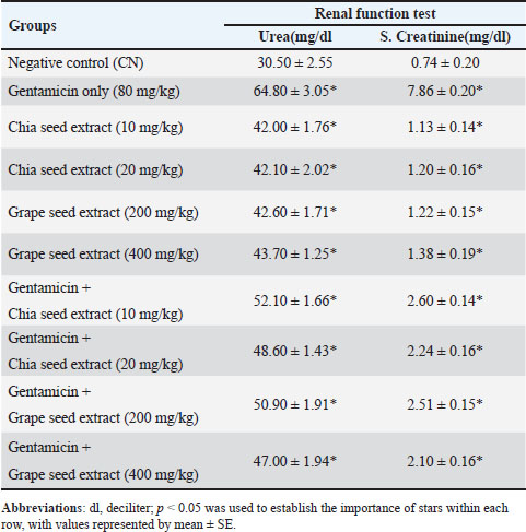
Serum ALT levels were significantly higher in the group treated with gentamicin (146.50 ± 22.46 U/l) than in the control group (63.90 ± 7.05 U/l). Potent hepatic injury was induced by gentamicin administration (p < 0.01). Similarly, the AST levels increased significantly in the gentamicin group (591.50 ± 25.49 U/l), again supporting hepatotoxicity (Table 3). Significant decreases in INF and GTF levels were observed following treatment with chia seed extract and grape seed extract compared with the gentamicin group. Grape seed extract (400 mg/kg) induced a better effect than CSE, as evidenced by a more pronounced decrease in enzyme levels. Renal function markers in response to treatment with gentamicin and plant extractsThe analysis of renal function markers showed significant (p < 0.05) alterations in both urea and serum creatinine levels across the experimental groups (Table). Specifically, urea levels, a critical indicator of kidney function, were significantly (p < 0.05) elevated in the gentamicin-treated group compared with the control group. To the negative control group, when gentamicin was combined with CSE (10 mg/kg and 20 mg/kg) and grape seed extract (200 mg/kg and 400 mg/kg), statistically significant (p < 0.05) reduction in serum urea and creatinine levels was observed indicating a partial protective effect against gentamicin-induced nephrotoxicity, but not a full reversal of gentamicin-induced nephrotoxicity (Table 4). Table 4. Renal function markers in response to Gentamicin and plant extract treatments.
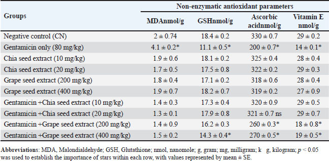
Attenuation of oxidative stress by gentamicin induced GSE and Chia SENonenzymatic antioxidant parametersGentamicin-mediated MDA expression was significantly (p < 0.05) higher compared with the negative control. Chia and grape seed extracts lowered MDA by 1.3–1.9 nmol/g, indicating protective effects. GSH levels decreased in the gentamicin group versus the CSE abated GSH levels, while grape seed extract exerted variable effects: a significant (p < 0.05) decrement at high dose. Ascorbic acid and vitamin E were similarly decreased by gentamicin, and the addition of chia and grape maintained these parameters in the range near the control values (Table 5). Table 5. Non-enzymatic antioxidant parameters in response to Gentamicin and plant extract treatments.
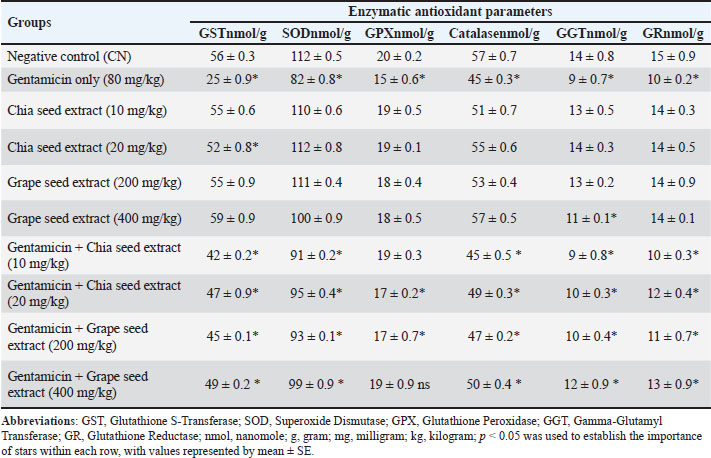
Impact of GSE and chia SE on antioxidant enzyme parametersGentamicin clearly decreased the activity of GST, SOD, GPX, and catalase in the treated groups, but both extracts (chia and grape seed) increased their levels maintained into normal level significantly (p < 0.05). GGT and GR activities were significantly decreased (p < 0.05) by gentamicin; however, both extracts protected the enzymes to a certain extent, with partial recovery in combined treatments (Table 6). Table 6. Enzymatic antioxidant parameters in response to Gentamicin and plant extract treatment.
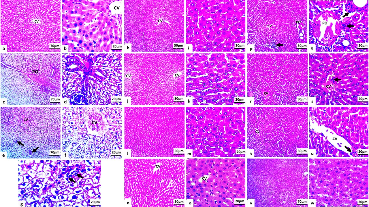
Impact of GSE and chia SE on the histopathologic alteration of gentamicinThe microscopic features of the liver in the control group (Fig. 1a,b) revealed normal histologic organization and intact morphology of the central vein, which was surrounded by plates of polygonal hepatocytes with intact cytoplasm and nucleus. The cells were separated by sinusoidal capillaries, and the portal area had normal morphology (Fig. 1a,b). In comparison to the control group, the rat treated with gentamicin presented alteration in the liver parenchyma and recorded severe lesions; the central vein, sinusoid, and portal vein underwent marked congestion (Fig. 1c–g) (Fig. 1b–f). The hepatocytes were enlarged, two to three times larger than normal, and looked like a balloon with eccentrically located nuclei (ballooning degeneration or hydropic degeneration). Neutrophils, infiltrated throughout the liver parenchyma (Fig. 1b–g). Regarding the treatment groups (Fig. 1h–o), for example, the group that was treated with only chia seed showed cellular swelling; enlargement of hepatocyte with centrally located nuclei that leads to narrowing of sinusoidal capillary in mild degree in G3 versus the G4 represented as the mild-to-moderate degree of swelling (Fig. 1h–k), also the G5 and G6 that treated with grape seed revealed mild-to-moderate cellular swelling; mild in G5 versus G6 showed mild-moderate cellular swelling (Fig. 1l–o). The addition of chia seed and grape seed improved the damage impact of gentamicin and showed moderate congestion of the central vein, sinusoid, and portal vein in G7, mild swelling of the hepatocyte, and mild infiltration of neutrophils in the liver parenchyma (Fig. 1p,q), in comparison to G8 that revealed mild-moderate congestion of the hepatic vasculature with mild swelling of cellular walls and minimal inflammatory cell reaction (Fig. 1r,s). The groups that were treated with grape seed attenuated the side effects of gentamicin significantly versus chia seed and showed only mild congestion of the central vein and mild cellular swelling with few inflammatory reactions in G9 (Fig. 1t,u), in comparison to the liver section in G10 that presented only mild swelling of hepatocytes (Fig. 1v,w).
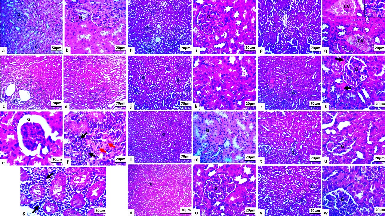
Fig. 1. Microscopic section of the liver of a rat; a and b: Normal histologic features of the liver parenchyma in the negative control group. c–g: Marked congestion in the hepatic vasculature in sections c and d. Severe hydropic degeneration of hepatocytes with moderate inflammatory reaction, indicated by a black arrow in G2. h and i: Mild cellular swelling in G3. j and k: Mild-to-moderate hepatocyte swelling in G4. l and m: Mild enlargement of G5 liver cells. n and o: Mild-moderate cellular swelling in G6 cells. p and q: Moderate congestion of the vasculature, mild hepatocyte swelling with mild neutrophil infiltration (black arrows) in G7. r and s: Mild-to-moderate vascular congestion with mild hepatocyte swelling in the G8 group. t and u: Mild central vein congestion and mild cellular swelling in G9. v and w: Mild hepatocyte swollen in G10, CV; central vein; PO; portal vein (hematoxylin and eosin stain). Microscopic features of the kidney (Fig. 2a,b) in the negative control group showed normal histologic structures of glomeruli and proximal and distal convoluted tubules without any evidence of inflammation in renal interstitial tissue (Fig. 2a,b). In the positive control group (G2), the gentamicin-induced alteration in the glomeruli was characterized by swelling with dilation of Bowman’s space. In a few sections, focal atrophy of the glomeruli was observed (Fig. 2). The proximal and distal convoluted tubules
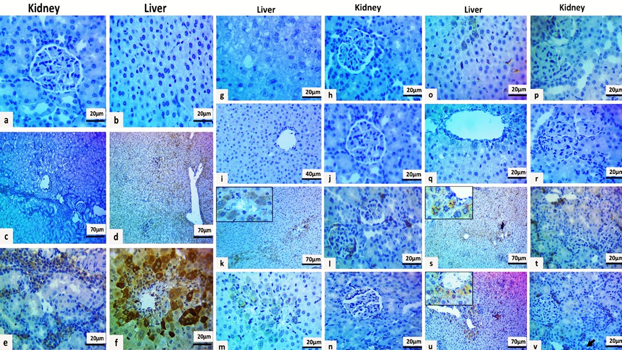
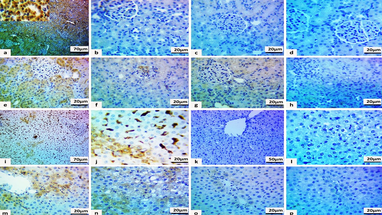
Fig. 2. Microscopic section of rat kidney exhibited; a and b: Normal histologic features of kidneys in the negative control group (G1). c–g: Marked degeneration of glomeruli (G) with focal atrophy of the tuft capillary in section e, marked renal tubule necrosis (red arrows) with neutrophils in the interstitial tissue (black arrows) in section G2. h and i: Normal histologic kidney structures in G3. j and k: Mild swelling of renal tubules in G4. l and m: Normal glomeruli and renal tubules in G5, respectively. n and o: Mild cellular swelling in G6. p and q: Mild-to-moderate swelling of renal tubules with vascular congestion (CV), mild inflammatory reaction (inset) in G7. r and s: Mild swollen tubules with interstitial exudate (black arrows) in G8. T–w: Moderate-to-mild cellular swelling of proximal and distal convoluted tubules in G9 and G10, respectively, (hematoxylin and eosin stain). showed degeneration, particularly swelling. In few sections, renal tubules showed all features of necrosis, including swollen renal tubules with eosinophilic cytoplasm and pyknotic nuclear changes (Fig. 2f) and marked interstitial neutrophil seen (Fig. 2c–g). The kidneys of rats treated only with chia and grape seed remained normal, with cellular swelling; in G3, the kidney parenchyma remained intact (Fig. 2h,i), versus G4, which showed mild swelling of proximal and distal convoluted tubules, while the renal corpuscles remained normal (Fig. 2j,k). In addition, all kidney sections in G5 showed normal histologic features (Fig. 2l,m), versus G6, which revealed mild tubular swelling with intact glomeruli (Fig. 2n,o). The administration of chia and grape seed for groups that were treated with gentamicin had a great impact in reducing the alteration in the kidney. In G7, the kidney revealed mildly swollen renal tubules with congestion of the renal vasculature and mild infiltration of neutrophils in the interstitial space (Fig. 2p,q) versus G8, which showed mild swelling in the epithelial lining of proximal and distal convoluted tubules with interstitial exudate and minimal infiltration of neutrophils (Fig. 2r,s). While grape seed improved the effect, it only showed moderate swelling in renal tubules in G9 (Fig. 2t,u), versus mildly swollen in G10 (Fig. 2v,w). The expression of the TNF-α and Caspase-3 (Fig. 3) in the liver and kidney differed significantly among the studied groups, reflecting the dynamic role of both extracted plants in attenuating the injurious effect of gentamicin. Regarding TNF-α expression in the control negative groups, there was no expression (score=0) compared with the control positive group, which was prominently expressed by diffuse-strong staining in inflammatory cells in the renal interstitial tissue (score=12) and inflammatory and liver parenchyma (score=12) (Fig. 3a–d). TNF-α expression was detected focally by weak positive expression in hepatocytes (score=1) with no positive cells in the kidney (score=0) in the 10 mg/kg chia seed-treated group (Fig. 3g,h), while no expression was seen in (score=0) in 20 mg/kg chia seed-treated group (score=0), which is an indicator of reducing inflammatory reaction (Please label or cite the figure for each interpretation Fig. 3i,j). Also, the rate of expression improved by using the chia seed to diffuse moderate staining in inflammatory and liver parenchyma with renal interstitial inflammatory cells (score=8) in the gentamicin + 10 mg/kg chia seed-treated group (Fig. 3k,l) versus the gentamicin + 20 mg/kg chia seed-treated group, in which the expression of TNF-α was focal-moderate positive expression in hepatocytes and renal interstitial (score=4 Fig. 3m,n). This proposes the improvement in inflammatory damage induced by gentamicin.
Fig. 3. Immunohistochemical sections of the liver and kidney for TNF-α expression in the studied groups; a and b: No immunostaining in the control group. c and d: Diffuse-strong staining expression in the control positive group. e and f: Diffuse-strong staining expression in the positive control group. g and h: Focal, weak positive expression in hepatocytes with no immunostaining in the kidney in the 10 mg/kg chia seed-treated group. i and j: No positive staining in the group treated with 20 mg/kg chia seed. k and l: Diffuse-moderate staining expression in the group treated with gentamicin + 10 mg/kg chia seed. m and n: focal-moderate positive expression in the gentamicin + 20 mg/kg chia seed-treated group. o–r: No immunostaining in the 200 mg/kg and 400 mg/kg GSE-treated groups, respectively. s and t: Focal-moderate staining expression in the gentamicin + 200 mg/kg GSE group. u and v: Focal-weak positive expression in the group treated with gentamicin + 400 mg/kg GSE. In the kidney section (Fig. 4), diffuse-strong positive Caspase-3 expression (score=12) was detected in the positive control group (Fig. 4a), suggesting increased damage and dead cells. Caspase 3 expression was not detected in GSE-treated groups (Fig. 4d), with weak expression in the chia seed groups (score=1 Fig. 4b,c). However, the addition of Chia seed reduced the number of dead cells and was seen as diffuse-moderate staining expression (score=8) in the gentamicin+10 mg/kg Chia seed treated group, Fig. 4e with diffuse-weak positive expression (score=4) in the gentamicin+20 mg/kg Chia seed treated group (Fig. 4f), in which indicator of improvement by Chia seed versus to the treated groups by GSE showed diffuse, weak-moderate staining expression (score=6) in the gentamicin+200 mg/kg GSE treated group (Fig. 4g) and focal-weak positive expression (score=2) in the gentamicin+400 mg/kg GSE treated group (Fig. 4h). This suggests a peak in reducing the activity of gentamicin.
Fig. 4. Immunohistochemical sections of the kidney and liver for Caspase-3 expression in the studied groups showed: a: Diffusestrong positive expression in the positive control group. b and c: Weak expression in the chia seed groups. d: No positive GSE staining in the GSE-treated groups. e: Diffuse-moderate staining expression in the group treated with gentamicin + 10 mg/kg chia seed. f: Diffuse-weak positive expression in the group treated with gentamicin + 20 mg/kg chia seed. g: Diffuse, weak-moderate staining expression in the gentamicin+200 mg/kg GSE-treated group. h: Focal-weak positive expression in the group treated with gentamicin + 400 mg/kg GSE. i and j: Diffuse-strong positive expression in the positive control group. k: No expression in the chia seed group. l: No positive GSE staining in the GSE-treated groups. m: Focal, moderate-to-strong staining expression in the group treated with gentamicin + 10 mg/kg chia seed. n: Diffuse-weak positive expression in the group treated with gentamicin + 20 mg/kg chia seed. o: Focal-moderate staining expression in the gentamicin + 200 mg/kg GSE-treated group. p: Focal-weak positive expression in the group treated with gentamicin + 400 mg/kg GSE. The expression of caspase-3 in the liver showed (Fig. 4) extreme expression in the control positive group (score=12 Fig. 4i,j). Expression was not detected in the chia seed and GSE groups (score=0 Fig. 4m,n). Treatment with different doses of chia seed reduced the dead cells by focal, moderate-to-strong staining expression (score=6) in gentamicin + 10 mg/kg to diffuse-weak positive expression (score=4) in gentamicin +20 mg/kg (Fig. 4k,l) in comparison to groups that were treated with GSE that improved the alteration into focal-moderate staining expression (score=4) in gentamicin + 200 mg/kg to focal-weak positive expression (score=2) in gentamicin + 400 mg/kg (Fig. 4o,p), suggesting the impact of GSE in reducing the oxidative stress of gentamicin. DiscussionIn the present study, gentamicin administration induced marked hepato-renal injury, as evidenced by altered liver and kidney function markers, increased oxidative stress, severe histopathological lesions, and enhanced expression of TNF-α and Caspase-3. These findings are consistent with the well-established toxic profile of gentamicin, which is mainly mediated through oxidative stress, inflammation, and apoptosis, particularly in the liver and kidneys, which are involved in drug metabolism and excretion (Gamaan et al., 2023; Moafa et al., 2023; Elbarbary et al., 2024; Desu et al., 2025; Klementa et al., 2025). Significant elevation in ALT, AST, ALP, and GGT, together with histopathological alterations including hepatocellular degeneration, vascular congestion, and inflammatory cell infiltration, reflected the hepatotoxic effect of gentamicin. Increased serum urea and creatinine levels and renal histopathological changes characterized by tubular degeneration, glomerular alterations, and interstitial inflammatory infiltration confirmed nephrotoxicity. These observations are in agreement with previous reports describing gentamicin-induced hepatic and renal injury as a consequence of ROS overproduction, membrane lipid peroxidation, mitochondrial dysfunction, and pro-inflammatory pathway activation (Abd Elwahab et al., 2021; Alkhedaide, 2024; Behvandi et al., 2025). Chia seed extract and RGSE attenuated gentamicin-induced toxicity; however, the co-administration of either extract improved biochemical parameters, reduced lipid peroxidation, restored antioxidant defenses, reduced histopathological damage, and decreased TNF-α and Caspase-3 expression (Mohr, 2008; Kothekar et al., 2020). These protective effects are biologically plausible because both extracts contain antioxidant phytochemicals that can reduce oxidative injury and limit downstream inflammatory and apoptotic responses. Previous studies have similarly reported the protective effects of grape seed-derived polyphenols and chia-derived bioactive compounds in experimental models of tissue injury (Salem and Salem, 2011; Abd Elwahab et al., 2021; Heo et al., 2022; Mojiri-Forushani et al., 2022; Chen et al., 2024). Importantly, this study provides a direct head-to-head comparison between CSE and RGSE under the same experimental conditions. The present head-to-head data suggest that RGSE provided stronger protection against histopathological and apoptotic injury, whereas CSE showed greater preservation of selected non-enzymatic antioxidant reserves, indicating complementary rather than uniformly superior effects. In the present model, RGSE was more effective in improving tissue architecture and suppressing Caspase-3 and TNF-α expression, especially at higher doses, suggesting stronger anti-apoptotic and anti-inflammatory activity. This finding may be related to the high polyphenolic and proanthocyanidin content of grape seed extract, which has been widely associated with potent free-radical scavenging, membrane stabilization, and apoptosis-related pathway modulation (Salem and Salem, 2011; Sur et al., 2023; Sivanandy et al., 2024; Madbouly et al., 2025). In contrast, CSE showed a more favorable effect on selected non-enzymatic antioxidant parameters, particularly glutathione, vitamin C, and vitamin E. This pattern may indicate that CSE contributes more strongly to the preservation of endogenous antioxidant reserves and redox balance than to maximal structural tissue protection. Such an effect may be explained by its content of omega-3 fatty acids and phenolic constituents, which have been associated with antioxidant and anti-inflammatory actions in previous studies (Senila et al., 2020; De Paula Dias Moreira et al., 2022; Chen et al., 2024). Therefore, the current findings suggest that the 2 extracts do not act identically; rather, they appear to exert overlapping but distinct protective mechanisms. The literature generally supports the protective potential of both extracts, but most previous studies have evaluated them separately and have focused mainly on antioxidant endpoints. Reports on grape seed extract frequently emphasize its strong polyphenolic antioxidant activity and capacity to improve renal and hepatic injury markers, including gentamicin-induced nephrotoxicity, in toxicological models (Salem and Salem, 2011; Abd Elwahab et al., 2021). In contrast, studies on chia seed have highlighted its nutritional and antioxidant properties, but direct evidence of its role in gentamicin-induced hepato-renal injury remains limited (Chen et al., 2024; Hasan et al., 2024). Therefore, this study extends the existing literature by offering a comparative assessment of CSE and RGSE using integrated biochemical, histopathological, and immunohistochemical endpoints in a single experimental model. Although both extracts improved renal function, urea remained relatively elevated in some treated groups despite the improvement in creatinine and tissue morphology. This may reflect incomplete functional recovery, persistent subclinical tubular dysfunction, or slower normalization of urea compared with other markers. Such partial recovery has also been reported in other nephrotoxicity studies, indicating that biochemical restoration may lag behind structural improvement (Mojiri-Forushani et al., 2022; Alkhedaide, 2024; Jitang Shah et al., 2024; Shah and Patani, 2024). This point is important because it suggests that longer treatment duration or different dosing strategies may be required for full functional normalization. This study has some limitations that should be acknowledged. First, the comparison between CSE and RGSE was not fully dose-equivalent because the administered doses differed substantially between extracts. Accordingly, the present data are more suitable for comparing efficacy under the tested conditions than for a strict potency comparison. Second, although GC–MS characterization was described, full phytochemical profiling and standardization of the extracts were not comprehensively established, which limits the precise attribution of the observed effects to specific active constituents. In addition, the study used a prophylactic coadministration design, which demonstrates protective potential but does not fully address whether these extracts can reverse already established organ injury. Overall, both CSE and RGSE mitigate gentamicin-induced hepatorenal toxicity through antioxidant, anti-inflammatory, and antiapoptotic mechanisms. However, their protective profiles were not identical: RGSE showed stronger protection against histopathological and apoptotic injury, whereas CSE showed better preservation of selected nonenzymatic antioxidant reserves. These results support the potential value of both extracts as natural adjunctive agents and provide a clearer comparative basis for future studies aimed at optimizing plant-based strategies against GP-associated organ damage. ConclusionChia seed extract and RGSE significantly attenuated gentamicin-induced hepatic and renal injury by modulating oxidative stress, inflammation, and apoptotic pathways. At the tested doses, RGSE was more effective in reducing histopathological damage and apoptotic marker expression, whereas CSE was more effective in preserving selected non-enzymatic antioxidant parameters, including GSH, vitamin C, and vitamin E. These findings indicate that both extracts provide protection through complementary mechanisms.” Also, harmonize the abstract conclusion with the same interpretation. At the tested doses, RGSE showed greater protection in terms of histopathological and apoptotic outcomes, whereas CSE was more effective in preserving selected nonenzymatic antioxidant parameters. AcknowledgmentWe thank the University of Sulaimani, especially the histopathology laboratory team of the College of Veterinary at Anwar Shexa Hospital, for IHC support. Conflict of interestThe authors have no conflicts of interest to declare FundingNone. Authors’ contributionRKM: study conception/design, experiments, data acquisition, and drafting; NAS and SMAH: supervision, methodology, and critical revision. SMAH: proposal concept, data analysis, and histopathological examination. Both authors have approved the final version of the manuscript. Data availabilityAll data are included in this manuscript. ReferencesAbd Elwahab, S.A., Mohamed, M.M., Abdel-Maksoud, A.M. and Sadek, E.M. 2021. Nephro-protective effects of grape and guava seeds extracts on gentamicin-induced nephrotoxicity in rats. J. Environ. Sci. 50, 333–357. Alkhedaide, A.Q. 2024. Impacts of gentamycin toxicity: nephroprotective role of guarana through different signaling pathways. Toxicol. Res. (Camb). 13, e167. Behvandi, M.M., Sabbagh, S., Rostami, R., Moradipour, A., Karami, M., Jafarian., Ashkan, J, Marjan, J., Leila, J. and Reza, N. 2025. Effect of gallic acid in mitigating hepatorenal injuries induced by gentamicin administration in male Wistar rats. Naunyn. Schmiedebergs. Naunyn-Schmiedeberg’s Arch. Pharmacol. 398(12), 18035–18047. Cani, P.D., Amar, J., Iglesias, M.A., Poggi, M., Knauf, C., Bastelica, D., Neyrinck, A.M., Fava, F., Tuohy, K.M., Chabo, C., Waget, A., Delmée, E., Cousin, B., Sulpice, T., Chamontin, B., Ferrières, J., Tanti, J.F., Gibson, G.R., Casteilla, L., Delzenne, N.M., Alessi, M.-C. and Burcelin, R. 2007. Metabolic endotoxemia initiates obesity and insulin resistance. Diabetes 56(7), 1761–1772. Chen, J., Wu, G., Zhu, L., Karrar, E. and Zhang, H. 2024. Functional activities of chia seed and mechanisms of action related to molecular targets. Food. Funct. 15, 1158–1169. Desu, H.L., Thougaard, E., Carney, B.N., Illiano, P., Plastini, M.J., Florimon, Y., Mini, A., Guastucci, C., Kang, B., Lee, J.K., Lambertsen, K.L. and Brambilla, R. 2025. TNFR2 signaling in oligodendrocyte precursor cells suppresses their immune-inflammatory function and detrimental microglia activation in CNS demyelinating disease. Brain Behav. Immun. 123, 81-98. De Paula, D.M., Bárbara, N.E., Vinícius. P., Brilhante. S.J., Renata. C.L.T., Luiz Carlos. M.L., Rodrigo. R.C., Vinícius. S.D., Helen Hermana. M.H., Frederico Augusto. R. B. and Hércia Stampini, D.M. 2022. Chia flour and oil ameliorate metabolic disorders in rats. Foods 11(3), 285. Downey, M.O., Mazza, M. and Krstic, M.P. 2007. Development of a stable extract for anthocyanins and flavonols from grape skin. Am. J. Enol. Vitic. 58, 358–364. Elbarbary, N.S., Ismail, E.A., El-Hamamsy, M.H., Ibrahim, M.Z., Elkholy, A.A. 2024. The DPP-4 inhibitor sitagliptin improves glycaemic control and early-stage diabetic nephropathy in adolescents with type 1 diabetes using the MiniMed 780G advanced hybrid closed-loop system: a randomised controlled trial. Diabetologia 67(12), 2637–2649. El Dabee, Y.A., Hassan, M.S.H., Mahmoud, H.I. and Hassan, H.M. 2024. Antioxidant properties of chia seed extract in wound healing. Minia J. Agric. Res. 44, 698–704. Farid, A., Mohamed, D., Mostafa, D., Tarek, R., Sherif, V. and Safwat, G. 2023. Novel grape seed extract nanoparticles attenuate amikacin-induced nephrotoxicity in rats. AMB. Express 13, 122–129. Gamaan, M.A., Zaky, H.S. and Ahmed, H.I. 2023. Gentamicin-induced nephrotoxicity: a mechanistic approach. Azhar Int. J. Pharm. Med. Sci. 3, 11–19. Hasan, A.F., Hameed, H.M., Hadid, M.A. and Tousson, E. 2024. Impact of chia (Salvia hispanica) seeds extract on Ehrlich ascites model-induced kidney toxicity in female mice. Asian J. Dairy Food Res. 1, 750–756. Hassan, S.M.A., Saeed, A.K., Rahim, O.O. and Mahmood, S.A.F. 2022. Alleviation of cisplatin-induced hepatotoxicity and nephrotoxicity by L-carnitine. Iran. J. Basic. Med. Sci. 25, 890–897. Heo, Y.R., Son, C.N., Baek, W.K. and Kim, S.H. 2022. Grape seed proanthocyanidin extract induces apoptotic and autophagic cell death in rheumatoid arthritis fibroblast-like synoviocytes. Arch. Rheumatol. 37, 393–403. Klementa, V., Petejova, N., Horak, P., Kurasova, E. and Zadrazil, J. 2025. Acute kidney injury due to gentamicin nephrotoxicity and specific miRNAs as biomarkers. Biomed. Pap. 169, 1–8. Kothekar, A.T., Divatia, J.V., Myatra, S.N., Patil, A., Nookala Krishnamurthy, M., Maheshwarappa, H.M., Siddiqui, S.S., Gurjar, M., Biswas, S. and Gota, V. 2020. Clinical pharmacokinetics of 3-h extended infusion of meropenem in adult patients with severe sepsis and septic shock: implications for empirical therapy against Gram-negative bacteria. Ann. Intensive Care. 10(1), 4–10. Layas, K., Chatterjee, P. and Pannala, A. 2023. Acute kidney injury: current and future therapies involving antioxidants and antioxidant formulations. Med. Res. Arch. 1–11. Madbouly, N.A., Ali, D.M. and Farid, A.A. 2025. Nanoparticles from grape seed extract inhibit inflammatory cytokines and ameliorate CCl4-induced hepatotoxicity. BMC. Complement. Med. Ther. 25, 270–276. Miao, W., Huang, R., Huang, X., Gao, F., Leng, X. and Li, Q. 2023. Physicochemical properties and in vivo hepatoprotective effect of polysaccharides from grape pomace. Antioxidants 12, 390–394. Moafa, A., Aldossary, S.A., Al Mohaini, M. and J. Alsalman, A. 2023. Protective effect of aspirin against gentamicin-induced hepatotoxicity in rats model. Biomed. Pharmacol. J. 16, 2293–2299. Mohr, J.F. Murray, B.E. 2008. Point: Vancomycin is not obsolete for the treatment of infection caused by methicillin-resistant Staphylococcus aureus. Clin. Infect. Dis. 46(2), 193-201. Mojiri-Forushani, H., Hemmati, A., Khanzadeh, A. and Zahedi, A. 2022. Effectiveness of grape seed extract in patients with nonalcoholic fatty liver: a randomized double-blind clinical study. Hepat. Mon. 22, e132309. Mukaromah, A.H., Suhartati, S., Amalia, A.A., Kamaruddin, M., Wiyarti, V.I. and Wardoyo, F.A. Effectiveness of grape seed extract on liver and heart organs in rats exposed to formaldehyde. In AIP Conf Proc, Melville, New York, NY, 2024 vol. 7, p 60008. Pam, P., Asemani, S., Azizi, M.H. and Jamilian, P. 2024. Chia seed supplementation and inflammatory biomarkers: a systematic review and meta-analysis. J. Nutr. Sci. 13, 85–91. Pan, S.C. and Rickard, T.C. 2018. Transfer of test-enhanced learning: Meta-analytic review and synthesis. Psychol. Bull. 144(7), 710–756. Rahimi Monfared, S., Valibeik, A., Tavakoli Dastjerd, N., Leila, J., Ashkan, J., Mohammad Nabi, M. and Hassan. A. 2024. Protective role of citronellol on antioxidant enzymes and oxidative damage induced by gentamicin. Mol. Biol. Rep. 51, 378–389. Rajput, S.A., Shaukat, A., Wu, K., Rajput, M.R., Baloch, M.D., Akhtar, R.W., Muhammad, A.R., Rafal, P., Ashraf, A. and Attala, F.E. 2021. Luteolin alleviates aflatoxin B1-induced apoptosis and oxidative stress via Nrf2 signaling pathway. Antioxidants 10, 1261–1268. Salem, N.A. and Salem, E.A. 2011. Renoprotective effect of grape seed extract against oxidative stress induced by gentamicin. Ren. Fail. 33, 824–832. Senila, L., Neag, E., Cadar, O., Kovacs, M.H., Becze, A. and Senila, M. 2020. Chemical, nutritional and antioxidant characteristics of different food seeds. Appl. Sci. 10, 1582–1589. Sivanandy, P., Manirajan, P., Wen Qi, O., Teng Khai, O,. Chun Wei, O., Wei Ying, N., Wadingasafi, NAN., Azhar, N.A., Nor Rohaizan, N.A. 2024. A systematic review of efficacy and safety of newer drugs approved from 2016 to 2023 for the treatment of complicated urinary tract infections. Ann Med. 56(1), 2403724. Shah, N.P. and Patani, P. 2024. The multifaceted health benefits of grape seed extract: a comprehensive review. Eurasian. J. Anal. Chem. 19, 429–434. Sur, A., Iflazoglu Mutlu, S., Tatli Seven, P., Aslan, A., Kizil, M., Kulaksiz, M., Hilmi Yaranoglu, M. and Esen, S. 2023. Effects of grape seed proanthocyanidin extract on drug-induced toxicity. Toxicol. Res. 39, 749–759. Thakur, M., Vasudeva, N., Sharma, S. and Datusalia, A.K. 2023. Plants and their bioactive compounds in multi-organ dysfunction syndrome. CNS. Neurol. Disord. Drug Targets 22, 1313–1334. Thy, M., Timsit, J.F. and De Montmollin, E. 2023. Aminoglycosides for treatment of resistant gram-negative infections. Antibiotics 12, 855–860. Tunç, M.A. 2020. Grape seed extract as feed additive against gentamicin toxicity in broilers. Atatürk Üniv. Vet. Sci. Fac. J. 15, 271–278. Zager, R.A. 2007. Subclinical gentamicin nephrotoxicity: role in endotoxin-driven TNF-alpha production. Am. J. Physiol. Renal. Physiol. 293, F43–F49. Zhang, D., Wu, J., Wang, Q., Liu, M., Li, W. and Wang, Y. 2024. Targeting the powerhouse: the mitochondrial perspective on gentamicin-induced nephrotoxicity. Arch Toxicol. 100(5), 1685–1697. Zhang, P., Chen, Q., Lao, J., Shi, J., Jia, C., Xialo, L. and Xin, H. 2025. Machine learning modeling for risk of acute kidney injury in patients receiving aminoglycosides. Front. Pharmacol. 16, 1538074. Supplementary MaterialGrape seed collection and preparation: In the middle of august of 2024, the grapes were taken manually from Sulaimani (Surdash, Sulaimani, Kurdistan Region, Iraq). The grapes were isolated from the skin, dried in the open air away from direct sunlight, and grounded into powder by the electrical grinder. The powders were stored in dark glass containers at −20⹅ until the use. After that sample had been washed with distilled water to remove adhering dirt and dust. Then they were dried in the shade away from direct sunlight under room temperature and humidity conditions. The dried aerial parts were ground into a fine powder using pistol, mortar and electrical blender, then it was stored in a closed airtight container at 4°C for further use. Chia seed collection and preparation: Chia seed were obtained from a local Kurdish market (Khaje Xanm, Sulaimani, Kurdistan Region, Iraq) and crushed by the electric grinder. Powders were kept in dark glass vessels at −20°C before used. Subsequently, that sample was rinsed with distilled water to remove settling dirt and dust. They were then air-dried under conditions typical of room temperature and humidity, out of direct sunlight. The aerial parts so dried were then pulverized to a fine powder using pistol, mortar and electrical blender and were stored in a closed air tightly container at 4°C for further use. | ||
| How to Cite this Article |
| Pubmed Style Majeed RK, Salih NA, Hassan SMA. Alleviation of gentamicin-induced injury in liver and kidney by impact of red grape seed and chia seed extraction. doi:10.5455/OVJ.2026.v16.i6.66 Web Style Majeed RK, Salih NA, Hassan SMA. Alleviation of gentamicin-induced injury in liver and kidney by impact of red grape seed and chia seed extraction. https://www.openveterinaryjournal.com/?mno=309473 [Access: June 28, 2026]. doi:10.5455/OVJ.2026.v16.i6.66 AMA (American Medical Association) Style Majeed RK, Salih NA, Hassan SMA. Alleviation of gentamicin-induced injury in liver and kidney by impact of red grape seed and chia seed extraction. doi:10.5455/OVJ.2026.v16.i6.66 Vancouver/ICMJE Style Majeed RK, Salih NA, Hassan SMA. Alleviation of gentamicin-induced injury in liver and kidney by impact of red grape seed and chia seed extraction. doi:10.5455/OVJ.2026.v16.i6.66 Harvard Style Majeed, R. K., Salih, . N. A. & Hassan, . S. M. A. (2026) Alleviation of gentamicin-induced injury in liver and kidney by impact of red grape seed and chia seed extraction. doi:10.5455/OVJ.2026.v16.i6.66 Turabian Style Majeed, Rebin Kanabi, Nadia Abdulkarim Salih, and Snur Mohammad Amin Hassan. 2026. Alleviation of gentamicin-induced injury in liver and kidney by impact of red grape seed and chia seed extraction. doi:10.5455/OVJ.2026.v16.i6.66 Chicago Style Majeed, Rebin Kanabi, Nadia Abdulkarim Salih, and Snur Mohammad Amin Hassan. "Alleviation of gentamicin-induced injury in liver and kidney by impact of red grape seed and chia seed extraction." doi:10.5455/OVJ.2026.v16.i6.66 MLA (The Modern Language Association) Style Majeed, Rebin Kanabi, Nadia Abdulkarim Salih, and Snur Mohammad Amin Hassan. "Alleviation of gentamicin-induced injury in liver and kidney by impact of red grape seed and chia seed extraction." doi:10.5455/OVJ.2026.v16.i6.66 APA (American Psychological Association) Style Majeed, R. K., Salih, . N. A. & Hassan, . S. M. A. (2026) Alleviation of gentamicin-induced injury in liver and kidney by impact of red grape seed and chia seed extraction. doi:10.5455/OVJ.2026.v16.i6.66 |


