





| Research Article | ||
Open Vet. J.. 2026; 16(6): 3528-3545 Open Veterinary Journal, (2026), Vol. 16(6): 3528-3545 Research Article A molecular survey of canine coronavirus among domestic dogs in Japan from 2019 to 2020Hitomi Kumano1†, Ichika Kitashin1 and Keisuke Nakagawa1,2,3,*†1Laboratory of Veterinary Microbiology, Joint Department of Veterinary Medicine, Gifu University, Yanagido, Japan 2Joint Graduate School of Veterinary Sciences, Gifu University, Yanagido, Japan 3Center for One Medicine Innovative Translational Research, Gifu University, Gifu, Japan †These authors contributed equally to this study. *Corresponding Author: Keisuke Nakagawa. Laboratory of Veterinary Microbiology, Joint Department of Veterinary Medicine, Gifu University, Yanagido, Japan. Email: nakagawa.keisuke.r0 [at] f.gifu-u.ac.jp Submitted: 06/02/2026 Revised: 22/04/2026 Accepted: 04/05/2026 Published: 05/06/2026 © 2025 Open Veterinary Journal
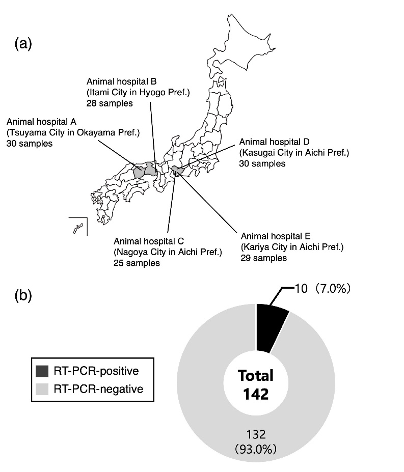
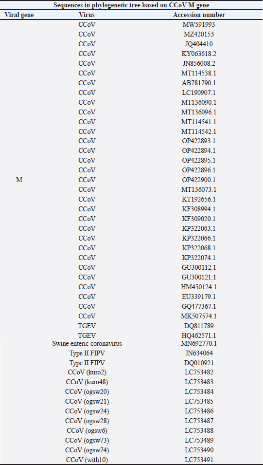
AbstractBackground: Canine coronavirus (CCoV) is an important enteric virus of dogs and is widely distributed worldwide. The virus shows considerable genetic diversity and is classified into genotypes of type I, type IIa, and type IIb. Although CCoV has been reported in various countries, information on its circulation and molecular characteristics in Japan remains limited. Aim: This study aimed to investigate the circulation and genetic characteristics of CCoV in domestic dogs in Japan between 2019 and 2020. Methods: Stool samples were collected from domestic dogs with or without diarrhea. Viral RNA was extracted from the samples and screened for CCoV by RT-PCR targeting the M gene. Positive samples were further analyzed by S gene-based reverse transcription polymerase chain reaction (RT-PCR) to determine viral genotypes, including type I, type IIa, and type IIb CCoVs. Selected PCR products were sequenced, and the genetic relationships between the detected strains and previously reported CCoVs were evaluated using phylogenetic analyses. Results: CCoV RNA was detected in a subset of the analyzed samples. Genotyping analysis identified type I, IIa, and IIb CCoVs among the positive samples. Sequence and phylogenetic analyses revealed that the detected strains clustered with previously reported CCoV lineages. These findings indicate that genetically diverse CCoVs are circulating in Japanese domestic dogs. Conclusion: This study demonstrates the CCoV circulation in domestic dogs in Japan and provides updated molecular information on circulating strains. Although the samples were collected from a limited number of regions and may not represent the nationwide situation, the results contribute to a better understanding of the molecular epidemiology and genetic diversity of CCoV in Japan. Keywords: Canine coronavirus, Domestic dogs, Molecular survey, Japan. IntroductionViral diarrhea has a high incidence in canine diseases, and various pathogens cause diarrhea in dogs. Canine coronavirus (CCoV) causes mild gastroenteritis in dogs worldwide (Licitra et al., 2014). Although the mortality rates of CCoV-derived diarrhea are not so high in adult dogs, some puppies die from CCoV infection, indicating that puppies have a higher risk for CCoV infection than do adult dogs. Although vaccines against CCoV are available worldwide, their effectiveness remains controversial (Tizard, 2020). Canine coronavirus (CCoV) is widely distributed in dog populations worldwide and remains difficult to control in many settings. CCoV belongs to the Alphacoronavirus genus in the Coronavirinae subfamily and Coronaviridae family (Pratelli, 2011). In addition to CCoV, the alphacoronavirus genus is composed of animal and human coronaviruses, such as feline coronavirus (FCoV), transmissible gastroenteritis virus (TGEV), and human coronavirus 229E. The viral genome enclosed by an envelope is a positive-sense single-stranded RNA that is approximately 29 kb in length. Upon entry into host cells, coronavirus genome expression is initiated by the translation of two large precursor polyproteins, pp1a and pp1ab, into 16 mature non-structural proteins, which are processed by viral proteinases. Many of these proteins play an essential role in viral RNA replication and transcription, leading to the translation of four canonical structural proteins: S, E, M, and N (Nakagawa et al., 2016). In addition to structural proteins, the viral genome encodes accessory proteins that suppress host innate immunity (Neuman et al., 2014; Fang et al., 2021). Among these viral proteins, the S protein is a critical component because it determines the host range, cell tropism, and pathogenicity (Regan et al., 2012). Based on differences in the sequence of the S gene encoding the S protein, CCoV is classified into type I and type II genotypes. Type I CCoV has a high level of identity in the nucleotide sequence of the S gene with FCoV (Tekes and Thiel, 2016), suggesting homologous recombination between FECoV and type II CCoV. Type II CCoV is further subclassified into two types, type Ⅱa and type Ⅱb (Decaro et al., 2009; Decaro et al., 2010; Licitra et al., 2014), based on the amino acid sequence of the N-terminal domain (NTD) of the S protein. Studies have shown that type Ⅱa CCoV has an NTD of the S protein consistent with the prototype CCoV, but type Ⅱb CCoV has a TGEV-like NTD in the S protein (Decaro et al., 2009), suggesting homologous recombination between TGEV and type Ⅱa CCoV. Hence, mutations and recombination in the S gene are involved in the promotion of CCoV genetic diversity. Several molecular surveys for CCoV in domestic dogs in Japan have shown the circulation of CCoV in domestic dogs (Bandai et al. 1999; Yachi and Mochizuki, 2006; Soma et al., 2011; Terada et al., 2014; Takano et al., 2016). Recently, canine-like coronaviruses have been detected in human patients with pneumonia, raising concerns about the potential zoonotic transmission of CCoV-related viruses (Lednicky et al., 2022; Vlasova et al., 2022). Because dogs live in close contact with humans, understanding the circulation and genetic characteristics of CCoV in domestic dogs is important from both veterinary and public health perspectives. Thus, the importance of continuous CCoV surveys in domestic dogs has been increasing. We conducted a molecular survey of CCoV in fecal samples from domestic dogs in Japan from 2019 to 2020 using reverse transcription polymerase chain reaction (RT-PCR) targeting CCoV genes. Our results indicate that the circulation of CCoV in domestic dogs in Japan is currently not as high as that in previous reports (Soma et al., 2011; Takano et al., 2016). Meanwhile, the CCoVs detected in this study in Japan are genetically close to those in several countries, especially Asian countries, suggesting that CCoVs prevailing in Asia could be transmitted to other Asian countries. The results of our study will advance a better understanding of the ecology of CCoV in domestic dogs and provide fundamental information to prevent CCoV in domestic dogs. Materials and MethodsSample collection and individual dog informationWe collected 142 feces samples from domestic dogs that were brought to animal hospitals in Aichi, Hyogo, and Okayama Prefectures in Japan from August 2019 to March 2020 (Fig. 1). Sample collection was conducted with the consent of each owner, and no invasive treatment was performed on the dogs. The Committee for Animal Research and Welfare of Gifu University approved the sampling methods (Approval Number 2019) and performed the sampling following the relevant guidelines and regulations. Information on individual domestic dogs (sex, age, breed, stool property, and immunization status) was obtained from the medical records of each animal hospital. The information for the samples is shown in Supplemental Table 1. The feces samples were diluted to 20% with phosphate-buffered saline and clarified by centrifugation at 750 × g for 10 min. The supernatants were collected and stored at -80°C until further use.
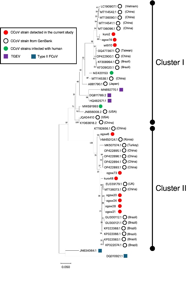
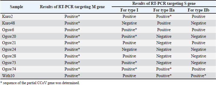
Fig. 1. Map of sampling regions and results for the detection of the CCoV M gene in stool samples. (a) Stool samples from domestic dogs were collected from 5 animal hospitals in Okayama, Hyogo, and Aichi Prefectures. The number of samples collected from the animal hospitals is shown. (b) The number of samples that tested positive and negative for RT-PCR to detect the CCoV M gene are shown. RNA extraction and RT-PCRRNAs from the specimens were extracted using the TRI reagent LS (Molecular Research Center, Inc, Cincinnati, OH, USA) and the Direct-zol RNA MiniPrep kit (Zymo Research, Irvine, CA, USA) according to the manufacturer’s instructions. cDNAs were synthesized using SuperScript Ⅲ reverse transcriptase (Thermo Fisher Scientific, Waltham, MA, USA) and random primers (Thermo Fisher Scientific). RT-PCR for detecting the CCoV M gene and genotyping the CCoV S gene were performed using Ex Taq HS (TaKaRa Bio, Shiga, Japan) under the cycling conditions and primers previously described (Pratelli et al., 1999; Erles and Brownlie, 2009)(Pratelli et al., 1999; Erles and Brownlie, 2009). Direct sequencingAll PCR products were separated on 1.0% agarose gels containing ethidium bromide and then purified using the NucleoSpin Gel and PCR Clean-up Kit (Macherey-Nagel, Duren, Germany). The purified PCR products were sequenced using the BigDye Terminator v3.1 software. A Cycle Sequencing kit (Thermo Fisher Scientific, Waltham, MA, USA) was used with an ABI Prism 3100 DNA analyzer (Thermo Fisher Scientific). Sequencing was performed using the forward and reverse primers used for the PCR reactions. The accession numbers of the determined CCoV sequences are listed in Supplemental Tables 4–7. Phylogenetic analysisSequence alignments and phylogenetic tree construction were performed using MEGA version Ⅺ software (Tamura et al., 2021). The phylogenetic tree was constructed using the maximum likelihood method with 1 000 bootstrap replicates. Ethical approvalThe Committee for Animal Research and Welfare of Gifu University (Approval Number 2019). ResultsCollection of feces from domestic dogs in Japan from 2019 to 2020Supplemental Fig. 1 shows an overview of the experimental design. A total of 142 samples from 5 animal hospitals in Okayama, Hyogo, and Aichi Prefectures were submitted to our laboratory from 2019 to 2020 (Fig. 1a). Samples from animal hospitals A, B, C, D, and E were 30, 28, 25, 30, and 29, respectively. We collected feces from domestic dogs that were taken to animal hospitals for their annual checkup or vaccinations, and obtained individual information (sex, age, breed, stool property, and immunization status) from the medical records of each animal hospital. Information for individual dogs is shown in Supplemental Table 1. Detection of the CCoV-M gene in fecal samples from domestic dogsAmong the 142 samples from domestic dogs, CCoV M genes were detected in 10 samples (7.0%) (Fig. 1b). Subsequently, we conducted genotyping using RT-PCR targeting the S gene of type I, Ⅱa, or Ⅱb (Erles and Brownlie, 2009). We found at least one of the genotypes of the S gene of CCoVs from all of the samples positive for the CCoV M gene, except for Ogsw24 (Supplemental Table 2). These results suggest that 3 genotypes of CCoV prevail among domestic dogs in Japan. In addition, some dogs were coinfected with a different genotype of CCoV (e.g., Kuro2, Ogsw6, and with10). Individual information for the dogs is shown in Supplemental Table 1 with the RT-PCR results. Phylogenetic analysis based on the partial CCoV M gene from domestic dogsTo explore the genetic relationship of CCoV strains detected from domestic dogs in Japan, a phylogenetic tree was drawn based on the ten determined partial sequences (215 bp) of the CCoV M genes (red circles) with those in other countries (white open circles) and other Alphacoronaviruses, TGEV (purple squares), and type II FCoV (blue squares) (Fig. 2). CCoVs were divided into two clusters in the phylogenetic tree based on the CCoV M genes. The CCoV M genes detected in this study were included in both clusters. Their genetic relationship was close to that of CCoVs in China, Korea, Vietnam, and Taiwan.
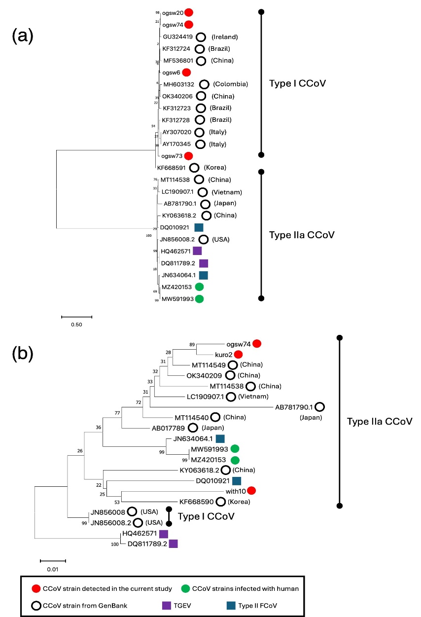
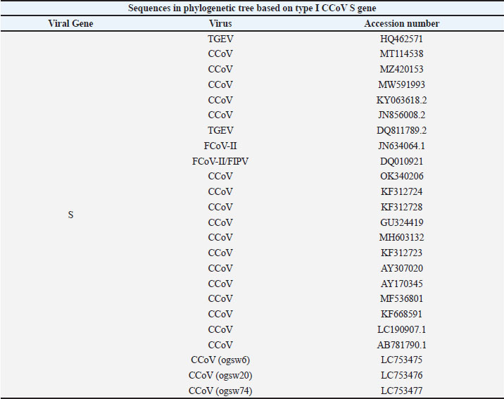
Fig. 2. Phylogenetic tree based on the partial CCoV M gene sequences detected in this study. The tree was generated using the maximum likelihood method (bootstrap value: 1 000 replicates) with MEGA version Ⅺ (Tamura et al., 2021). The phylogenetic tree was constructed based on 10 determined partial sequences (215 bp) of the CCoV M gene obtained in this study (red circles), together with corresponding sequences from other countries (open circles) and other alphacoronaviruses, including TGEV (purple squares) and type II FCoV (blue squares). Phylogenetic analysis based on the partial CCoV-S Ⅰ and Ⅱa genes detected from domestic dogsNext, we drew a phylogenetic tree based on the 4 determined sequences (292 bp) of the partial type I CCoV S genes (red circles) with those of CCoVs in other countries (white open circles) and other Alphacoronaviruses, TGEV (purple squares), and type Ⅱ FCoV (blue squares) (Fig. 3a). In the phylogenetic tree, the CCoVs were clearly divided into genotypes I and Ⅱa CCoVs. The type I CCoV strains detected in the current study were clustered with type I CCoVs in other countries. We found that they were genetically close to type I CCoV in China, Ireland, Brazil, Colombia, and Italy (Nucleotide idesntity: >95%).
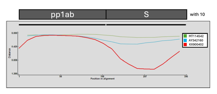
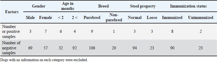
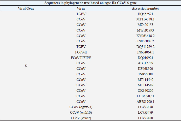
Fig. 3. Phylogenetic trees based on partial S gene sequences of type I or type Ⅱa. (a and b) Trees were generated using the maximum-likelihood method (bootstrap value: 1 000 replicates) with MEGA version Ⅺ (Tamura et al., 2021). The phylogenetic trees in panels (a) and (b) were constructed based on different partial S gene sequences of type I and IIa CCoVs. In panel (a), the phylogenetic tree was based on four determined partial sequences (292 bp) of the type I CCoV S gene obtained in this study (red circles), together with corresponding sequences of CCoVs from other countries (open circles) and other alphacoronaviruses, including TGEV (purple squares) and type II FCoV (blue squares). In panel (b), the phylogenetic tree was based on three determined partial sequences (495 bp) of the type IIa CCoV S gene obtained in this study (red circles), together with corresponding sequences from other countries (open circles) and other alphacoronaviruses, including TGEV (purple squares) and type II FCoV (blue squares). Furthermore, we generated a phylogenetic tree based on the three determined sequences (495 bp) of the partial type Ⅱa CCoV S genes (red circles) with those determined in other countries (white open circles) and other Alphacoronaviruses, TGEV (purple squares), and type Ⅱ FCoV (blue squares) (Fig. 3b). The three type Ⅱa CCoVs detected in the current study existed in the cluster of type Ⅱa and were genetically close to type Ⅱa CCoVs in Asian countries, such as China, Vietnam, and Korea (nucleotide identity: >94%). DiscussionReports of animal coronaviruses, including CCoV-like, FCoV-like, and porcine deltacoronavirus-like viruses, causing infection in humans have been accumulating (Lednicky et al., 2021; Lednicky et al., 2022; Vlasova et al., 2022). These reports emphasize the importance of continuous surveys of coronaviruses in animals for the health of both animals and humans. In the current study, we performed molecular epidemiology for CCoV in the feces of domestic dogs in Japan between 2019 and 2020. As a result, we found that 10 out of 142 feces samples (about 7%) were positive for the CCoV M gene, showing a quite low positive rate compared with previously reported rates (Soma et al., 2011; Takano et al., 2016). Our data suggest a low level of risk for the transmission of CCoV to humans in Japan. Although we obtained individual information on the dogs (sex, age in years, breed, stool property, and immunization status) (Supplemental Table 3), we could not conduct a statistical analysis to assess the risk factors of CCoV infection in domestic dogs because of the small number of samples positive for CCoV. First, we found that 3 out of 94 normal stool samples (approximately 3.1%) and 3 out of 26 loose stool samples (approximately 11.5%) were positive for CCoV. These positive rates for CCoV are quite low compared with the positive rates in previous surveys of CCoV in healthy and diarrheic dogs in Japan during the periods from 2007 to 2009 and 2011 to 2014 (Soma et al., 2011; Takano et al., 2016). Second, 8 out of 98 immunized dogs (8.2%) were positive for CCoV, whereas 2 out of 25 unimmunized dogs (8.0%) were positive, showing a low level of CCoV circulation even in unimmunized dogs. Taken together, these data suggest that CCoV circulation in domestic dogs in Japan has been controlled in recent years. According to a phylogenetic tree based on partial CCoV M genes (Fig. 2), two clusters of CCoVs were generated, and the CCoVs in the current study existed in both clusters. They were genetically close to CCoVs in China, Taiwan, Vietnam, and Korea, suggesting the potential interaction of CCoVs among Asian countries. Notably, kuro2 and ogsw74 generated an independent cluster with a high bootstrap value. This suggests that some of the current CCoVs in Japan could undergo independent evolution. From the samples positive for the CCoV M gene, we successfully amplified and determined partial S genes of types Ⅰ and Ⅱa CCoV. In a phylogenetic tree based on the partial types I and IIa CCoV S genes (Fig. 3a), the type I CCoVs in the current study generated a cluster with type I CCoVs in other countries, indicating that the determined sequences were indeed derived from type I CCoV. Interestingly, the type I CCoVs determined in the current study were genetically close to not only CCoVs in Asian countries but also CCoVs in Ireland, Brazil, Colombia, and Italy. These results may be attributed to the fact that domestic dogs that could be infected with CCoV travel with humans via airplanes or ships to many countries. In contrast to type Ⅰ CCoV, we found that type Ⅱa CCoVs were genetically close to those in Asian countries, China, Vietnam, and Korea (Fig. 3b). Taken together, the phylogenetic trees suggest the potential interaction of types Ⅰ and Ⅱa CCoVs among several countries, especially those in Asia. We could determine the sequence of the RT-PCR product from within 10 that was amplified by RT-PCR with primers, CEPol-1 and TGSP-2, targeting between the pp1ab and S genes of type Ⅱb CCoV (Erles and Brownlie, 2009) . Phylogenetic tree analysis based on the determined gene showed that with10 is classified into type Ⅱa CCoV (Supplemental Fig. 2). Recombination analysis was performed using a similarity plot generated by RDP5 to examine the possibility of recombination (Martin et al. 2021) A similarity plot between the CCoV pp1ab and S gene of with10, comparing MT114542 (type Ⅱa CCoV), AY342160 (type Ⅱa CCoV), and KX900402 (TGEV), showed that with10 was similar to type Ⅱa CCoV, but not to TGEV (Supplemental Fig. 3). Notably, in terms of comparison between with10 and TGEV, the similarity score dropped from the C-terminal region of pp1ab to the N-terminal region of the S gene and then recovered, implying that the primers for detecting the pp1ab and S genes of type Ⅱb CCoV also may bind to those of type Ⅱa CCoVs. To accumulate correct information on the circulation of type Ⅱb CCoV by RT-PCR, redesign of primers specifically binding to the N-terminl region of the S gene of Ⅱb CCoV may be required. In the current study, we found a relatively low molecular detection rate of CCoV in domestic dogs in Japan compared with previous reports (Soma et al., 2011; Takano et al., 2016), suggesting a limited current burden of CCoV in the sampled dog population. However, the results should not be interpreted as representing the nationwide situation because the samples analyzed in this study were collected from limited regions of Japan. The CCoV strains detected in this study were genetically close to CCoVs reported in Asian countries, including China, Korea, Vietnam, and Taiwan. Our results suggest that genetically related CCoVs are circulating across Asia. Continuous surveillance of CCoV in domestic dogs is important for understanding its circulation and genetic diversity and considering its potential relevance to animal and public health. AcknowledgmentsWe are grateful to Yoshiaki Kobe, Yusuke Hanada, Saeka Hanada, Naoki Watanabe, Taku Yasuda, and Hironori Doi for sampling stools from domestic dogs. FundingThis work was funded by Grants-in-Aid for Scientific Research from the Japan Society for the Promotion of Science (numbers 19K23706 and 23K05552) and a grant from the Ministry of Education, Culture, Sports, Science, and Technology, Japan, for the Joint Research Program of the Research Center for Zoonosis Control, Hokkaido University. Authors’ contributionsH.K. and K.N. equally contributed to this study. H.K. and K.N. coordinated sample collection, RNA extraction, RT-PCR assays, and sequencing experiments, and drafted the initial manuscript. I.K. organized and curated the experimental data and performed phylogenetic analyses, including the construction and interpretation of phylogenetic trees. K.N. conceived and designed the study, supervised the experiments, interpreted the data, and critically revised the manuscript. All authors discussed the results and approved the final version of the manuscript. Conflicts of InterestThe authors have no conflicts of interest to declare. Data availabilityThe generated nucleotide sequence data have been deposited in the GenBank database under the accession numbers listed in Supplementary Tables 4–7. All other data supporting this study’s findings are available within the article and its supplementary materials. ReferencesBandai, C., Ishiguro, S., Masuya, N., Hohdatsu, T. and Mochizuki, M. 1999. Canine coronavirus infections in Japan: virological and epidemiological aspects. J. Vet. Med. Sci. 61, 731–736. Decaro, N., Mari, V., Campolo, M., Lorusso, A., Camero, M., Elia, G., Martella, V., Cordioli, P., Enjuanes, L. and Buonavoglia, C. 2009. Recombinant Canine Coronaviruses Related to Transmissible Gastroenteritis Virus of Swine Are Circulating in Dogs. J. Virol. 83, 1532–1537. Decaro, N., Mari, V., Elia, G., Addie, D.D., Camero, M., Lucente, M.S., Martella, V. and Buonavoglia, C. 2010. Recombinant canine coronaviruses in dogs, Europe. Emerg. Infect. Dis. 16, 41–47. Erles, K. and Brownlie, J. 2009. Sequence analysis of divergent canine coronavirus strains present in a UK dog population. Virus Res. 141, 21–25. Fang, P., Fang, L., Zhang, H., Xia, S. and Xiao, S. 2021. Functions of coronavirus accessory proteins: overview of the state of the art. Viruses 13, 1139. Lednicky, J.A., Tagliamonte, M.S., White, S.K., Blohm, G.M., Alam, M.M., Iovine, N.M., Salemi, M., Mavian, C. and Morris, J.G. 2022. Isolation of a novel recombinant canine coronavirus from a visitor to Haiti: further Evidence of Transmission of Coronaviruses of Zoonotic Origin to Humans. Clin. Infect. Dis. 75, e1184–e1187. Lednicky, J.A., Tagliamonte, M.S., White, S.K., Elbadry, M.A., Alam, M.M., Stephenson, C.J., Bonny, T.S., Loeb, J.C., Telisma, T., Chavannes, S., Ostrov, D.A., Mavian, C., Beau De Rochars, V.M., Salemi, M. and Morris, J.G. 2021. Independent infections of porcine deltacoronavirus among Haitian children. Nature 600, 133–137; doi:10.1007/s00501-020-0050-3 Licitra, B.N., Duhamel, G.E. and Whittaker, G.R. 2014. Canine enteric coronaviruses: emerging viral pathogens with distinct recombinant spike proteins. Viruses 6, 3363–3376. Martin, D.P., Varsani, A., Roumagnac, P., Botha, G., Maslamoney, S., Schwab, T., Kelz, Z., Kumar, V. and Murrell, B. 2021. RDP5: a computer program for analyzing recombination in, and removing signals of recombination from, nucleotide sequence datasets. Virus. Evol. 7, 87; doi:10.1016/j.veaa087 Nakagawa, K., Lokugamage, K. G., and Makino, S. (2016). Viral and cellular mRNA translation in coronavirus-infected cells. Adv. Virus. Res. 96, 165–192 Neuman, B.W., Chamberlain, P., Bowden, F. and Joseph, J. 2014. Atlas of coronavirus replicase structure. Virus. Res. 194, 49–66. Pratelli, A. 2011. The evolutionary processes of canine coronaviruses. Adv. Virol. 2011, 562831. Pratelli, A., Tempesta, M., Greco, G., Martella, V. and Buonavoglia, C. 1999. Development of a nested PCR assay for the detection of canine coronavirus. J. Virol. Methods 80, 11–15. Regan, A.D., Millet, J.K., Tse, L.P.V., Chillag, Z., Rinaldi, V.D., Licitra, B.N., Dubovi, E.J., Town, C.D. and Whittaker, G.R. 2012. Characterization of a recombinant canine coronavirus with a distinct receptor-binding (S1) domain. Virology 430, 90–99. Soma, T., Ohinata, T., Ishii, H., Takahashi, T., Taharaguchi, S. and Hara, M. 2011. Detection and genotyping of canine coronavirus RNA in diarrheic dogs in Japan. Res. Vet. Sci. 90, 205–207. Takano, T., Yamashita, S., Murata-Ohkubo, M., Satoh, K., Doki, T. and Hohdatsu, T. 2016. Prevalence of canine coronavirus (CCoV) in dog in Japan: detection of CCoV RNA and retrospective serological analysis. J. Vet. Med. Sci. 78, 341–345. Tamura, K., Stecher, G. and Kumar, S. 2021. MEGA11: molecular Evolutionary Genetics Analysis Version 11. Mol. Biol. Evol. 38, 3022–3027. Tekes, G. and Thiel, H.J. 2016. Feline coronaviruses: pathogenesis of feline infectious peritonitis. Adv. Virus Res. 96, 193–218. Terada, Y., Matsui, N., Noguchi, K., Kuwata, R., Shimoda, H., Soma, T., Mochizuki, M. and Maeda, K. 2014. Emergence of pathogenic coronaviruses in cats by homologous recombination between feline and canine coronaviruses. PLos One 9, e106534. Tizard, I.R. 2020. Vaccination against coronaviruses in domestic animals. Vaccine 38, 5123–5130. Vlasova, A.N., Diaz, A., Damtie, D., Xiu, L., Toh, T.H., Lee, J.S.Y., Saif, L.J. and Gray, G.C. 2022. Novel canine coronavirus isolated from a hospitalized patient with pneumonia in East Malaysia. Clin. Infect. Dis. 74, 446–454. Yachi, A. and Mochizuki, M. 2006. Survey of dogs in Japan for group 2 canine coronavirus infection. J. Clin. Microbiol. 44, 2615–2618.
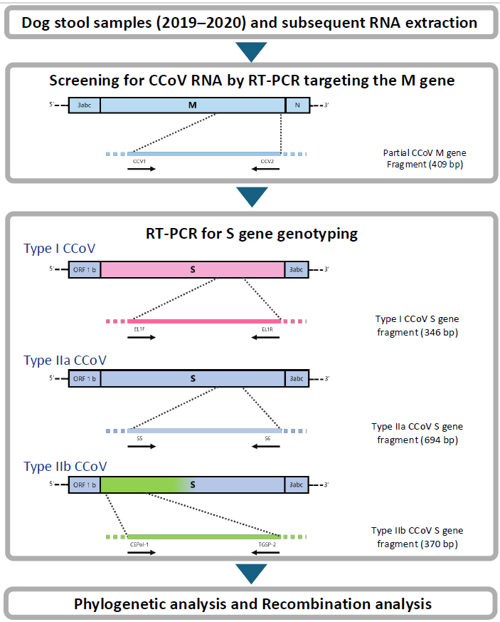
Fig. S1. Schematic overview of the workflow used for molecular detection and characterization of canine coronavirus (CCoV). Stool samples collected from domestic dogs between 2019 and 2020 were subjected to RNA extraction, followed by RT-PCR targeting the CCoV M gene for initial screening. Positive samples were further analyzed by S gene-based RT-PCR to determine viral genotypes (type I, type IIa, and type IIb). PCR products were sequenced and used for phylogenetic analysis. In addition, recombination analysis was performed to assess the genetic characteristics of the detected strains.
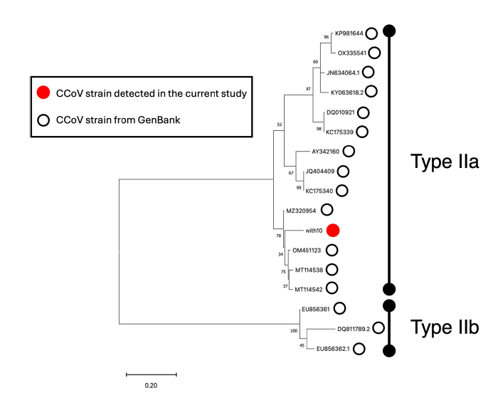
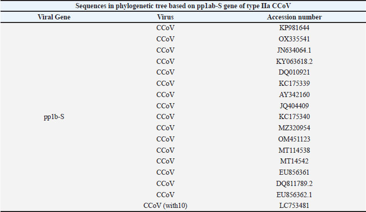
Fig. S2. Phylogenetic tree based on sequences of the partial pp1b-S gene of with10 CCoV detected by RT-PCR for detection of type IIb CCoV. The tree was generated by the maximum likelihood method (bootstrap value: 1,000 replicates) using MEGA version Ⅺ (Tamura et al. 2021).
Fig. S3. Similarity plot of sequences of pp1b-S gene from with10 against type Ⅱa CCoVs and TGEV. Similarity plot of with10 against type IIa CCoV strains (MT114542 and AY342160) and TGEV strain (KX900402) was drawn by RDP5 (Martin et al. 2021). Supplemental Table 1. Individual information on investigated domestic dogs in the current study.
Supplemental Table 2. Overview of results of RT-PCR targeting CCoV genes in stool samples from domestic dogs.
Supplemental Table 3. The results of detection for CCoV M gene by factors of domestic dogs. Dogs with no information on each category were excluded.
Supplemental Table 4. Accession numbers of viruses for phylogenetic tree based on M gene of CCoV.
Supplemental Table 5. Accession numbers of viruses for phylogenetic tree based on S gene of type I CCoV.
Supplemental Table 6. Accession numbers of viruses for phylogenetic tree based on S gene of type Ⅱa CCoV.
Supplemental Table 7. Accession numbers of viruses for phylogenetic.
| ||

| How to Cite this Article |
| Pubmed Style Kumano H, Kitashin I, Nakagawa K. A molecular survey of canine coronavirus among domestic dogs in Japan from 2019 to 2020. Open Vet. J.. 2026; 16(6): 3528-3545. doi:10.5455/OVJ.2026.v16.i6.23 Web Style Kumano H, Kitashin I, Nakagawa K. A molecular survey of canine coronavirus among domestic dogs in Japan from 2019 to 2020. https://www.openveterinaryjournal.com/?mno=309510 [Access: June 26, 2026]. doi:10.5455/OVJ.2026.v16.i6.23 AMA (American Medical Association) Style Kumano H, Kitashin I, Nakagawa K. A molecular survey of canine coronavirus among domestic dogs in Japan from 2019 to 2020. Open Vet. J.. 2026; 16(6): 3528-3545. doi:10.5455/OVJ.2026.v16.i6.23 Vancouver/ICMJE Style Kumano H, Kitashin I, Nakagawa K. A molecular survey of canine coronavirus among domestic dogs in Japan from 2019 to 2020. Open Vet. J.. (2026), [cited June 26, 2026]; 16(6): 3528-3545. doi:10.5455/OVJ.2026.v16.i6.23 Harvard Style Kumano, H., Kitashin, . I. & Nakagawa, . K. (2026) A molecular survey of canine coronavirus among domestic dogs in Japan from 2019 to 2020. Open Vet. J., 16 (6), 3528-3545. doi:10.5455/OVJ.2026.v16.i6.23 Turabian Style Kumano, Hitomi, Ichika Kitashin, and Keisuke Nakagawa. 2026. A molecular survey of canine coronavirus among domestic dogs in Japan from 2019 to 2020. Open Veterinary Journal, 16 (6), 3528-3545. doi:10.5455/OVJ.2026.v16.i6.23 Chicago Style Kumano, Hitomi, Ichika Kitashin, and Keisuke Nakagawa. "A molecular survey of canine coronavirus among domestic dogs in Japan from 2019 to 2020." Open Veterinary Journal 16 (2026), 3528-3545. doi:10.5455/OVJ.2026.v16.i6.23 MLA (The Modern Language Association) Style Kumano, Hitomi, Ichika Kitashin, and Keisuke Nakagawa. "A molecular survey of canine coronavirus among domestic dogs in Japan from 2019 to 2020." Open Veterinary Journal 16.6 (2026), 3528-3545. Print. doi:10.5455/OVJ.2026.v16.i6.23 APA (American Psychological Association) Style Kumano, H., Kitashin, . I. & Nakagawa, . K. (2026) A molecular survey of canine coronavirus among domestic dogs in Japan from 2019 to 2020. Open Veterinary Journal, 16 (6), 3528-3545. doi:10.5455/OVJ.2026.v16.i6.23 |


