





| Case Report | ||
Open Vet. J.. 2026; 16(6): 3601-3605 Open Veterinary Journal, (2026), Vol. 16(6): 3601-3605 Case Report Clinical and pathological findings of metastatic hemangiosarcoma in a DobermanShahrzad Azizi1, Setayesh Sadat Naseri1, Mahdieh Rezaei2, Reza Kheirandish1, Mohammad Mehdi Oloumi2, Saeed Hassanzadeh1 and Sina Delshad1*1Department of Pathobiology, Faculty of Veterinary Medicine, Shahid Bahonar University of Kerman, Kerman, Iran 2Department of Clinical Sciences, Faculty of Veterinary Medicine, Shahid Bahonar University of Kerman, Kerman, Iran *Corresponding Author: Sina Delshad. Department of Pathobiology, Faculty of Veterinary Medicine, Shahid Bahonar University of Kerman, Kerman, Iran. Email: sinadelshad [at] vet.uk.ac.ir Submitted: 08/02/2026 Revised: 25/04/2026 Accepted: 05/05/2026 Published: 05/06/2026 © 2025 Open Veterinary Journal
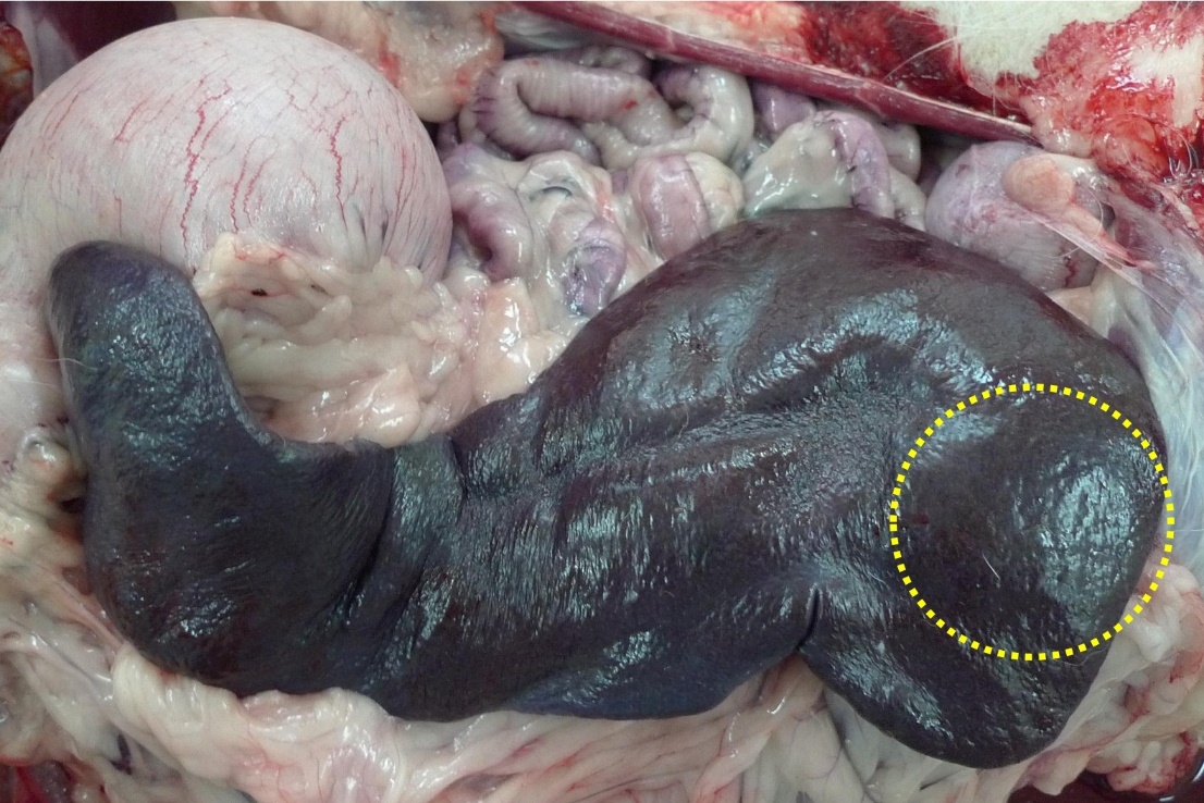
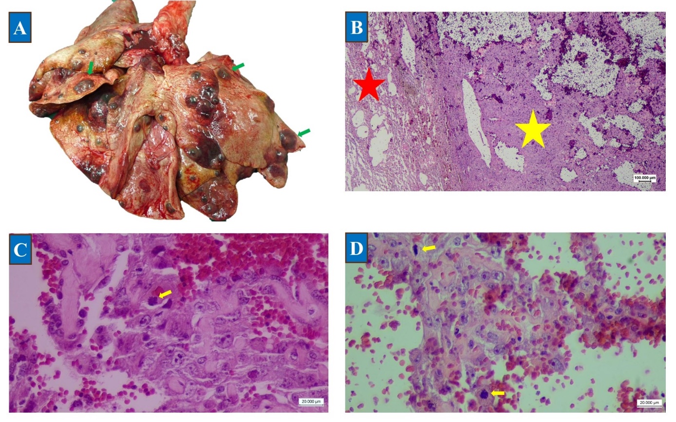
AbstractBackground: Canine hemangiosarcoma (HSA) is a highly malignant tumor originating from endothelial cells that frequently affects the spleen with rapid metastatic spread. Although metastases commonly involve the lungs and liver, brain involvement is rare. Case Description: Splenic HSA with extensive metastasis in a 7-year-old male Doberman is described. The dog was referred with lethargy, anorexia, and walking incoordination. Radiographic and ultrasonographic examination confirmed liver and lung metastases. Necropsy revealed multiple dark-red nodules in the spleen, lungs, and central nervous system, including the cerebrum and cerebellum. Conclusion: In the histopathologic investigation, HSA was diagnosed. Irregular vascular channels lined the proliferative endothelial cells containing erythrocytes, and pleomorphism was observed. Because canine HSA is analogous to human angiosarcoma, further studies are needed to explore targeted therapies that could enhance survival in both species. Keywords: Dog, Hemangiosarcoma, Metastasis, Pathology. IntroductionHemangiosarcoma (HSA) is a malignant tumor originating from the endothelial cells lining blood vessels. It includes two types: non-visceral and visceral HSA. Non-visceral HSAs can affect skin and muscle tissues, whereas visceral HSAs are often found in the spleen, liver, heart, and other organs (Mullin and Clifford, 2020). In veterinary science, splenic HSA is particularly common in dogs, with a prevalence rate of 50%–65%. Although the exact cellular origin remains unclear, recent investigations indicate that it develops from pluripotent bone marrow progenitor cells (Kim et al., 2015). Chronic infections, such as those caused by Bartonella, may also contribute to its pathogenesis (Brown et al., 1985; Smith, 2003). Certain dog breeds, particularly larger varieties such as Golden Retrievers and German Shepherds, exhibit a heightened susceptibility to HSA (Davies and Taylor, 2020; De Nardi et al., 2023). Splenic HSA is known for its highly aggressive nature in dogs. The "two-thirds rule" serves as a statistical guideline to estimate the rate of malignancy in splenic neoplasms, with two-thirds of neoplasms diagnosed in the spleen being malignant (Hammond and Pesillo-Crosby, 2008; Davies and Taylor, 2020) cellular origins may indeed come from bone marrow cells, although this remains an area of ongoing investigation (Mullin and Clifford, 2020; Griffin et al., 2021). In this study, we describe the pathological findings of metastatic HSA in a male Doberman dog. Case DetailsA 7-year-old male Doberman was referred to the teaching veterinary hospital of Shahid Bahonar University of Kerman, Kerman, Iran, with complaints of lethargy, anorexia, and difficulty in walking. Clinical examination revealed abdominal pain and splenomegaly. Cerebellar involvement was confirmed through the hypermetric gait neurological examination of the animal. Radiography and ultrasonography revealed the involvement of visceral organs, particularly the lungs and spleen. Euthanasia was chosen because of the extensive nature of the lesions and evidence of metastasis to some organs. At necropsy, the spleen was markedly enlarged and congested. Multiple well-demarcated nodules of various sizes with soft texture were observed in the spleen (Fig. 1). The lungs showed extensive involvement with multiple red-to-black nodules ranging in diameter from approximately 0.5 to 3 cm (Fig. 2A). Metastatic lesions were also observed in the cerebral cortex and cerebellum (Fig. 3A).
Fig. 1. A 7-year-old male Doberman dog with a primary HSA mass in the spleen (circle). Histopathological examination revealed that the neoplastic nodules in the lungs and brain were composed of numerous thin-walled, irregular blood vessels lined by a single layer of endothelial cells, and, less commonly, two layers of endothelial cells. Some vessels had a cavernous pattern. The lining cells had a spindle or ovoid shape with vesicular to hyperchromatic nuclei and prominent nucleoli. Endothelial cells bulged into the vascular lumens, and erythrocytes filled the lumens. The malignant cells were pleomorphic, and the mitotic figures were frequently visible (Figs. 2B–D, 3B–E).
Fig. 2. (A) Metastatic HSA in the lungs with multifocal masses in all lobes (green arrows). (B) Histological views of the lungs, including the normal portion of lung tissue (red star) and the metastatic portion (yellow star). (C, D) Mitotic figures were often visible (yellow arrows) (H&E, 40× and 100×).
Fig. 3. (A) Metastatic HSA in the brain and cerebellum (green arrows). (B, C) Subgross photomicrographs reveal neoplastic nodules and cavernous channels in the cerebellum and brain. (D, E) Neoplastic nodules with irregular blood vessels, spindle-shaped endothelial cells, hyperchromatic nuclei, and pleomorphic malignant cells filled with erythrocytes (H&E, 40× and 100×). Ethical approvalNot needed for this study. DiscussionHSA is a highly aggressive and malignant tumor. Hemorrhage and necrosis characterize this neoplasm, which exhibits capillary, cavernous, or solid morphologies. Neoplastic cells delineate irregular blood-filled vascular compartments (Fosmire et al., 2004). The spleen, heart, and subcutaneous tissue are the principal sites of occurrence for HSA (Schultheiss, 2004). Despite the insidious nature of tumor development, they may precipitate acute complications resulting from hemorrhage into body cavities. Histopathologically, HSA bears resemblance to human angiosarcomas, displaying high mortality rates within the first year after diagnosis (Antonescu, 2014). The presented case of metastatic HSA in a Doberman describes the aggressive and highly metastatic nature of HSA, as shown in the literature. Neurological involvement, which was evidenced by cerebellar metastases and a hypermetric gait, closely resembled the brain metastases reported by Yan et al. (2024). The histopathological examination in all cases consistently indicated the formation of irregular vascular channels by neoplastic endothelial cells. HSA are mostly dark red on gross examination because of substantial blood accumulation within the tumor tissue, although some cases may appear as solid white-tan masses (Valli et al., 2016; De Nardi et al., 2023). Gross diagnosis of HSA is difficult because it can be confused with other tumors, such as hematomas, hemangiomas, or other sarcomas. However, the presence of metastases is the most reliable sign of HSA in cases of dark-red splenic masses (Valli et al., 2016). The metastases in this case, primarily to the lungs and brain, reflect the hematogenous dissemination typical of HSA, as seen in Sharma et al.’s (2012) and Karabağlı et al.’s (2011) studies. All reports highlight the disease’s rapid progression and limited treatment options (De Nardi et al., 2023). This case illustrates the clinical and pathological variability of HSA and emphasizes the importance of comprehensive diagnostics to capture its diverse presentations and provide management decisions. The clinical manifestations of splenic HSA include lethargy, generalized weakness, and pallor, with the potential for abdominal pain and distension. Tumor rupture can cause hypovolemic shock and instant death (Hammond and Pesillo-Crosby, 2008). Laboratory analyses may reveal anemia, thrombocytopenia, and coagulation abnormalities (Masyr et al., 2021). Metastatic spread is frequently observed at the time of diagnosis (Story et al., 2020; Griffin et al., 2021; De Nardi et al., 2023). Death from visceral HSA is usually a result of tumor metastasis rather than local tumor recurrence (Smith, 2003) prognosis of splenic HSA in dogs is unfavorable because of the high metastatic capacity of the tumor and the high rate of metastasis at the time of diagnosis (Mullin and Clifford, 2020; Rozolen et al., 2021). The ontogeny of canine HSA remains unclear; however, certain breeds have demonstrated a higher predisposition, implying that genetic factors are involved (Tonomura et al., 2015). Environmental etiologies, such as exposure to ionizing radiation, have also been proposed, whereas infectious causation remains ambiguous (Varanat et al., 2011). As previously posited, the cellular origin appears to stem from a bone marrow progenitor rather than endothelial cells. These progenitor cells retain stem cell-like characteristics, facilitating tumorigenesis across diverse organ systems. Heterogeneity characterizes HSA, encompassing angiogenic, inflammatory, and connective tissue subtypes. The evolution of angiosarcoma in humans parallels this model (Gorden et al., 2014). Splenic injuries can be diagnosed through abdominal ultrasound, thoracic radiography, and fluid analysis. Hematological evaluations, including complete blood counts, may assist in the diagnosis of malignancy, although they do not provide definitive evidence of malignancy (Yankin et al., 2020). Although ultrasound can detect splenic lesions, advanced imaging modalities, such as computed tomography or magnetic resonance imaging, may offer more precise staging of the condition. Blood contamination often compromised fine-needle aspiration cytology, making it less effective. Histopathological analysis remains the most reliable diagnostic approach (Maronezi et al., 2022; De Nardi et al., 2023). Surgical intervention is considered the primary treatment for HSA, frequently accompanied by chemotherapy to enhance survival prospects. Although various chemotherapy regimens are available, none have shown significant differences in OS outcomes. The prognostic outlook for splenic HSA remains poor due to the high rates of metastasis present at the time of diagnosis (Marconato et al., 2019; Faulhaber et al., 2021). Conflict of interestThe authors declare no conflict of interest. FundingThis study received no specific grant. Authors' contributionsShahrzad Azizi: Methodology, project administration, data analyses, and writing of the original draft; Setayesh Sadat Naseri, Mahdieh Rezaei, Reza Kheirandish, Mohammad Mehdi Oloumi, and Saeed Hassanzadeh: Methodology and project administration; Sina Delshad: Writing, review, and editing, writing of the original draft, supervision, formal analysis, conceptualization, and supervision. Data availabilityAll data supporting the findings of this study are available within the manuscript. ReferencesAntonescu, C. 2014. Malignant vascular tumors—an update. Modern. Pathol. 27(1), S30–S38. Brown, N.O., Patnaik, A.K. and Macewen, E.G. 1985. Canine hemangiosarcoma: retrospective analysis of 104 cases. J. Am. Vet. Med. Assoc. 186(1), 56–58. Davies, O. and Taylor, A.J. 2020. Refining the “double two‐thirds” rule: genotype‐based breed grouping and clinical presentation help predict the diagnosis of canine splenic mass lesions in 288 dogs. Vet. Comparative Oncol. 18(4), 548–558. De Nardi, A.B., De Oliveira Massoco Salles Gomes, C., Fonseca-Alves, C.E., De Paiva, F.N., Linhares, L.C.M., Carra, G.J.U., Dos Santos Horta, R., Ruiz Sueiro, F.A., Jark, P.C., Nishiya, A.T., De Carvalho Vasconcellos, C.H., Ubukata, R., Batschinski, K., Sobral, R.A., Fernandes, S.C., Biondi, L.R., De Francisco Strefezzi, R., Matera, J.M., Rangel, M.M.M., Dos Anjos, D.S., Brunner, C.H.M., Laufer-Amorim, R., Cadrobbi, K.G., Cirillo, J.V., Martins, M.C., De Paula Reis Filho, N., Silva Lessa, D.F., Portela, R., Scarpa Carneiro, C., Ricci Lucas, S.R., Fukumasu, H., Feliciano, M.A.R., Gomes Quitzan, J. and Dagli, M.L.Z. 2023. Diagnosis, prognosis, and treatment of canine hemangiosarcoma: a review based on a consensus organized by the Brazilian association of veterinary oncology, ABROVET. Cancers 15(7), 2025. Faulhaber, E.A., Janik, E. and Thamm, D.H. 2021. Adjuvant carboplatin for treatment of splenic hemangiosarcoma in dogs: retrospective evaluation of 18 cases (2011‐2016) and comparison with doxorubicin‐based chemotherapy. J. Vet. Internal. Med. 35(4), 1929–1934. Fosmire, S.P., Dickerson, E.B., Scott, A.M., Bianco, S.R., Pettengill, M.J., Meylemans, H., Padilla, M., Frazer-Abel, A.A., Akhtar, N., Getzy, D.M., Wojcieszyn, J., Breen, M., Helfand, S.C. and Modiano, J.F. 2004. Canine malignant hemangiosarcoma as a model of primitive angiogenic endothelium. Lab. Invest. 84(5), 562–572. Gorden, B.H., Saha, J., Khammanivong, A., Schwartz, G.K. and Dickerson, E.B. 2014. Lysosomal drug sequestration as a mechanism of drug resistance in vascular sarcoma cells marked by high CSF-1R expression. Vascular Cell 6, 1–14. Griffin, M.A., Culp, W.T.N. and Rebhun, R.B. 2021. Canine and feline haemangiosarcoma. Vet. Rec. 189(9), no-no. Hammond, T.N. and Pesillo-Crosby, S.A. 2008. Prevalence of hemangiosarcoma in anemic dogs with a splenic mass and hemoperitoneum requiring a transfusion: 71 cases (2003–2005). J. Am. Vet. Med. Assoc. 232(4), 553–558. Karabağlı, G., Düzgün, O., Yıldar, E., Erdoğan. and Gürel, A. 2011. A hemangiosarcoma case in a dog. İstanbul Üniversitesi. Veteriner. Fakültesi. Dergisi. 37(2), 161–165. Kim, J.H., Graef, A., Dickerson, E. and Modiano, J. 2015. Pathobiology of hemangiosarcoma in dogs: research advances and future perspectives. Vet. Sci. 2(4), 388–405. Marconato, L., Chalfon, C., Finotello, R., Polton, G., Vasconi, M.E., Annoni, M., Stefanello, D., Mesto, P., Capitani, O., Agnoli, C., Amati, M. and Sabattini, S. 2019. Adjuvant anthracycline‐based vs metronomic chemotherapy vs no medical treatment for dogs with metastatic splenic hemangiosarcoma: a multi‐institutional retrospective study of the Italian Society of Veterinary Oncology. Vet. Comparative. Oncol. 17(4), 537–544. Maronezi, M.C., Carneiro, R.K., Da Cruz, I.C.K., De Oliveira, A.P.L., De Nardi, A.B., Pavan, L., Del’Aguila-Silva, P., Uscategui, R.A.R. and Feliciano, M.A.R. 2022. Accuracy of B-mode ultrasound and ARFI elastography in predicting malignancy of canine splenic lesions. Sci. Rep. 12(1), 4252. Masyr, A.R., Rendahl, A.K., Winter, A.L., Borgatti, A. and Modiano, J.F. 2021. Retrospective evaluation of thrombocytopenia and tumor stage as prognostic indicators in dogs with splenic hemangiosarcoma. J. Am. Vet. Med. Assoc. 258(6), 630–637. Mullin, C. and Clifford, C. 2020. Miscellaneous tumors. In Withrow and MacEwen’s small animal clinical oncology. Eds., David M. Vail, Douglas H. Thamm, and Julius M. Liptak. 6th ed. Rozolen, J.M., Teodoro, T.G.W., Sobral, R.A., Sueiro, F.A.R., Laufer-Amorim, R., Elias, F. and Fonseca-Alves, C.E. 2021. Investigation of prognostic value of claudin-5, PSMA, and Ki67 expression in canine splenic hemangiosarcoma. Animals 11(8), 2406. Schultheiss, P.C. 2004. A retrospective study of visceral and nonvisceral hemangiosarcoma and hemangiomas in domestic animals. J. Vet. Diagnostic. Invest. 16(6), 522–526. Sharma, D. 2012. Hemangiosarcoma in a geriatric Labrador retriever. Can. Vet. J. 53(8), 889. Smith, A.N. 2003. Hemangiosarcoma in dogs and cats. Vet. Clinics. Small. Anim. Pract. 33(3), 533–552. Story, A.L., Wavreille, V., Abrams, B., Egan, A., Cray, M. and Selmic, L.E. 2020. Outcomes of 43 small breed dogs treated for splenic hemangiosarcoma. Vet. Surg. 49(6), 1154–1163. Tonomura, N., Elvers, I., Thomas, R., Megquier, K., Turner-Maier, J., Howald, C., Sarver, A.L., Swofford, R., Frantz, A.M., Ito, D., Mauceli, E., Arendt, M., Noh, H.J., Koltookian, M., Biagi, T., Fryc, S., Williams, C., Avery, A.C., Kim, J.H., Barber, L., Burgess, K., Lander, E.S., Karlsson, E.K., Azuma, C., Modiano, J.F., Breen, M. and Lindblad-Toh, K. 2015. Genome-wide association study identifies shared risk loci common to two malignancies in golden retrievers. PLos Genet. 11(2), e1004922. Valli, V.E., Bienzle, D. and Meuten, D.J. 2016. Tumors of the hemolymphatic system. Tumors in Domestic Animals. Ames, IA: John Wiley & Sons Inc, pp: 203–321. Varanat, M., Maggi, R.G., Linder, K.E. and Breitschwerdt, E.B. 2011. Molecular prevalence of Bartonella, Babesia, and Hemotropic mycoplasma sp. in dogs with splenic disease. J. Vet. Internal Med. 25(6), 1284–1291. Yan, J., Evered, C., Raheb, S., Lillie, B. and Fonfara, S. 2024. Ventricular cardiac hemangiosarcoma with brain metastases in a dog. J. Vet. Cardiol. 55, 32–37. Yankin, I., Nemanic, S., Funes, S., De Morais, H., Gorman, E. and Ruaux, C. 2020. Clinical relevance of splenic nodules or heterogeneous splenic parenchyma assessed by cytologic evaluation of fine‐needle samples in 125 dogs (2011‐2015). J. Vet. Internal. Med. 34(1), 125–131. | ||

| How to Cite this Article |
| Pubmed Style Azizi S, Naseri SS, Rezaei M, Kheirandish R, Oloumi MM, Hassanzadeh S, Delshad S. Clinical and pathological findings of metastatic hemangiosarcoma in a Doberman. Open Vet. J.. 2026; 16(6): 3601-3605. doi:10.5455/OVJ.2026.v16.i6.32 Web Style Azizi S, Naseri SS, Rezaei M, Kheirandish R, Oloumi MM, Hassanzadeh S, Delshad S. Clinical and pathological findings of metastatic hemangiosarcoma in a Doberman. https://www.openveterinaryjournal.com/?mno=309770 [Access: June 26, 2026]. doi:10.5455/OVJ.2026.v16.i6.32 AMA (American Medical Association) Style Azizi S, Naseri SS, Rezaei M, Kheirandish R, Oloumi MM, Hassanzadeh S, Delshad S. Clinical and pathological findings of metastatic hemangiosarcoma in a Doberman. Open Vet. J.. 2026; 16(6): 3601-3605. doi:10.5455/OVJ.2026.v16.i6.32 Vancouver/ICMJE Style Azizi S, Naseri SS, Rezaei M, Kheirandish R, Oloumi MM, Hassanzadeh S, Delshad S. Clinical and pathological findings of metastatic hemangiosarcoma in a Doberman. Open Vet. J.. (2026), [cited June 26, 2026]; 16(6): 3601-3605. doi:10.5455/OVJ.2026.v16.i6.32 Harvard Style Azizi, S., Naseri, . S. S., Rezaei, . M., Kheirandish, . R., Oloumi, . M. M., Hassanzadeh, . S. & Delshad, . S. (2026) Clinical and pathological findings of metastatic hemangiosarcoma in a Doberman. Open Vet. J., 16 (6), 3601-3605. doi:10.5455/OVJ.2026.v16.i6.32 Turabian Style Azizi, Shahrzad, Setayesh Sadat Naseri, Mahdieh Rezaei, Reza Kheirandish, Mohammad Mehdi Oloumi, Saeed Hassanzadeh, and Sina Delshad. 2026. Clinical and pathological findings of metastatic hemangiosarcoma in a Doberman. Open Veterinary Journal, 16 (6), 3601-3605. doi:10.5455/OVJ.2026.v16.i6.32 Chicago Style Azizi, Shahrzad, Setayesh Sadat Naseri, Mahdieh Rezaei, Reza Kheirandish, Mohammad Mehdi Oloumi, Saeed Hassanzadeh, and Sina Delshad. "Clinical and pathological findings of metastatic hemangiosarcoma in a Doberman." Open Veterinary Journal 16 (2026), 3601-3605. doi:10.5455/OVJ.2026.v16.i6.32 MLA (The Modern Language Association) Style Azizi, Shahrzad, Setayesh Sadat Naseri, Mahdieh Rezaei, Reza Kheirandish, Mohammad Mehdi Oloumi, Saeed Hassanzadeh, and Sina Delshad. "Clinical and pathological findings of metastatic hemangiosarcoma in a Doberman." Open Veterinary Journal 16.6 (2026), 3601-3605. Print. doi:10.5455/OVJ.2026.v16.i6.32 APA (American Psychological Association) Style Azizi, S., Naseri, . S. S., Rezaei, . M., Kheirandish, . R., Oloumi, . M. M., Hassanzadeh, . S. & Delshad, . S. (2026) Clinical and pathological findings of metastatic hemangiosarcoma in a Doberman. Open Veterinary Journal, 16 (6), 3601-3605. doi:10.5455/OVJ.2026.v16.i6.32 |


