





| Research Article | ||
Open Vet. J.. 2026; 16(6): 3749-3758 Open Veterinary Journal, (2026), Vol. 16(6): 3749-3758 Research Article In vitro evaluation of culture surface coating on the phagocytic activity of murine macrophagesMoe Ikeda and Akikazu Sakudo*School of Veterinary Medicine, Okayama University of Science, Imabari, Japan *Corresponding Author: Akikazu Sakudo. School of Veterinary Medicine, Okayama University of Science, Imabari, Japan. Email: akikazusakudo [at] gmail.com Submitted: 15/02/2026 Revised: 05/05/2026 Accepted: 14/05/2026 Published: 16/06/2026 © 2025 Open Veterinary Journal
AbstractBackground: Macrophage phagocytosis is a fundamental component of innate immunity in animals and plays a critical role in host defense against bacterial infection, inflammation, and tissue homeostasis. In vitro macrophage assays are widely used in veterinary immunology research. The biochemical and biophysical conditions used in the culturing of cells are known to influence macrophage phagocytosis. However, the effect of the surface coating substrate of culture dishes on phagocytic activity in vitro remains unclear. Aim: This study aimed to evaluate how commonly used glass surface coatings affect macrophage phagocytic activity and to identify an optimal in vitro culture condition suitable for veterinary immunology research. Methods: Non-coated (NC), poly-L-lysine-coated (PL), and collagen-coated (CC) glass-bottom dishes were compared as culture surface coating substrates for RAW264.7 macrophages. Lipopolysaccharide-stimulated RAW264.7 cells were treated with fluorescent latex beads, and phagocytic activity was subsequently quantified by assessing the proportion of bead-positive cells, intracellular bead load, and composite phagocytic index after 3 and 24 hours. Results: All surface coating substrates supported similar adhesion and comparable phagocytic performance at 3 hours. However, at 24 hours, the cells cultured on NC dishes showed a significantly higher proportion of phagocytic activity, greater bead internalization per cell, and the highest phagocytic index by comparison to those cultured on PL or CC dishes. Conclusion: These findings demonstrate that the culture surface coating significantly influences macrophage phagocytic activity during prolonged incubation. NC glass provides the most appropriate and least confounding substrate for in vitro macrophage phagocytosis assays and should be preferentially considered in veterinary immunology. Keywords: Macrophage, Phagocytosis, RAW264.7, Surface coating substrate, Veterinary immunology. IntroductionMacrophages are central effector cells of the innate immune system, where they play an important role in host defense and inflammation regulation through a combination of cytokine production and phagocytosis (Hirayama et al., 2018). In veterinary medicine, macrophage-mediated phagocytosis is essential for protection against bacterial and parasitic infections in livestock and companion animals, and is a key determinant of disease outcome, vaccine efficacy, and inflammatory pathology. Macrophage-mediated phagocytosis not only removes microbes and cell debris but also initiates downstream pro- and anti-inflammatory signalling cascades (Aderem and Underhill, 1999; Hirayama et al., 2018; Lim et al., 2017). Thus, phagocytosis acts as a key effector in the innate immune signalling network (Underhill and Ozinsky, 2002). Crucially, phagocytic uptake of microbes, apoptotic cells, and synthetic particles is coupled to cytoskeletal remodelling and receptor-mediated signalling. As a result, the phagocytic process is highly sensitive to the physical and biochemical properties of the microenvironment (Underhill and Ozinsky, 2002; Fu and Harrison, 2021). Several lines of evidence demonstrate that substrate and particle surface chemistry modulate macrophage phagocytosis. For example, chitosan-coated glass culture dishes were shown to enhance macrophage mobility, cytoplasm spreading, and bacterial phagocytosis by comparison to non-coated (NC) glass dishes, leading to efficient clearance of surface-associated bacteria (Gu et al., 2014). Moreover, chitosan-coated nanocarriers are readily internalized by macrophages via actin-dependent phagocytosis, while mannose functionalization does not alter phagocytic uptake efficiency (Coya et al., 2019). Phosphatidylserine (PS), a representative “eat-me” signal exposed on apoptotic cells, increases recognition and phagocytosis of PS-coated single-walled carbon nanotubes by RAW264.7 cells (Konduru et al., 2009). In contrast, layers of hydrophilic polyethylene glycol (PEG) reduce protein adsorption and macrophage uptake (Zahr et al., 2006). Indeed, PEGylated core-shell nanoparticles show three-fold lower levels of internalization compared to their positively or negatively charged counterparts (Zahr et al., 2006). More recently, anionic hybrid microparticles of poly(lactic-co-glycolic) acid nanoparticles combined with lipid nanoparticles were shown to be taken up three-fold more efficiently than those with cationic formulations, indicating the importance of surface charge in shaping macrophage-particle interactions (Maghrebi et al., 2020). Biomimetic strategies using living cell-derived membrane coatings showed that silica nanoparticles coated with RAW264.7 cell-derived membrane components displayed reduced phagocytosis by RAW264.7 and enhanced uptake by HeLa cells (Mizuta et al., 2024). Thus, detailed studies into how particle or coating chemistry modulates macrophage uptake have been undertaken (Shen and Horbett, 2001; Septiadi et al., 2020). However, comparatively little attention has been paid to an elementary aspect of phagocytosis assays, namely the nature of the culture surface substrate on which macrophages themselves are adhered. Indeed, to improve cell attachment and spreading, a surface coating substrate is often used in phagocytosis assays (Hudalla and Murphy, 2011). This coating may comprise extracellular matrix (ECM) proteins or synthetic polymers. Collagen, a major fibrillar ECM component, not only provides structural support but also regulates adhesion, survival, migration, and differentiation via integrin signalling and cytoskeletal organization (Chua and Lim, 2023). In mesenchymal stem cells, for example, collagen-coated (CC) surfaces enhance adhesion and stress resistance and promote lineage-specific differentiation (Somaiah et al., 2015). By contrast, poly-L-lysine is a synthetic, polycationic coating that increases the density of positively charged sites on glass or plastic, thereby enhancing electrostatic interactions with the negatively charged plasma membrane and associated proteins (Mazia et al., 1975; Schwieger and Blume, 2007; Gorman et al., 2020). RAW264.7 cells, a murine monocyte-macrophage-like cell line, are widely used as an in vitro model to quantify macrophage functions, including phagocytosis, nitric oxide production, and polarization (Aderem and Underhill, 1999). This cell line is often used as a standardized experimental model in veterinary immunology, zoonotic infection research, and translational studies to understand host-pathogen interactions relevant to animal health. However, no comparative studies have been conducted to investigate how commonly used dish surface coatings, such as NC, poly-L-lysine–coated (PL), or CC glass, differentially affect macrophage phagocytosis in the widely used RAW264.7 model. In the present study, macrophage phagocytosis assays were performed to clarify how commonly used culture surface coatings influence macrophage function and to establish an optimal experimental condition for reproducible in vitro phagocytosis assays applicable to veterinary immunology research. Specifically, RAW264.7 murine macrophage-like cells were cultured on NC, PL, or CC glass-bottom dishes, stimulated with lipopolysaccharide (LPS), exposed to fluorescent latex beads, and analyzed at 3 and 24 hours by quantifying the percentage of bead-positive cells, the intracellular bead load, and the phagocytic index. Using this three-condition comparative in vitro system, we found that phagocytic activity was comparable among NC, PL, and CC substrates at 3 hours, whereas at 24 hours, NC glass-bottom dishes supported a significantly higher percentage of phagocytic cells, greater bead internalization per cell, and a higher phagocytic index than PL or CC glass-bottom dishes. These findings highlight the methodological importance of culture surface selection and support the use of NC glass-bottom dishes as a simple and reliable platform for in vitro macrophage phagocytosis assays in veterinary immunology research. Materials and MethodsCell line and culture conditionsRAW264.7 cells, a murine macrophage-like cell line (American Type Culture Collection TIB-71; ATCC, Manassas, VA), were maintained in Dulbecco’s Modified Eagle Medium (4.5 g/l glucose, Catalog No. 08458-16; Nacalai Tesque, Inc., Kyoto, Japan) supplemented with 10% heat-inactivated fetal calf serum (HyClone® fetal bovine serum, Catalog No. SH30396.03; Thermo Fisher Scientific, Waltham, MA) as well as 100 Unit/ml penicillin and 100 μg/ml streptomycin (penicillin-streptomycin mixed solution, Catalog No.26253-84; Nacalai Tesque). Cells were cultured at 37°C in a humidified atmosphere containing 5% CO₂. Adhered cells were detached by brief exposure to 0.5 ml of 2.5 g/l trypsin-1 mM Ethylenediaminetetraacetic acid (EDTA) solution (Catalog No. 35554-64; Nacalai Tesque) for 5 minutes at 37°C, followed by gentle scraping using a cell scraper (Catalog No.9010-320; Asahi Glass Co., Ltd., Tokyo, Japan). Detached cells were resuspended in complete medium and adjusted to a final cell density of approximately 6.0 × 10⁴ cells/ml using a cell counter plate (Catalog No. 2-7124-01; AS ONE Corp., Osaka, Japan). Measurement of pseudopodiaAn aliquot of the cell suspension (6.0 × 10⁴ cells/ml) was seeded into φ35-mm glass-bottom dishes (dish diameter: φ35-mm; glass diameter: φ14-mm; Matsunami Glass Ind., Ltd., Osaka, Japan) and a No.1S glass thickness (0.16–0.19 mm) with one of three surface conditions: NC (D11130H), PL (D11131H), and CC (D11134H). After incubation of cells on NC, PL, or CC dishes for 24 hours, phase-contrast microscopic images of the RAW264.7 cells were acquired and imported into ImageJ software (version 1.49; National Institutes of Health, Bethesda, MD). Random microscopic fields were analyzed, and the number of cells in each field was counted. In addition, the length of each pseudopodium was measured using the line tool in ImageJ from the two-dimensional phase-contrast images. Measurements of pseudopodia were made from the edge of the cell body to the tip of the pseudopodium. When branched processes were present, only the longest branch was measured. Quantitative comparisons were then performed. For each condition, at least 30 cells per field were analyzed across five independent fields, resulting in a total of more than 150 cells evaluated per condition. Phagocytosis assaysAn aliquot of the cell suspension (6.0 × 10⁴ cells/ml) was seeded into the above φ35-mm glass-bottom dishes (Matsunami Glass Ind., Ltd.) with one of three surface conditions: NC, PL, or CC. After incubation of cells on NC, PL, or CC dishes for 24 hours, RAW264.7 cells were exposed to Fluoresbrite YG Carboxylate Microspheres (diameter 1.53 μm, coefficient of variation 4%, Catalog No.09719-10; Polysciences, Inc., Warrington, PA) in the culture medium containing a final concentration of 0.0026% of solid latex. To activate macrophages, LPS from Escherichia coli O111 (Catalog No.125-05181; FUJIFILM Wako Pure Chemical Corp., Osaka, Japan) was added simultaneously to a final concentration of 1 µg/ml. Cells were incubated with beads and LPS for either 3 or 24 hours at 37°C in a 5% CO₂ incubator. At each time point, dishes were gently washed three times with Dulbecco’s phosphate-buffered saline (Catalog No. 14190-144; Thermo Fisher Scientific) to remove non-internalized or loosely adherent beads. The cells were then stained by adding 10 µg/ml of Hoechst 33342 dye (Catalog No.346-07951; Dojindo Laboratories, Kumamoto, Japan) and incubated for 15 minutes at 37°C. After staining, cells were washed once with phosphate-buffered saline (PBS). A small amount of PBS (0.5 ml) was retained in each dish to prevent drying during image acquisition. Image acquisition and quantitative analysis of phagocytosisFluorescence and phase-contrast images were acquired using a fluorescence microscope equipped with appropriate filter sets and channels (Biozero BZ-8100; KEYENCE Corp., Osaka, Japan) with a 20× objective lens (numerical aperture, 0.45). For each dish, random fields of view were selected to avoid sampling bias. Images were captured using three channels, corresponding to phase-contrast imaging, Hoechst 33342 fluorescence (excitation, 360/40 nm; dichroic mirror, 400 nm; emission, 460/50 nm), and Fluoresbrite YG fluorescence (excitation, 470/40 nm; dichroic mirror, 495 nm; emission, 535/50 nm) at a resolution of 2,720 × 2,048 pixels, with exposure time automatically determined by the imaging system. The images were overlaid to generate composites. A minimum of 150 cells using images acquired from randomly selected fields were analyzed for each condition in each independent experiment. Phagocytic activity was measured using previously reported methods (Uraki et al., 2010). Briefly, the following parameters were determined: (1) Total cell number: the total number of Hoechst 33342-positive nuclei per field. (2) Number of phagocytic cells: the number of cells containing at least one internalized fluorescent bead. (3) Total number of internalized beads: the sum of all fluorescent beads observed within the phagocytic cells. From these values, three factors of phagocytic activity were calculated:
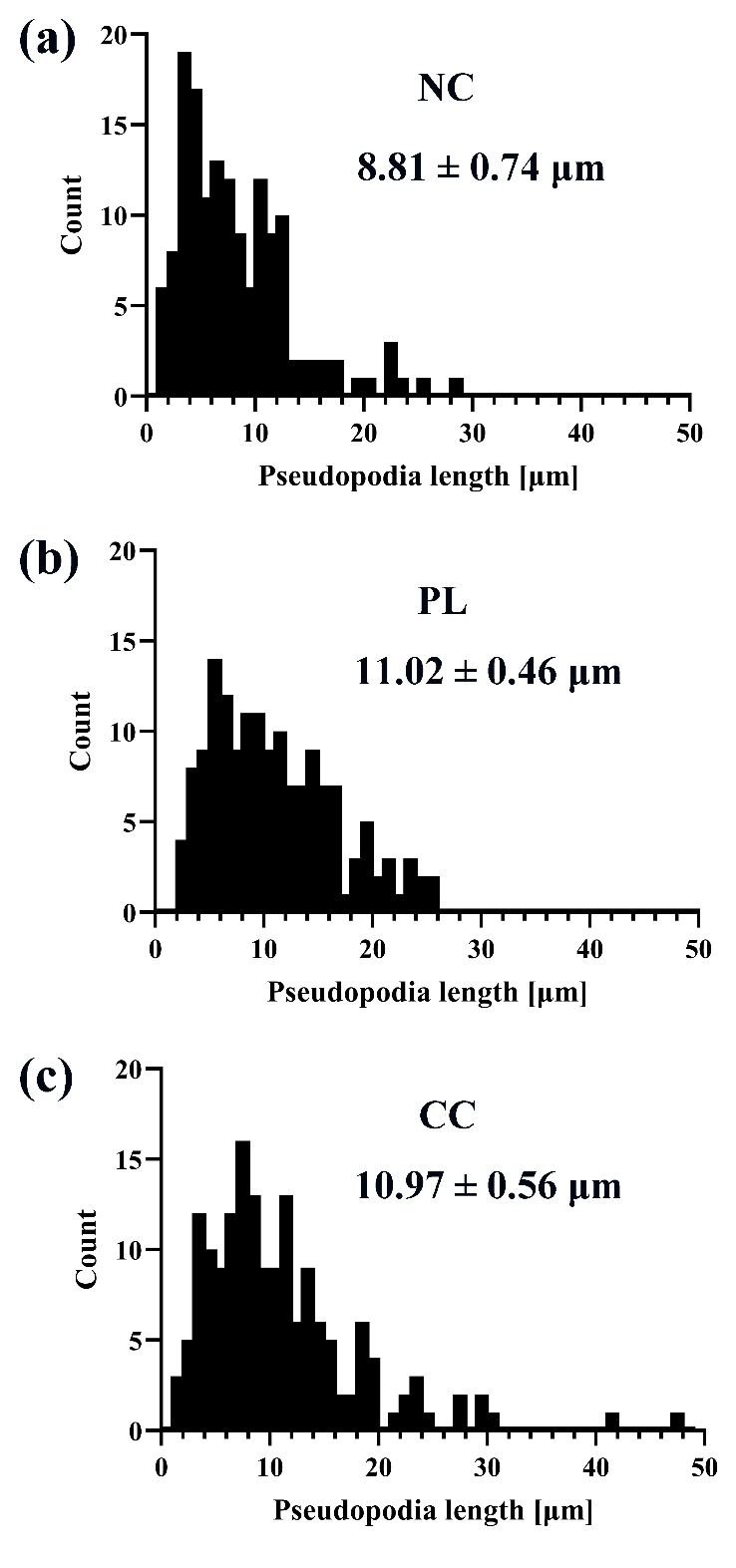
Statistical analysesStatistical analyses were performed using GraphPad Prism (version 7.02; GraphPad Prism Software Inc., La Jolla, CA). Differences among treatment groups were evaluated by one-way analysis of variance, followed by Dunnett’s post-hoc test for multiple comparisons against the NC group. A p value < 0.05 was considered statistically significant. All data are shown as the average ± standard error of the mean (SEM). Ethical approvalNot needed for this study. ResultsRAW264.7 cells were spread on three types of glass-bottom dishes (NC, PL, or CC) to examine whether the surface coating influences phagocytosis. First, the length of pseudopodia was measured under each culture condition (Fig. 1). The mean pseudopodia length was longer in cells cultured on PL (11.02 ± 0.46 µm) or CC (10.97 ± 0.56 µm) compared to NC (8.81 ± 0.74 µm). The number of analyzed pseudopodia was 151 for NC, 156 for PL, and 165 for CC. Here, differences of PL or CC where p < 0.05(*) versus NC were both considered significant (NC vs. PL: p=0.0183; NC vs. CC: p=0.0196).
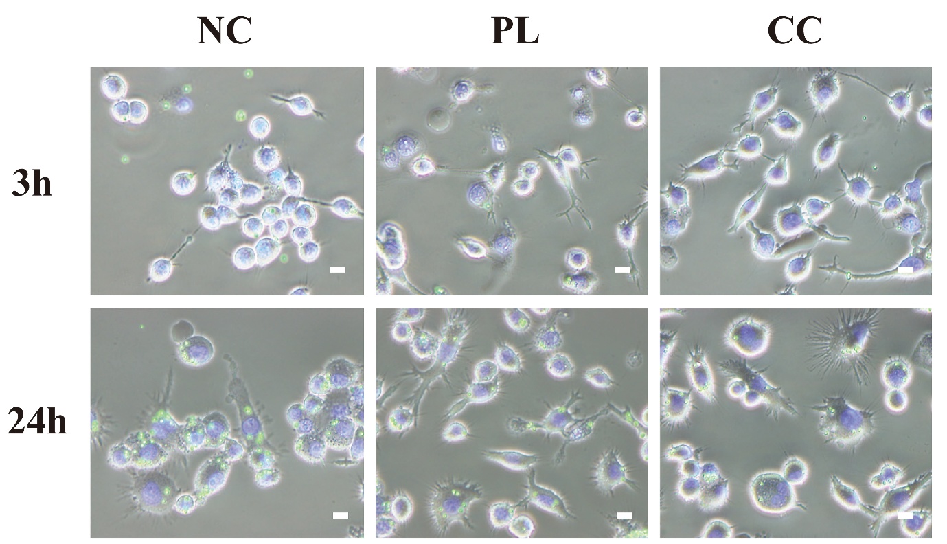
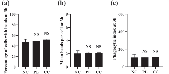
Fig. 1. Pseudopodia length of RAW264.7 cells cultured on different coated substrates. Phase-contrast images of RAW264.7 cells cultured on NC (a), PL (b), or CC (c) glass-bottom dishes were analyzed using ImageJ software. At least 150 pseudopodia were measured in each group. Distribution of pseudopodia length of RAW264.7 cells cultured on NC, PL, or CC glass-bottom dishes are shown. The average ± SEM of pseudopodia length is also shown for each group (N=151 for NC, N=156 for PL, N=165 for CC). Next, RAW264.7 cells were spread on the three types of glass-bottom surface-coated dishes to examine whether the surface coating influenced phagocytic activity. To evaluate phagocytic activity, RAW264.7 cells were incubated with fluorescent beads in the presence of LPS. Phase-contrast and fluorescence imaging showed fluorescent bead uptake was detectable under all three conditions, with numerous intracellular Fluoresbrite YG microspheres being observed in Hoechst 33342-positive cells after 3 hours, but particularly at the 24 hours time point (Fig. 2). At the early time point of 3 hours exposure, quantitative analysis demonstrated that RAW264.7 cells cultured on NC, PL, and CC dishes had internalized beads (Fig. 3). At this time point, a similar number of bead-positive cells were observed in each of the three groups. The percentage of phagocytic cells was 46.95% ± 5.60% in NC, 49.48% ± 1.92% in PL, and 52.05% ± 1.02% in CC (N=3 each). The mean number of internalized beads per bead-positive cell was 2.04 ± 0.43 in NC, 2.15 ± 0.09 in PL, and 2.10 ± 0.08 in CC (N=3 each). The phagocytic index was 105.60 ± 36.01 in NC, 107.90 ± 2.80 in PL, and 109.70 ± 2.44 in CC (N=3 each). In addition, the percentage of phagocytic cells (p=0.6081), the mean number of internalized beads per bead-positive cell (p=0.9591), and the derived phagocytic index (p=0.9903) did not differ significantly among the three coating conditions. Statistical analysis indicated no significant coating substrate-dependent effect at 3 hours.
Fig. 2. Representative fluorescence microscopic images of RAW264.7 cells cultured on different coated substrates at 3 and 24 hours. Representative overlay images of RAW264.7 cells cultured on NC, PL or CC glass-bottom dishes. Cells were treated with fluorescent latex beads in the presence of LPS and observed by fluorescent microscopy after an additional 3 or 24 hours incubation. Nuclei were stained with Hoechst 33342 (blue). Internalized Fluoresbrite YG beads (green) are readily observed. Phase-contrast microscopic images were overlaid. Scale bar, 10 µm.
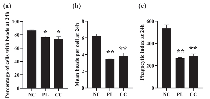
Fig. 3. Quantitative analysis of phagocytosis by RAW264.7 cells at 3 hours on different surface coating substrates. RAW264.7 cells were cultured on NC, PL or CC glass-bottom dishes, treated with fluorescent latex beads and LPS, and analyzed after 3 hours of incubation (N=3 for each group). (a) Percentage of phagocytic cells, defined as the proportion of cells containing at least one internalized fluorescent bead. (b) Mean number of internalized beads per phagocytic (bead-positive) cell. (c) Phagocytic index, calculated as the product of the percentage of phagocytic cells and the mean number of beads per phagocytic cell. NS, not significant by comparison to NC. By contrast, however, significant differences were observed after 24 hours of bead exposure. The proportion of bead-positive cells was significantly higher in the NC group than in the PL or CC groups at the 24 hours time point (NC, 86.64% ± 0.77%; PL, 76.48% ± 1.62%; CC, 73.76% ± 3.62%; N=3 each) (NC vs. PL: p=0.0377; NC vs. CC: p=0.0140) (Fig. 4). Moreover, the mean number of beads per phagocytic cell was significantly higher in the NC group compared to the PL and CC group (NC, 6.20 ± 0.26; PL, 3.47 ± 0.01; CC, 3.87 ± 0.31; N=3 each) (NC vs. PL: p=0.0003; NC vs. CC: p=0.0008). Overall, these results show a significantly elevated phagocytic index for cells cultured on NC glass-bottom dishes compared with those cultured on PL or CC glass-bottom dishes (NC, 536.60 ± 30.52; PL, 265.70 ± 6.95; CC, 288.90 ± 16.79; N=3 each) (NC vs. PL: p=0.0002; NC vs. CC: p=0.0003).
Fig. 4. Quantitative analysis of phagocytosis by RAW264.7 cells at 24 hours on different surface coated substrates. RAW264.7 cells were cultured on NC, PL, or CC glass-bottom dishes, stimulated with fluorescent latex beads and LPS, and analyzed after 24 hours incubation (N=3 for each group). (a) Percentage of phagocytic cells, defined as the proportion of cells containing at least one internalized fluorescent bead. (b) Mean number of internalized beads per phagocytic (bead-positive) cell. (c) Phagocytic index, calculated as the product of the percentage of phagocytic cells and the mean number of beads per phagocytic cell. Differences where p < 0.05(*) and p < 0.01(**) versus NC were considered significant. Taken together, RAW264.7 cells showed measurable bead uptake on all three coating substrates, whereas the NC group exhibited the highest values for the proportion of bead-positive cells, bead number per phagocytic cell, and phagocytic index at 24 hours. DiscussionCoating-dependent effects on phagocytosis and mechanical implicationsIn this study, we aimed to determine the most suitable surface coating substrate to provide a robust and physiologically interpretable platform for assessing macrophage phagocytosis. Our strategy was to culture RAW264.7 cells on NC, PL, or CC glass-bottom dishes and then determine phagocytic activity by quantifying the internalization of fluorescent latex beads. Specifically, the proportion of bead-positive cells, bead load per phagocytic cell, and a composite phagocytic index at early (3 hours) and later (24 hours) time points were measured. At the early time point (3 hours), cells cultured on NC, PL, or CC glass-bottom dishes gave similar results in the phagocytic assay. In a previous report, macrophages were also reported to show phagocytosis of Mycobacterium bovis or opsonized erythrocytes on uncoated glass coverslips or polystyrene culture plates within 1 or 2 hours, respectively (Platt and Fineran, 2015). Furthermore, a study using RAW264.7 cells on uncoated culture plates together with LPS treatment showed them to display phagocytic activity during prolonged incubation (4 hours) (Taciak et al., 2018). Here, we carried out a systematic study to investigate the effect of the coating surface of the culture dish on phagocytosis of RAW264.7 cells. Phagocytosis is an actin-related, receptor-triggered process that competes for cytoskeletal and signalling resources shared with adhesion and mechanotransduction pathways (May and Machesky, 2001). Although early phagocytic cup formation is primarily governed by acute receptor binding and inflammatory priming, longer-term adhesion-dependent stabilization of the actin architecture, membrane tension, and receptor availability can affect the efficiency of serial phagocytosis and downstream trafficking (Flannagan et al., 2012). Thus, adhesion influenced by the culture surface coating substrate has the potential to influence phagocytic activity over the long term, even when early uptake remains unchanged. Integrin-mediated adhesion is a key regulator of this process. Collagen binds multiple β1 and β2 integrins and reorganizes the cytoskeleton architecture (Chua and Lim, 2023). In mesenchymal stem cells, CC substrates promote cell adhesion and lineage commitment via integrin signalling (Somaiah et al., 2015). In RAW264.7 macrophages, FcγR-mediated phagocytosis is driven by remodeling of the actin cytoskeleton, characterized by the assembly of F-actin-rich phagocytic cups that facilitate efficient particle engulfment (Greuber and Pendergast, 2012). Therefore, adhesion induced by collagen may compete with phagocytosis activity. Similarly, although poly-L-lysine is often regarded as a biologically inactive attachment factor, it increases surface positive charge density and thereby enhances electrostatic interactions with the negatively charged surfaces (Schwieger and Blume, 2007). Given that carboxylated beads are negatively charged, a PL can promote the adhesion of both cells and beads to the surface substrate. With the carboxylated beads tightly bound to the dish, they may be inefficiently lifted and engulfed by phagocytosis. However, not all previous studies are fully consistent with the present findings. For example, CC surfaces have been reported to enhance macrophage spreading and bacterial phagocytosis compared with NC glass, suggesting that some surface modifications can enhance, rather than reduce, phagocytic function (Gu et al., 2014). This apparent discrepancy may reflect methodological differences, including differences in cell type, beads, coating conditions, target identity, or opsonization state, and assay time point. Intriguingly, cells cultured on PL and CC had a pseudopodial length of 1.344-fold and 1.338-fold greater than those of cells cultured on NC, respectively. These observations indicate that PL and CC substrates promote pseudopodial extension. Pseudopodia were longest on PL surfaces, whereas phagocytic activity was highest under the NC condition. This finding indicates that extension of pseudopodia or enhanced cell spreading does not necessarily translate into increased phagocytic activity. Under the experimental conditions used in this study, the length of the pseudopodia did not directly correlate with efficient bead engulfment. Thus, strong adhesion or cytoskeletal stabilization may impede the dynamic actin remodeling required for efficient phagocytosis. In addition, coating-dependent differences in integrin signaling and cytoskeletal organization, possibly accompanied by some changes in macrophage polarization during 24 hours culture (Cha et al., 2017), may also contribute to the reduced phagocytic activity observed on PL and CC surfaces. It should be noted that phagocytic activity was similar among NC, PL, and CC at 3 hours but different at 24 hours, suggesting that coating-dependent effects became evident only during prolonged incubation. This non-correlation between greater pseudopodial extension and lower phagocytic activity on PL and CC surfaces could be tested in future studies by time-resolved live-cell actin imaging, integrin-blocking experiments, and pharmacological perturbation assay of actin polymerization, as well as measurements of adhesion strength (e.g., detachment assays or traction force microscopy). These interpretations are consistent with the widely used protocols for Fcγ receptor-mediated phagocytosis in which murine macrophages are plated on acid-washed glass coverslips. Indeed, a surface coating is not required for macrophage attachment or growth, supporting the notion that NC glass provides optimal adhesion for robust phagocytic assays (Platt and Fineran, 2015). In summary, this study provides an explanation as to why substrate-dependent effects only become apparent at the 24 hours time point. Although early phagocytic activity is dominated by LPS priming and receptor engagement, longer incubation allows adhesion-dependent reorganization of actin dynamics that is crucial for efficient bead engulfment (May and Machesky, 2001; Flannagan et al., 2012). These findings suggest that substrate effects on macrophage phagocytosis reflect time-dependent differences in adhesion-linked actin remodeling rather than simple differences in attachment alone. However, this interpretation cannot yet be generalized to non-bead targets or primary macrophages. Limitations and future directionsSeveral limitations of this study should be acknowledged when interpreting these results. First, all experiments were performed using a single immortalized murine macrophage-like cell line, RAW264.7. Although this cell line is widely used in vitro model for studying immune function and gives high levels of reproducibility, it does not fully recapitulate the heterogeneity, receptor expression profiles, or mechanosensitivity of primary macrophages or tissue-resident macrophage subsets (Chamberlain et al., 2009; Berghaus et al., 2010). Therefore, the surface coating substrate-dependent effects observed in RAW264.7 cells in the present study may differ from those observed in primary cells or in vivo settings. Second, phagocytosis was assessed using uniform, carboxylated fluorescent latex beads under LPS stimulation, which represents a simplified model of phagocytosis. Under physiological conditions, macrophages encounter a variety of targets, including opsonized bacteria, apoptotic cells, and immune complexes. Each target is recognized by a distinct receptor, which induces different downstream signalling pathways. Thus, the influence of substrate coatings may vary depending on target type, opsonization state, and inflammatory response. Third, our analysis relied on imaging acquisition at 3 and 24 hours, which does not capture the dynamic processes of particle binding, internalization, recycling of receptors, or intracellular trafficking and degradation of beads. Differences in bead retention or degradation efficiency among targets could therefore contribute to the change in phagocytic activity. Fourth, although this study focused on commonly used surface coating substrate conditions, we did not measure adhesion strength, integrin engagement, cytoskeletal tension, or signalling processes. Thus, the underlying mechanism of the observed phagocytosis remains poorly defined. Finally, the experiments were conducted using a surface coating substrate on glass under two-dimensional culture conditions. This does not accurately reflect the diverse milieus encountered by macrophages in tissues. These limitations may also partly explain the internal inconsistencies observed in the present dataset. Specifically, the use of endpoint image analysis, uniform carboxylated beads, and a fixed LPS stimulation protocol may have contributed to the apparent differences between the 3 and 24 hours results and to the lack of correlation between pseudopodial extension and phagocytic efficiency. Together, these limitations indicate that our findings should be interpreted as a methodological and comparative analysis within a defined in vitro system, rather than as a comprehensive description of macrophage phagocytosis. In conclusion, to facilitate replication and comparison across laboratories, future studies should standardize key assay conditions. Specifically, assays should use both early and late time points (e.g., 3 and 24 hours), a standardized bead type and size, LPS concentration, and coating substrate identity and product source. As a follow-up experiment, RAW264.7 cells could be cultured on NC, PL, and CC dishes under otherwise identical conditions and challenged with the same 1.53 μm carboxylated beads used in the present study, with phagocytosis quantified at 3 and 24 hours under a fixed LPS concentration. This approach would establish whether NC still gave the highest phagocytic efficiency under standardized assay conditions at 24 hours. If the present interpretation is correct, NC should give similar results to CC and PL coatings at the 3 hours time point. ConclusionThis study demonstrates that the physical nature of the culture surface coating substrate is a critical determinant of RAW264.7 macrophage phagocytic performance. From a veterinary research perspective, these findings highlight the importance of standardizing in vitro macrophage assay conditions when evaluating innate immune responses relevant to animal infectious diseases, vaccine development, and host-pathogen interactions. In brief, the results of the present study show that early phagocytic activity of RAW264.7 cells at 3 hours was comparable for cells cultured on NC, PL, and CC glass-bottom dishes. Differences, however, were observed after a longer time course of 24 hours. Here, NC glass clearly supported the highest phagocytic efficiency, as reflected by a greater proportion of bead-positive cells, a higher bead load per phagocytic cell, and a superior phagocytic index compared with cells cultured on PL and CC glass-bottom dishes. These findings indicate that, under the present experimental conditions using LPS-stimulated RAW264.7 cells and freely suspended latex beads, NC glass-bottom dishes provide a simple, reproducible, and physiologically relevant platform for macrophage phagocytosis assays and are particularly suitable for veterinary immunology studies, where methodological consistency is essential and should be reflected in standard operating procedures to support translational interpretation. AcknowledgmentsNone. Conflicts of interestThe authors declare that there is no conflict of interest. FundingThis research was partly supported by JSPS KAKENHI Grant Numbers 23K26099 and 24K07444. Authors' contributionsAll authors participated in data analysis, manuscript drafting, and editing. Consent to assume responsibility for all aspects of this study. Data availabilityAll data were provided in the manuscript. ReferencesAderem, A. and Underhill, D.M. 1999. Mechanisms of phagocytosis in macrophages. Annu. Rev. Immunol. 17, 593–623; doi:10.1146/annurev.immunol.17.1.593 Berghaus, L.J., Moore, J.N., Hurley, D.J., Vandenplas, M.L., Fortes, B.P., Wolfert, M.A. and Boons, G.J. 2010. Innate immune responses of primary murine macrophage-lineage cells and RAW 264.7 cells to ligands of Toll-like receptors 2, 3, and 4. Comp. Immunol. Microbiol. Infect. Dis. 33(5), 443–454; doi:10.1016/j.cimid.2009.07.001 Cha, B.H., Shin, S.R., Leijten, J., Li, Y.C., Singh, S., Liu, J.C., Annabi, N., Abdi, R., Dokmeci, M.R., Vrana, N.E., Ghaemmaghami, A.M. and Khademhosseini, A. 2017. Integrin-mediated interactions control macrophage polarization in 3D hydrogels. Adv. Healthc. Mater. 6(21), 1700289; doi:10.1002/adhm.201700289 Chamberlain, L.M., Godek, M.L., Gonzalez‐Juarrero, M. and Grainger, D.W. 2009. Phenotypic non-equivalence of murine (monocyte-) macrophage cells in biomaterial and inflammatory models. J. Biomed. Mater. Res. A 88(4), 858–871; doi:10.1002/jbm.a.31930 Chua, P. and Lim, W.K. 2023. The strategic uses of collagen in adherent cell cultures. Cell Biol. Int. 47(2), 367–373; doi:10.1002/cbin.11966 Coya, J.M., De Matteis, L., Giraud-Gatineau, A., Biton, A., Serrano-Sevilla, I., Danckaert, A. et al. 2019. Tri-mannose grafting of chitosan nanocarriers remodels the macrophage response to bacterial infection. J. Nanobiotechnol. 17(1), 15; doi:10.1186/s12951-018-0439-x Flannagan, R.S., Jaumouillé, V. and Grinstein, S. 2012. The cell biology of phagocytosis. Annu. Rev. Pathol. 7, 61–98; doi:10.1146/annurev-pathol-011811-132445 Fu, Y.L. and Harrison, R.E. 2021. Microbial phagocytic receptors and their potential involvement in cytokine induction in macrophages. Front. Immunol. 12, 662063; doi:10.3389/fimmu.2021.662063 Gorman, A., Hossain, K.R., Cornelius, F. and Clarke, R.J. 2020. Penetration of phospholipid membranes by poly-l-lysine depends on cholesterol and phospholipid composition. Biochim. Biophys. Acta (BBA). Biomembranes 1862(2), 183128; doi:10.1016/j.bbamem.2019.183128 Greuber, E.K. and Pendergast, A.M. 2012. Abl family kinases regulate FcgammaR-mediated phagocytosis in murine macrophages. J. Immunol. 189(11), 5382–5392; doi:10.4049/jimmunol.1200974 Gu, Y., Zhang, W., Wang, H. and Lee, W.Y. 2014. Chitosan surface enhances the mobility, cytoplasm spreading, and phagocytosis of macrophages. Colloids. Surf. B. Biointerfaces. 117, 42–50; doi:10.1016/j.colsurfb.2014.01.051 Hirayama, D., Iida, T. and Nakase, H. 2018. The phagocytic function of macrophage-enforcing innate immunity and tissue homeostasis. Int. J. Mol. Sci. 19(1), 92; doi:10.3390/ijms19010092 Hudalla, G.A. and Murphy, W.L. 2011. Chemically well-defined self-assembled monolayers for cell culture: toward mimicking the natural ECM. Soft Matter 7(20), 9561–9571; doi:10.1039/C1SM05596H Konduru, N.V., Tyurina, Y.Y., Feng, W., Basova, L.V., Belikova, N.A., Bayir, H., Clark, K., Rubin, M., Stolz, D., Vallhov, H., Scheynius, A., Witasp, E., Fadeel, B., Kichambare, P.D., Star, A., Kisin, E.R., Murray, A.R., Shvedova, A.A. and Kagan, V.E. 2009. Phosphatidylserine targets single-walled carbon nanotubes to professional phagocytes in vitro and in vivo. PLos One 4(2), 4398; doi:10.1371/journal.pone.0004398 Lim, J.J., Grinstein, S. and Roth, Z. 2017. Diversity and versatility of phagocytosis: roles in innate immunity, tissue remodeling, and homeostasis. Front. Cell. Infect. Microbiol. 7, 191; doi:10.3389/fcimb.2017.00191 Maghrebi, S., Jambhrunkar, M., Joyce, P. and Prestidge, C.A. 2020. Engineering PLGA-lipid hybrid microparticles for enhanced macrophage uptake. ACS Appl. Bio. Mater. 3(7), 4159–4167; doi:10.1021/acsabm.0c00251 May, R.C. and Machesky, L.M. 2001. Phagocytosis and the actin cytoskeleton. J. Cell Sci. 114(6), 1061–1077; doi:10.1242/jcs.114.6.1061 Mazia, D., Schatten, G. and Sale, W. 1975. Adhesion of cells to surfaces coated with polylysine. Applications to electron microscopy. J. Cell. Biol. 66(1), 198–200; doi:10.1083/jcb.66.1.198 Mizuta, R., Kanao, E., Ukyo, K., Kuwada, S., Sawada, S.I., Ishihama, Y., Akiyoshi, K. and Sasaki, Y. 2024. A Direct approach for living biomembrane printing on a nanoparticle. Nano. Lett. 24(41), 12907–12914; doi:10.1021/acs.nanolett.4c03293 Platt, N. and Fineran, P. 2015. Measuring the phagocytic activity of cells. Methods Cell Biol. 126, 287–304; doi:10.1016/bs.mcb.2014.10.025 Schwieger, C. and Blume, A. 2007. Interaction of poly(L-lysines) with negatively charged membranes: an FT-IR and DSC study. Eur. Biophys. J. 36(4-5), 437–450; doi:10.1007/s00249-006-0080-8 Septiadi, D., Lee, A., Spuch‐Calvar, M., Moore, T.L., Spiaggia, G., Abdussalam, W., Rodriguez‐Lorenzo, L., Taladriz‐Blanco, P., Rothen‐Rutishauser, B. and Petri‐Fink, A. 2020. Particle surfaces to study macrophage adherence, migration, and clearance. Adv. Funct. Mater. 30, 2002630; doi:10.1002/adfm.202002630 Shen, M. and Horbett, T.A. 2001. The effects of surface chemistry and adsorbed proteins on monocyte/macrophage adhesion to chemically modified polystyrene surfaces. J. Biomed. Mater. Res. 57(3), 336–345. Somaiah, C., Kumar, A., Mawrie, D., Sharma, A., Patil, S.D., Bhattacharyya, J., Swaminathan, R. and Jaganathan, B.G. 2015. Collagen promotes higher adhesion, survival and proliferation of mesenchymal stem cells. PLos One 10(12), 145068; doi:10.1371/journal.pone.0145068 Taciak, B., Białasek, M., Braniewska, A., Sas, Z., Sawicka, P., Kiraga, Ł., Rygiel, T. and Król, M. 2018. Evaluation of phenotypic and functional stability of RAW 264.7 cell line through serial passages. PLos One 13(6), e0198943; doi:10.1371/journal.pone.0198943 Underhill, D.M. and Ozinsky, A. 2002. Phagocytosis of microbes: complexity in action. Annu. Rev. Immunol. 20, 825–852; doi:10.1146/annurev.immunol.20.103001.114744 Uraki, R., Sakudo, A., Ando, S., Kitani, H. and Onodera, T. 2010. Enhancement of phagocytotic activity by prion protein in PrP-deficient macrophage cells. Int. J. Mol. Med. 26(4), 527–532; doi:10.3892/ijmm_00000495 Zahr, A.S., Davis, C.A. and Pishko, M.V. 2006. Macrophage uptake of core-shell nanoparticles surface modified with poly(ethylene glycol). Langmuir 22(19), 8178–8185; doi:10.1021/la060951b | ||
| How to Cite this Article |
| Pubmed Style Ikeda M, Sakudo A. In vitro evaluation of culture surface coating on the phagocytic activity of murine macrophages. Open Vet. J.. 2026; 16(6): 3749-3758. doi:10.5455/OVJ.2026.v16.i6.46 Web Style Ikeda M, Sakudo A. In vitro evaluation of culture surface coating on the phagocytic activity of murine macrophages. https://www.openveterinaryjournal.com/?mno=310595 [Access: June 26, 2026]. doi:10.5455/OVJ.2026.v16.i6.46 AMA (American Medical Association) Style Ikeda M, Sakudo A. In vitro evaluation of culture surface coating on the phagocytic activity of murine macrophages. Open Vet. J.. 2026; 16(6): 3749-3758. doi:10.5455/OVJ.2026.v16.i6.46 Vancouver/ICMJE Style Ikeda M, Sakudo A. In vitro evaluation of culture surface coating on the phagocytic activity of murine macrophages. Open Vet. J.. (2026), [cited June 26, 2026]; 16(6): 3749-3758. doi:10.5455/OVJ.2026.v16.i6.46 Harvard Style Ikeda, M. & Sakudo, . A. (2026) In vitro evaluation of culture surface coating on the phagocytic activity of murine macrophages. Open Vet. J., 16 (6), 3749-3758. doi:10.5455/OVJ.2026.v16.i6.46 Turabian Style Ikeda, Moe, and Akikazu Sakudo. 2026. In vitro evaluation of culture surface coating on the phagocytic activity of murine macrophages. Open Veterinary Journal, 16 (6), 3749-3758. doi:10.5455/OVJ.2026.v16.i6.46 Chicago Style Ikeda, Moe, and Akikazu Sakudo. "In vitro evaluation of culture surface coating on the phagocytic activity of murine macrophages." Open Veterinary Journal 16 (2026), 3749-3758. doi:10.5455/OVJ.2026.v16.i6.46 MLA (The Modern Language Association) Style Ikeda, Moe, and Akikazu Sakudo. "In vitro evaluation of culture surface coating on the phagocytic activity of murine macrophages." Open Veterinary Journal 16.6 (2026), 3749-3758. Print. doi:10.5455/OVJ.2026.v16.i6.46 APA (American Psychological Association) Style Ikeda, M. & Sakudo, . A. (2026) In vitro evaluation of culture surface coating on the phagocytic activity of murine macrophages. Open Veterinary Journal, 16 (6), 3749-3758. doi:10.5455/OVJ.2026.v16.i6.46 |


