





| Research Article | ||
Open Vet. J.. 2026; 16(6): 3444-3455 Open Veterinary Journal, (2026), Vol. 16(6): 3444-3455 Research Article Silencing TLR4 attenuates stress-induced leukocyte redistribution in rats: A longitudinal mixed-effects analysisSaja N. Ali and Salwan M. Abdulateef*Department of Biology, College of Education for Pure Sciences, University of Anbar, Anbar, Iraq *Corresponding Author: Salwan M. Abdulateef. Department of Biology, College of Education for Pure Sciences, University of Anbar, Anbar, Iraq Email: ag.salwan.mahmood [at] uoanbar.edu.iq Submitted: 16/02/2026 Revised: 27/04/2026 Accepted: 07/05/2026 Published: 05/06/2026 © 2025 Open Veterinary Journal
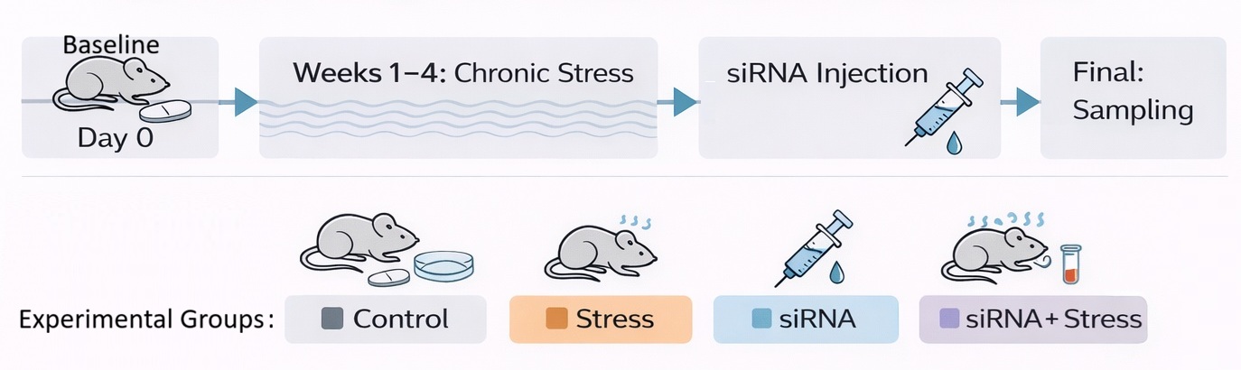
AbstractBackground: Psychosocial stress activates innate immune pathways, leading to leukocyte trafficking alterations and systemic immune dysregulation. Toll-like receptor 4 (TLR4) plays a central role in mediating the activation of stress-induced immune cells. Aim: This study investigated the effect of siRNA-mediated TLR4 silencing on stress-induced hematological alterations in rats using an unpredictable vibration–noise stress model. Methods: Forty adult male Wistar rats were randomly assigned to 4 groups: control, stress, siRNA, and siRNA + stress (n=10 per group). Chronic stress was induced daily for 4 weeks, followed by the intraperitoneal administration of TLR4-targeting siRNA (0.5 µg/g body weight). Hematological responses were assessed longitudinally using a mixed-effects linear model. Results: Chronic stress significantly increased the number of circulating neutrophils and the neutrophil-to-lymphocyte ratio (N/L), with the number of neutrophils increasing from 2.18 ×10³/µl to 4.51 ×10³/µl and N/L increasing from 0.39 to 0.85 (p < 0.001). TLR4 silencing markedly attenuated these responses, reducing the neutrophil count to 1.90 ×10³/µl and restoring the N/L ratio to baseline levels. Significant treatment × time interactions were observed for neutrophils (ηp²=0.566, p < 0.001) and N/L (ηp²=0.467, p < 0.001), whereas erythrocytic parameters remained unchanged. Conclusion: TLR4 signaling plays a mechanistic role in stress-induced leukocyte redistribution. Targeted TLR4 silencing effectively restores immune homeostasis, highlighting the role of innate immune pathways in stress adaptation. However, the absence of molecular validation of TLR4 silencing represents a limitation of this study. Keywords: Innate immune signaling, Leukocyte redistribution, Neutrophil–lymphocyte ratio, Stress-induced inflammation, TLR4 silencing. IntroductionPsychological stress disrupts homeostasis and induces integrated neuroendocrine–immune adaptations. Prolonged or repeated exposure to stress, particularly under chronic conditions, activates the hypothalamic–pituitary–adrenal (HPA) axis and alters glucocorticoid signaling, with far-reaching effects on immune function and inflammation at both the peripheral and central levels. Modern neurobiology recognizes stress responses as dynamic adaptive processes; however, persistent activation may lead to inflammatory dysregulation and increased susceptibility to affective and stress-related disorders (Mcewen and Akil, 2020). Pattern recognition receptors (PRRs) of the innate immune system play a central role in translating stress-induced signals into inflammatory responses at the molecular level. Among these receptors, Toll-like receptor 4 (TLR4) acts as a key mediator of inflammatory signaling through activation of NF-κB and subsequent cytokine production. Emerging evidence indicates that stress can activate TLR4-dependent pathways, thereby linking stress exposure to neuroinflammation and behavioral vulnerability. Thus, TLR4 signaling represents a critical interface between immune responsiveness and neuronal stress circuitry (Liu et al., 2014). In addition, TLR4 signaling has been implicated in stress-induced neuroinflammatory responses, with experimental evidence supporting its mechanistic role in stress-associated immune activation (Anwar et al., 2013). Leukocyte redistribution, characterized by increased circulating neutrophils and alterations in the neutrophil-to-lymphocyte (N/L) ratio, is a key systemic feature of stress-induced immune modulation. Glucocorticoids and catecholamines regulate leukocyte trafficking between the blood, lymphoid tissues, and peripheral organs (Ince et al., 2019). These hematological changes reflect dynamic immune redistribution and are widely recognized as integrative biomarkers of inflammatory status and physiological stress adaptation (Dhabhar, 2014). Such changes are particularly informative in longitudinal studies, where immune trajectories may reflect treatment-induced modulation over time. Recent advances in gene silencing technologies have enabled the targeted investigation of inflammatory mediators. Small interfering RNA (siRNA)-mediated sequence-specific post-transcriptional gene suppression provides a robust approach to modulate inflammatory pathways and investigate causal mechanisms. Given the involvement of TLR4 in stress-induced inflammatory responses, siRNA targeting this receptor represents a potential biological strategy to attenuate SNS activation and associated hematological alterations (Whitehead et al., 2009; Lu et al., 2020; Li et al., 2021). Stress-induced immune responses involve a complex network of cytokines, neuroimmune signaling pathways, and inflammatory mediators in addition to leukocyte redistribution. Pro-inflammatory cytokines, such as interleukin-6 (IL-6), tumor necrosis factor-alpha (TNF-α), and interleukin-1 beta (IL-1β), play pivotal roles in mediating communication between the central nervous system and the peripheral immune system. These mediators contribute to systemic inflammatory regulation during stress exposure and are closely linked to TLR4-dependent signaling cascades, further reinforcing the integration of neuroendocrine and immune responses under stress conditions. Although evidence for the involvement of TLR4 in stress-induced immune activation is increasing, its mechanistic role in leukocyte redistribution and the extent to which its inhibition can normalize immune trajectories remain unclear. Therefore, this study used a longitudinal mixed-effects design to investigate the effect of siRNA-mediated TLR4 silencing on stress-induced leukocyte redistribution in rats. Materials and MethodsExperimental animals and housing conditionsThe study was conducted using 40 adult male Wistar rats (weight, 180–220 g) obtained from an accredited laboratory animal facility. To minimize variability associated with sex-related hormonal fluctuations and to ensure consistency in stress and immune response measurements, only male rats were used. The animals were housed in standard polypropylene cages under controlled environmental conditions (22°C ± 2°C, 50%–60% relative humidity, 12 hours light/12 hours dark cycle). Laboratory chow and water were provided ad libitum. To ensure physiological stability and reduce baseline stress, all animals were acclimated to the housing conditions for 2 weeks before the initiation of experimental procedures. Experimental designAnimals were randomly allocated into four equal groups (n=10 per group): control (control), stress (stress), siRNA-treated (siRNA), and siRNA plus stress (siRNA + stress). The control group received neither stress exposure nor siRNA treatment. The stress group was subjected to the stress protocol without siRNA administration. To evaluate its potential to modulate stress-induced immune alterations, the siRNA group received siRNA administration without stress exposure, whereas the siRNA + stress group received siRNA administration following the chronic stress phase. These group designations were used consistently throughout the manuscript. The sample size was determined based on previous studies investigating stress-induced immune alterations and was considered sufficient to detect biologically meaningful differences among groups. Figure 1 shows a schematic overview of the experimental workflow, including treatment allocation, stress induction phase, and post-stress siRNA intervention.
Fig. 1. Schematic representation of the experimental design and timeline of TLR4 siRNA intervention in a model of chronic stress. Forty adult male Wistar rats (n=10 per group) were divided into 4 groups: control, stress, siRNA, and siRNA + stress. Chronic stress was applied for 4 weeks, followed by the intraperitoneal administration of TLR4-targeting siRNA. The hematological responses were assessed longitudinally at predefined stages. Silencing of TLR4 gene using siRNASmall interfering RNA (siRNA) specifically targeting TLR4 was obtained from a commercial supplier (Bioneer Corporation, Daejeon, South Korea). Lyophilized siRNA was reconstituted in sterile RNase-free solution according to the manufacturer’s instructions. For in vivo delivery, siRNA was complexed with a linear polyethylenimine-based transfection reagent (in vivo-jetPEI, Polyplus-transfection SA, France) following the manufacturer’s protocol to enhance stability and cellular uptake. The siRNA/jetPEI complexes were prepared under sterile conditions immediately before administration. The complexes were intraperitoneally injected at a dose of 0.5 µg/g body weight using sterile syringes under aseptic conditions. Intraperitoneal injection is a widely used route for systemic delivery in rodent models, allowing efficient distribution and uptake of nucleic acid-based therapeutics. Animals in the control and stress groups received sterile saline in equivalent volumes to control for injection-related effects. siRNA-mediated gene silencing is a sequence-specific post-transcriptional mechanism widely used to modulate inflammatory signaling pathways (Whitehead et al., 2009; Kanasty et al., 2013; Schroder et al., 2018). Experimental stress modelStress was induced using an unpredictable vibration–noise (aversive threat) paradigm, a model commonly employed to evoke psychological stress responses in rodents (Buynitsky and Mostofsky, 2009; Campos et al., 2013). Animals were transferred daily to a closed wooden box (1 m × 1 m) and exposed to aversive stimuli consisting of irregular mechanical shaking to generate vibrational disturbances, along with repeated knocking on the box walls from multiple directions to produce unpredictable auditory cues. To establish a chronic stress model, stress exposure was applied for approximately 30 minutes per day over a 4-week period. Following completion of the stress phase, animals assigned to siRNA-treated groups received staged intraperitoneal injections of TLR4-targeting siRNA to evaluate post-stress immunomodulatory effects. Stress sessions were conducted at the same time each day and performed by the same investigator to ensure procedural consistency and minimize variability. The apparatus was cleaned between sessions, eliminating residual odor cues, and the animals were continuously monitored during exposure to prevent injury or overheating. Immediately after each session, the animals were returned to their cages to allow recovery. This paradigm activates neuroendocrine stress pathways and induces measurable immune alterations (Buynitsky and Mostofsky, 2009). Hematological assessmentBlood samples were collected at 3 experimental stages (baseline, day 2, and final stage) to assess longitudinal hematological responses. Samples were obtained from the tail vein into ethylenediaminetetraacetic acid tubes and analyzed using an automated hematology analyzer calibrated daily according to standard procedures. The analysis included the total white blood cell (WBC) count, differential leukocyte counts (neutrophils, lymphocytes, and monocytes), and erythrocytic parameters (RBC, Hb, HCT, MCV, MCH, and MCHC). ELISASerum concentrations of TNF-α, IL-6, and IL-1β (and IL-10, where applicable) were quantified using commercially available rat-specific sandwich ELISA kits (Elabscience Biotechnology Inc., Houston, TX, USA; Cat. Nos. E-EL-R2856, E-EL-R0015, and E-EL-R0012, respectively). Blood samples were collected in plain tubes, allowed to clot at room temperature for 20 to 30 minutes, and centrifuged at 1500 to 2000 × g for 10 to 15 minutes. The serum was aliquoted and stored at −80 °C until analysis. All samples and standards were assayed in duplicate according to the manufacturer’s instructions. Absorbance was measured at 450 nm using a microplate reader, and standard curves generated using a four-parameter logistic regression model were used to calculate cytokine concentrations (pg/ml). At the final time point, cytokine levels were assessed to evaluate cumulative inflammatory responses following repeated stress exposure and TLR4 silencing. Neutrophil-to-lymphocyte ratio (N/L ratio)The N/L ratio was calculated as an index of systemic inflammatory status and physiological stress response. The ratio was calculated as follows:
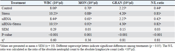
Neutrophil and lymphocyte counts were obtained from differential leukocyte analysis and expressed as cells ×10³/µl. The N/L ratio is widely recognized as an integrative biomarker reflecting stress-induced immune redistribution and inflammatory balance (Zahorec, 2001). Laboratory quality controlQuality control procedures were implemented to ensure the accuracy and reliability of hematological measurements. The analyzer was calibrated daily using standard control samples according to laboratory protocols. To verify the analytical precision, 10% of the samples were analyzed in duplicate. Hemolyzed or clotted samples were excluded to prevent measurement bias. Blood sampling was performed at standardized times to minimize circadian variation and ensure consistency across measurements. Statistical analysisData were analyzed using the SAS software (SAS Institute Inc., Cary, NC). Results are presented as mean ± SEM, and statistical significance was set at p < 0.05. For comparisons among the 4 experimental groups at each sampling stage, one-way analysis of variance (ANOVA) was performed separately for each variable. When significant differences were detected, Tukey’s honest significant difference post hoc test was used for pairwise comparisons. The effect size is reported as eta squared (η²). Longitudinal changes across experimental stages were analyzed using a linear mixed-effects model with fixed effects of treatment, time, and treatment × time interaction as fixed effects, and animal identity as a random effect to account for repeated measurements. The estimated marginal means were calculated where appropriate, and multiple comparisons were adjusted using the Holm method. The effect size of the interaction terms was expressed as the partial eta squared (ηp²), representing the magnitude of the treatment × time interaction effect. The neutrophil-to-lymphocyte (N/L) ratio was analyzed using the same approach as that used for other hematological variables. Cytokine data obtained at the final sampling point were analyzed using one-way analysis of variance (ANOVA) followed by Tukey’s honestly significant difference (HSD) test. Ethical approvalThe University of Anbar Ethical Approval Committee approved all experimental procedures (October 24, 2025). This study was conducted in accordance with international guidelines for the care and use of laboratory animals, and all efforts were made to minimize pain, distress, and unnecessary suffering throughout the experimental period. ResultsAs shown in Table 1, the experimental groups represent stress versus non-stress conditions rather than treatment effects at baseline. Significant differences were observed in the WBC count, monocytes, granulocytes, and N/L ratio (p < 0.001 for all variables). The total WBC count was significantly higher in the Stress (10.25 × 10³/µl) and siRNA + Stress (10.55 × 10³/µl) groups than in the Control (8.06 × 10³/µl) and siRNA (8.44 × 10³/µl) groups. Table 1. Leukocyte profile and inflammatory index in rats after initial stress exposure (Stage 1).
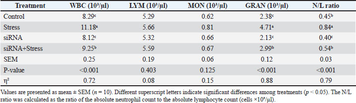
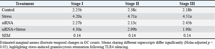
Monocyte counts were also significantly affected by treatment, with higher values observed in the Stress (0.88 × 10³/µl) and siRNA + Stress (0.92 × 10³/µl) groups than in the Control (0.70 × 10³/µl) and siRNA (0.61 × 10³/µl). Similarly, granulocyte counts were significantly increased in the Stress (4.20 × 10³/µl) and siRNA + Stress (4.30 × 10³/µl) groups relative to the Control (2.25 × 10³/µl) and siRNA (2.27 × 10³/µl). The N/L ratio showed a comparable pattern, with significantly higher values in Stress (0.81) and siRNA + Stress (0.82) compared with Control (0.44) and siRNA (0.42). As shown in Table 2, significant treatment effects were observed for WBC count, granulocytes, and the N/L ratio (p < 0.001), whereas lymphocyte and monocyte counts were not significantly affected. The WBC count was highest in the stress group (11.18 × 10³/µl), followed by the siRNA + stress group (9.25 × 10³/µl), whereas the control (8.29 × 10³/µl) and siRNA (8.12 × 10³/µl) groups remained within comparable ranges. Granulocyte counts were also significantly higher in the stress group (4.71 × 10³/µl) compared with the control (2.38 × 10³/µl) and siRNA (2.13 × 10³/µl). The siRNA + Stress group showed lower granulocyte values (2.99 × 10³/µl) than the Stress group. The N/L ratio exhibited a similar pattern, with the highest value observed in the Stress group (0.84), followed by siRNA + Stress (0.54), while lower values were observed in the Control (0.45) and siRNA (0.40). Table 2. Leukocyte dynamics and early immunomodulatory effects of TLR4 silencing during continued stress exposure (Stage 2).
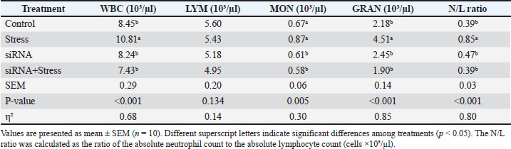
As shown in Table 3, significant differences in WBC count, monocytes, granulocytes, and the N/L ratio were observed among treatments. The WBC count remained higher in the stress group (10.81 × 10³/µl) compared with Control (8.45 × 10³/µl) and siRNA (8.24 × 10³/µl), whereas the siRNA + stress group showed lower values (7.43 × 10³/µl). Granulocyte counts were also highest in the stress group (4.51 × 10³/µl), while the siRNA + stress group exhibited lower values (1.90 × 10³/µl) compared with the control (2.18 × 10³/µl) and siRNA (2.45 × 10³/µl). The N/L ratio followed a similar pattern, remaining elevated in the Stress group (0.85), whereas lower values were observed in the Control (0.39), siRNA (0.47), and siRNA + Stress (0.39) groups. Table 3. Leukocyte homeostasis restoration following TLR4 silencing under repeated stress conditions (Stage 3).
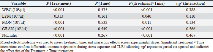
The mixed-effects analysis showed that Treatment × Time interactions were highly significant for granulocytes and N/L ratio (p < 0.001), confirming different changes in the immune status between treatments. The interaction effect was highest for granulocytes (ηp²=0.566), which denotes a pronounced treatment-related change from baseline to follow-up. The N/L ratio also showed a strong interaction (ηp²=0.467), indicating fluctuations in SI condition. These data suggest that rather than linear shifts across time as a result of stress exposure and TLR4 knockdown, opposing immune pathways are created. As shown in Table 4, the mixed-effects model revealed significant treatment effects for WBC count, monocyte count, granulocyte count, and N/L ratio (p < 0.001), whereas lymphocyte count was not significantly affected. No significant main effects of time were observed (p > 0.05). However, significant treatment × time interactions were detected for all variables, including WBC (p < 0.001), lymphocyte count (p=0.040), monocyte count (p=0.011), granulocyte count (p < 0.001), and N/L ratio (p < 0.001). The largest interaction effects were observed for granulocytes (ηp²=0.566) and the N/L ratio (ηp²=0.467), whereas smaller effects were noted for WBCs (ηp²=0.388), monocytes (ηp²=0.134), and lymphocytes (ηp²=0.110). Table 4. Longitudinal mixed-effect analysis of leukocyte redistribution and inflammatory index.
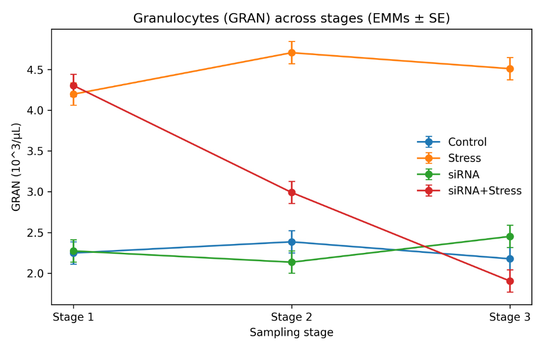
Figure 2 illustrates the temporal changes in GC counts across experimental stages. Granulocyte levels remained consistently elevated in the Stress group across all stages, with significantly higher values at Stage II and maintained at Stage III. In contrast, the siRNA + Stress group showed a progressive decrease in GC counts over time, declining from Stage II to Stage 3, where the lowest values were observed. Granulocyte counts in the siRNA group remained relatively stable and were comparable to controls across all stages. The control group exhibited minimal variation throughout the experimental period. These temporal patterns are consistent with the significant treatment × time interaction observed for granulocytes.
Fig. 2. Temporal changes in circulating granulocytes during sstress exposure and TLR4 silencing. Line graph illustrating the granulocyte trajectories across the experimental stages. Stress exposure induced sustained granulocytosis, whereas TLR4 silencing progressively reduced granulocyte counts and restored immune balance Table 5. Table 5. Estimated marginal means of circulating granulocytes across experimental stages.
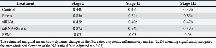
As shown in Table 6, the N/L ratio significantly differed among treatments across all experimental stages. At Stage 1, the highest values were observed in the siRNA + Stress (0.82) and Stress (0.81) groups, compared with the Control (0.44) and siRNA (0.42). At Stage 2, the N/L ratio remained highest in the stress group (0.84), whereas the siRNA + stress group showed lower values (0.54). The control (0.45) and siRNA (0.40) groups maintained lower ratios. At Stage 3, the N/L ratio remained elevated in the Stress group (0.85), whereas siRNA+Stress (0.39) was comparable to Control (0.39). The siRNA group exhibited intermediate values (0.47). Table 6. Estimated marginal means of neutrophil-to-lymphocyte ratio across experimental stages.
The temporal variation in the N/L ratio across experimental stages is shown in Figure 3. The stress group remained elevated across all stages, with consistently higher values than the other groups. In contrast, the siRNA+Stress group showed a progressive decrease in the N/L ratio from Stage II to Stage III. The siRNA group maintained relatively low and stable values across all stages, whereas the control group exhibited minimal variation throughout the experimental period. These temporal patterns are consistent with the significant treatment × time interaction observed for the N/L ratio.
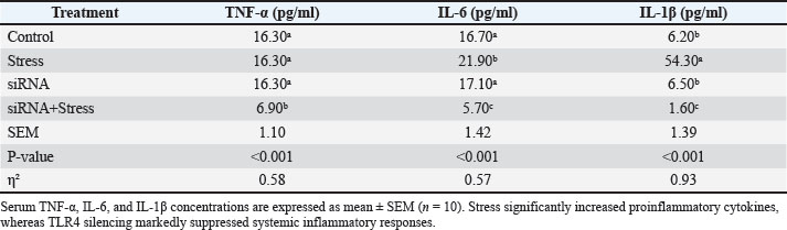
Fig. 3. Trajectory of Neutrophil-to-Lymphocyte Ratio During Repeated Stress and TLR4 Silencing. Line graph showing the temporal variation in the N/L ratio. Stress increased the systemic inflammatory status, whereas TLR4 silencing restored the ratio to baseline values. As shown in Table 7, there were significant differences among treatments for TNF-α, IL-6, and IL-1β (p < 0.001 for all variables). The IL-6 concentration was highest in the stress group (21.90 pg/ml) and lowest in the siRNA + stress group (5.70 pg/ml), whereas the control (16.70 pg/ml) and siRNA (17.10 pg/ml) groups showed intermediate values. IL-1β levels were significantly higher in the stress group (54.30 pg/ml) than in the control (6.20 pg/ml) and siRNA (6.50 pg/ml) groups, whereas the lowest value was observed in the siRNA + stress group (1.60 pg/ml). Similarly, TNF-α levels were lower in the siRNA + stress group (6.90 pg/ml) than in the control and stress groups (16.30 pg/ml). Table 7. Serum proinflammatory cytokine responses following stress exposure and TLR4 silencing.
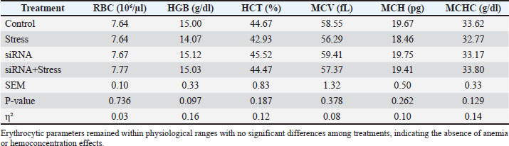
The erythrocytic parameters did not differ significantly among treatments (p > 0.05) (Table 8). The RBC counts ranged from 7.64 ×10⁶/µl in the Control and Stress groups to 7.77 ×10⁶/µl in the siRNA + stress group. Hemoglobin values varied from 14.07 g/dl in the stress group to 15.12 g/dl in the siRNA group, whereas hematocrit ranged from 42.93% to 45.52% across treatments. MCV values ranged from 56.29 fL (stress) to 59.41 fL (siRNA), MCH from 18.46 to 19.75 pg, and MCHC from 32.77 to 33.80 g/dl. Table 8. Red blood cell indices during initial stress exposure (Stage 1).
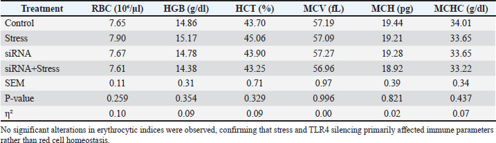
As shown in Table 9, no significant differences in erythrocytic indices were observed among treatments (p > 0.05). The RBC counts ranged from 7.61 ×10⁶/µl in the siRNA + stress group to 7.90 ×10⁶/µl in the stress group. Hemoglobin values ranged from 14.38 g/dl (siRNA+Stress) to 15.17 g/dl (Stress), while hematocrit values ranged from 43.25% to 45.06% across treatments. MCV, MCH, and MCHC values ranged from 56.96 to 57.27 fL, 18.92 to 19.44 pg, and 33.22 to 34.01 g/dl, respectively. Table 9. Red blood cell indices during continuous stress exposure (Stage 2).
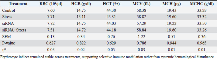
As shown in Table 10, there were no significant differences among treatments for erythrocytic indices (p > 0.05). The RBC counts ranged from 7.51 ×10⁶/µl in the siRNA+Stress group to 7.72 ×10⁶/µl in the siRNA group, with values comparable to the Control. Hemoglobin values ranged from 14.72 to 15.11 g/dl, while hematocrit values ranged from 44.03% to 45.31% across treatments. MCV, MCH, and MCHC values ranged from 57.29 to 58.84 fL, 19.22 to 19.60 pg, and 33.26 to 33.50 g/dl, respectively. Table 10. Red blood cell indices following repeated stress exposure (Stage 3).
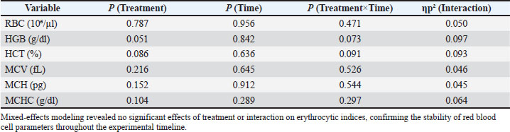
No significant effects of treatment, time, or their interaction were observed for erythrocytic indices (p > 0.05 for all variables) (Table 11). The RBC count was not significantly affected by treatment (p=0.787), time (p=0.956), or treatment × time interaction (p=0.471), with a small interaction effect size (ηp²=0.050). Hemoglobin showed a borderline treatment effect (p=0.051), whereas time (p=0.842) and interaction (p=0.073) effects were not significant. Similarly, Statistical analysis indicated that treatment (p=0.086), time (p=0.636), and their interaction (p=0.091) did not significantly influence hematocrit. No significant effects were observed for MCV (treatment, p=0.216; time, p=0.645; interaction, p=0.526), MCH (p=0.152; interaction, p=0.544), or MCHC (p=0.104; interaction, p=0.297). The interaction effect sizes were consistently small (partial η²=0.045–0.097). Table 11. Longitudinal mixed-effects analysis of erythrocytic parameters.
DiscussionThe sustained mobilization of leukocytes, characterized by concurrent leukocytosis, granulocytosis, and a persistent increase in the N/L ratio, indicates systemic inflammatory activation. These findings are consistent with established models of stress physiology, where activation of the hypothalamic–pituitary–adrenal axis and sympathetic nervous system promotes neutrophil demargination and leukocyte redistribution from lymphoid tissues to the circulation, thereby enhancing innate immune readiness (Dhabhar, 2000, 2014; Mcewen and Akil, 2020). The persistence of elevated granulocyte levels across the experimental stages suggests that the observed pattern reflects a transient adaptive response rather than sustained neuroendocrine stimulation or inflammatory priming. Chronic or unpredictable stress has been shown to maintain prolonged immune activation and elevated inflammatory tone, thereby increasing susceptibility to stress-related disorders (Engler et al., 2004; Russell and Lightman, 2019; White, 2024). The significant elevation of the N/L ratio further supports the presence of systemic inflammatory activation and highlights its utility as an integrative biomarker of stress physiology. This index reflects both neutrophil mobilization and lymphocyte redistribution and has gained increasing attention as a sensitive marker of neuroendocrine–immune interactions and inflammatory imbalance (Slavich, 2020). Elevated N/L ratio values are associated with sympathetic activation and glucocorticoid-mediated lymphocyte trafficking, indicating a shift toward innate immune predominance during stress exposure (Powell et al., 2021). The reduction in the N/L ratio following TLR4 silencing suggests that modulation of inflammatory signaling contributes to the observed changes in leukocyte distribution. One of the major findings of the present study is that TLR4 siRNA attenuated stress-induced granulocytosis and reduced the N/L ratio toward baseline levels, thereby implicating innate immune receptor signaling in stress-associated hematological alterations. TLR4 is a key pattern recognition receptor involved in NF-κB-dependent inflammatory signaling and cytokine production (Lu et al., 2020). Psychological stress can activate TLR4-dependent pathways, linking neuroendocrine stress signaling with peripheral immune activation and neuroimmune sensitization (Souza-Junior and Batistela, 2022). The observed reduction in granulocyte counts in the siRNA+Stress group suggests that innate immune cell mobilization is decreased, with partial restoration of immune balance. Although the efficiency of siRNA delivery is a known limitation in vivo, previous studies have demonstrated that systemically administered siRNA can exert biological effects through cellular uptake mechanisms and enhanced tissue permeability under inflammatory conditions (Whitehead et al., 2009; Kanasty et al., 2013). Therefore, the immunomodulatory effects observed in the present study support the biological activity of the administered siRNA. These findings further support the role of innate immune receptors, particularly TLR4, in mediating stress-induced immune activation beyond the classical neuroendocrine pathways. The cytokine results further support the mechanistic interpretation of these findings. Stress exposure was associated with increased IL-6 and IL-1β levels, both of which play central roles in inflammatory signaling and neuroimmune crosstalk. IL-6 is a key mediator of stress-related inflammatory responses and systemic immune regulation, whereas IL-1β is critically involved in neuroinflammatory activation and behavioral susceptibility (Calcia et al., 2016; Slavich, 2020). The reduction in TNF-α, IL-6, and IL-1β levels following TLR4 silencing indicates that TLR4-dependent signaling contributes to the regulation of proinflammatory pathways during stress. This finding is consistent with neuroimmune models proposing that repeated stress enhances cytokine signaling through innate immune receptors, leading to systemic inflammatory dysregulation (Dantzer et al., 2008; Furman et al., 2019). In this context, neutrophils represent dynamic regulators of inflammatory homeostasis, integrating neuroendocrine signals and contributing to downstream inflammatory responses (Kolaczkowska and Kubes, 2013; Silvestre-Roig et al., 2019; Haykin and Rolls, 2021). Neutrophils are also functionally heterogeneous and can be rapidly recruited during stress-induced immune activation (Palomino-Segura et al., 2023). Recent work has emphasized neutrophil plasticity and functional heterogeneity in the regulation of inflammatory responses (Goodman et al., 2024), suggesting that stress-induced neutrophilia reflects not only increased mobilization but also functional reprogramming toward innate immune dominance. The observed reduction in granulocyte counts following TLR4 silencing further supports the involvement of innate immune receptor signaling in neutrophil recruitment and activation during stress. Although significant immune alterations were observed, erythrocytic indices remained unchanged across treatments and experimental stages, indicating that stress exposure primarily affected leukocyte dynamics rather than red blood cell parameters. This selective response is consistent with previous findings indicating that stress predominantly influences immune regulation and inflammatory signaling without markedly altering erythrocytic homeostasis (Mcewen and Akil, 2020). The absence of significant changes in erythrocytic indices further supports the interpretation that the observed hematological responses are primarily driven by immune redistribution rather than changes in RBC physiology. Our findings agree with previous reports of stress-induced neutrophilia and inflammatory activation in rodent models (Frank et al., 2016; Fonken et al. 2016; White, 2024). Stress enhances immune signaling through TLR4-mediated pathways, thereby contributing to neuroimmune sensitization and behavioral vulnerability (Souza-Junior and Batistela, 2022; Schramm and Waisman, 2022; Zhao et al., 2025). The current study extends these observations by demonstrating that TLR4 silencing attenuates systemic inflammatory responses and modulates leukocyte trafficking dynamics over time in a longitudinal framework. While previous studies have primarily focused on neuroinflammatory or behavioral outcomes, the present findings highlight a systemic hematological perspective, where leukocyte redistribution represents a measurable downstream effect of TLR4-mediated stress signaling. The observed temporal changes highlight progressive immune adjustment following TLR4 silencing, a feature not typically captured in cross-sectional stress studies. Mixed-effects modeling revealed distinct immune trajectories between stressed and TLR4-silenced animals, indicating a shift in leukocyte dynamics over time rather than a transient suppression effect. These findings highlight the dynamic nature of stress–immune interactions and are consistent with evidence that stress-induced immune alterations can be modulated through upstream inflammatory signaling pathways (Ménard et al., 2017). Furthermore, accumulating evidence supports the role of bidirectional brain–immune communication in determining vulnerability and resilience to stress (Kuhn et al., 2024), reinforcing the role of neuroimmune signaling in stress adaptation. Overall, the present findings highlight the role of TLR4 in linking psychological stress to systemic inflammatory responses and leukocyte redistribution. The attenuation of granulocytosis, reduction in N/L ratio, and decrease in pro-inflammatory cytokines following TLR4 silencing provide evidence for the involvement of innate immune receptor signaling in stress-induced immune alterations. These findings offer mechanistic insight into the regulation of inflammatory responses under stress conditions. Given the increasing recognition of neuroimmune pathways in stress-related disorders, TLR4 signaling modulation may represent a potential strategy for mitigating stress-associated inflammatory dysregulation. Several limitations should be considered when interpreting the results. First, molecular validation of TLR4 silencing was not performed and should be confirmed at the mRNA and protein levels in future studies. Second, behavioral assessments, which may have provided additional insight into stress responsiveness, were not included. Third, the use of a single sex limits the generalizability of the findings, and future studies should address potential sex-specific differences in stress–immune interactions. In addition, the absence of immediate post-stress baseline measurements before siRNA administration represents a limitation in evaluating initial group comparability. ConclusionThis longitudinal study demonstrates that repeated psychological stress induces sustained leukocyte redistribution, characterized by granulocytosis and an increased N/L ratio, reflecting systemic inflammatory activation. TLR4 silencing significantly attenuated these alterations, as evidenced by reduced granulocyte counts, normalization of the N/L ratio, and suppression of proinflammatory cytokines. These findings indicate that TLR4-dependent signaling contributes to stress-induced immune response regulation and leukocyte trafficking. The longitudinal design further highlights that modulation of this pathway alters immune trajectories over time, supporting innate immune signaling in shaping stress-related inflammatory dynamics. Overall, this study provides evidence that targeting TLR4 signaling can modulate stress-associated immune alterations and may represent a potential approach for mitigating stress-related inflammatory dysregulation. AcknowledgmentsThe authors would like to express their sincere appreciation to the College of Education for Pure Sciences, University of Anbar, Iraq, for providing the necessary time and institutional support that facilitated this study’s completion. Conflict of interestThe authors declare that they have no known competing financial or personal interests that could have influenced the work reported in this paper. FundingNot applicable. Authors’ ContributionConceptualization: S. M. Abdulateef. Methodology: S. M. Abdulateef, S. N. Ali. Investigation: S. M. Abdulateef, S. N. Ali. Data curation: S. M. Abdulateef. Formal analysis: S. M. Abdulateef. Validation: S. M. Abdulateef, S. N. Ali. Resources: S. N. Ali. Writing – original draft: S. M. Abdulateef. Writing – review & editing: S. M. Abdulateef, S. N. Ali. Supervision: S. M. Abdulateef. Data availabilityUpon reasonable request, the datasets of this study can be obtained from the corresponding author. ReferencesAnwar, M.A., Basith, S. and Choi, S. 2013. Negative regulatory approaches to the attenuation of Toll-like receptor signaling. Exp Mol. Med. 45(2), e11; doi:10.1038/emm.2013.28 Buynitsky, T. and Mostofsky, D.I. 2009. Restraint stress in biobehavioral research: recent developments. Neurosci. &. Biobehavioral. Rev. 33(7), 1089–1098; doi:10.1016/j.neubiorev.2009.05.004 Calcia, M.A., Bonsall, D.R., Bloomfield, P.S., Selvaraj, S., Barichello, T. and Howes, O.D. 2016. Stress and neuroinflammation: a systematic review of the effects of stress on microglia and the implications for mental illness. Brain. Behav. Immunity. 54, 62–75; doi:10.1016/j.bbi.2015.12.012 Campos, A.C., Fogaça, M.V., Aguiar, D.C. and Guimarães, F.S. 2013. Animal models of anxiety disorders and stress. Revista Brasileira De Psiquiatria 35(Suppl 2), S101–S111; doi:10.1590/1516-4446-2013-1139 Dantzer, R., O'Connor, J.C., Freund, G.G., Johnson, R.W. and Kelley, K.W. 2008. From inflammation to sickness and depression: when the immune system subjugates the brain. Nature. Rev. Neurosci. 9(1), 46–56; doi:10.1038/nrn2297 Dhabhar, F.S. 2000. Acute stress enhances while chronic stress suppresses skin immunity: the role of stress hormones and leukocyte trafficking. Ann. N. Y. Acad. Sci. 917, 876–893; doi:10.1111/j.1749-6632.2000.tb05454.x Dhabhar, F.S. 2014. Effects of stress on immune function: the good, the bad, and the beautiful. Immunologic Res. 58(2–3), 193–210; doi:10.1007/s12026-014-8517-0 Engler, H., Bailey, M.T., Engler, A. and Sheridan, J.F. 2004. Effects of repeated social stress on leukocyte distribution, function, and psychosocial behavior in rats. J. Neuroimmunology 148(1–2), 106–115; doi:10.1016/j.jneuroim.2003.11.011 Fonken, L.K., Weber, M.D., Daut, R.A., Kitt, M.M., Frank, M.G., Watkins, L.R. and Maier, S.F. 2016. Stress-induced neuroinflammatory priming is time of day dependent. Psychoneuroendocrinology 66, 82–90; doi:10.1016/j.psyneuen.2016.01.006 Frank, M.G., Weber, M.D., Watkins, L.R. and Maier, S.F. 2016. Stress-induced neuroinflammatory priming: a liability factor in the etiology of psychiatric disorders. Neurobiol. Stress 4, 62–70; doi:10.1016/j.ynstr.2015.12.004 Furman, D., Campisi, J., Verdin, E., Carrera-Bastos, P., Targ, S., Franceschi, C., Ferrucci, L., Gilroy, D.W., Fasano, A., Miller, G.W., Miller, A.H., Mantovani, A., Weyand, C.M., Barzilai, N., Goronzy, J.J., Rando, T.A., Effros, R.B., Lucia, A., Kleinstreuer, N. and Slavich, G.M. 2019. Chronic inflammation in the etiology of disease across the life span. Nature Med. 25(12), 1822–1832; doi:10.1038/s41591-019-0675-0 Goodman, E.J., Disabato, D.J., Sheridan, J.F. and Godbout, J.P. 2024. Novel microglial transcriptional signatures promote social and cognitive deficits following repeated social defeat. Commun. Biol. 7, 1199; doi:10.1038/s42003-024-06898-9 Haykin, H. and Rolls, A. 2021. The neuroimmune response during stress: a physiological perspective. Immunity 54(9), 1933–1947; doi:10.1016/j.immuni.2021.08.023 Ince, L.M., Weber, J. and Scheiermann, C. 2019. Control of leukocyte trafficking by stress-associated hormones. Front. Immunol. 9, 3143; doi:10.3389/fimmu.2018.03143 Kanasty, R., Dorkin, J.R., Vegas, A. and Anderson, D. 2013. Delivery materials for siRNA therapeutics. Nature. Mater. 12, 967–977; doi:10.1038/nmat3765 Kolaczkowska, E. and Kubes, P. 2013. Neutrophil recruitment and function in health and inflammation. Nature Rev. Immunol. 13, 159–175; doi:10.1038/nri3399 Kuhn, A.M., Bosis, K.E. and Wohleb, E.S. 2024. Looking back to move forward: research in stress, behavior, and immune function. Neuroimmunomodulation 31(1), 211–229; doi:10.1159/000541592 Li, W., Zhang, X., Huang, Z., Chen, W., Shi, H. and Wang, Y. 2021. TLR4 gene silencing using siRNA attenuates inflammatory injury in experimental acute lung injury. Int. Immunopharmacology. 96, 107595; doi:10.1016/j.intimp.2021.107595 Liu, J.J., Buisman-Pijlman, F.T.A. and Hutchinson, M.R. 2014. Toll-like receptor 4: innate immune regulator of neuroimmune and neuroendocrine interactions in stress and major depressive disorder. Front. Neurosci. 8, 309; doi:10.3389/fnins.2014.00309 Lu, Y., -C.., Yeh, W., -C.. and Ohashi, P.S. 2020. LPS/TLR4 signal transduction pathway. Nature Rev. Immunol. 20(9), 545–558; doi:10.1038/s41577-020-0313-2 Mcewen, B.S. and Akil, H. 2020. Revisiting the stress concept: implications for affective disorders. J. Neurosci. 40(1), 12–21; doi:10.1523/JNEUROSCI.0733-19.2019 Ménard, C., Pfau, M.L., Hodes, G.E. and Russo, S.J. 2017. Immune and neuroendocrine mechanisms of stress vulnerability and resilience. Neuropsychopharmacology 42(1), 62–80; doi:10.1038/npp.2016.90 Palomino-Segura, M., Sicilia, J., Ballesteros, I. and Hidalgo, A. 2023. Strategies of neutrophil diversification. Nature. Immunol. 24(4), 515–525; doi:10.1038/s41590-023-01452-x Powell, N.D., Tarr, A.J. and Sheridan, J.F. 2021. Psychosocial stress and the immune system. Nature Rev. Immunol. 21(9), 531–545; doi:10.1038/s41577-021-00524-0 Russell, G. and Lightman, S. 2019. The human stress response. Nature Rev. Endocrinol. 15(9), 525–534; doi:10.1038/s41574-019-0228-0 Schramm, E. and Waisman, A. 2022. Microglia as central protagonists in the chronic stress response. Neurol. Neuroimmunology &. Neuroinflammation 9(6), e200023; doi:10.1212/NXI.0000000000200023 Schroder, A.R., Shieh, J.J. and Irvine, D.J. 2018. In vivo delivery of siRNA.In SiRNA Delivery Methods (Methods in Molecular Biology). New York, NY: Humana, vol. 1364; doi:10.1007/978-1-4939-6993-7_12 Silvestre-Roig, C., Fridlender, Z.G., Glogauer, M. and Scapini, P. 2019. Neutrophil diversity in health and disease. Trends. Immunol. 40(7), 565–583; doi:10.1016/j.it.2019.04.012 Slavich, G.M. 2020. Social safety theory: a biologically based evolutionary perspective on life stress, health, and behavior. Annu. Rev. Clin. Psychol. 16, 265–295; doi:10.1146/annurev-clinpsy-032816-045159 Souza-Junior, F.J.C., Cunha, L.C. and Lisboa, S.F. 2022. Toll-like receptor 4 in the interface between neuroimmune response and behavioral alterations caused by stress. Explor. Neuroprot. Ther. 2, 182–209; doi:10.37349/ent.2022.00028 White, A.G., Elias, E., Orozco, A., Robinson, S.A. and Manners, M.T. 2024. Chronic stress-induced neuroinflammation: relevance of rodent models to human disease. Int. J. Mol. Sci. 25(10), 5085; doi:10.3390/ijms25105085 Whitehead, K.A., Langer, R. and Anderson, D.G. 2009. Knocking down barriers: advances in siRNA delivery. Nature. Rev. Drug. Discovery. 8(2), 129–138; doi:10.1038/nrd2742 Zahorec, R. 2001. Ratio of neutrophil to lymphocyte counts—Rapid and simple parameter of systemic inflammation and stress in critically ill. Bratislavské. Lekárske. Listy. 102(1), 5–14. Zhao, K., Zhang, Y., Yang, S., Xiang, L., Wu, S., Dong, J., Li, H., Yu, H. and Hu, W. 2025. Neuroinflammation and stress-induced pathophysiology in major depressive disorder: mechanisms and therapeutic implications. Front. Cell. Neurosci. 19, 1538026; doi:10.3389/fncel.2025.1538026 | ||


| How to Cite this Article |
| Pubmed Style Ali SN, Abdulateef SM. Silencing TLR4 attenuates stress-induced leukocyte redistribution in rats: A longitudinal mixed-effects analysis. Open Vet. J.. 2026; 16(6): 3444-3455. doi:10.5455/OVJ.2026.v16.i6.15 Web Style Ali SN, Abdulateef SM. Silencing TLR4 attenuates stress-induced leukocyte redistribution in rats: A longitudinal mixed-effects analysis. https://www.openveterinaryjournal.com/?mno=310796 [Access: June 26, 2026]. doi:10.5455/OVJ.2026.v16.i6.15 AMA (American Medical Association) Style Ali SN, Abdulateef SM. Silencing TLR4 attenuates stress-induced leukocyte redistribution in rats: A longitudinal mixed-effects analysis. Open Vet. J.. 2026; 16(6): 3444-3455. doi:10.5455/OVJ.2026.v16.i6.15 Vancouver/ICMJE Style Ali SN, Abdulateef SM. Silencing TLR4 attenuates stress-induced leukocyte redistribution in rats: A longitudinal mixed-effects analysis. Open Vet. J.. (2026), [cited June 26, 2026]; 16(6): 3444-3455. doi:10.5455/OVJ.2026.v16.i6.15 Harvard Style Ali, S. N. & Abdulateef, . S. M. (2026) Silencing TLR4 attenuates stress-induced leukocyte redistribution in rats: A longitudinal mixed-effects analysis. Open Vet. J., 16 (6), 3444-3455. doi:10.5455/OVJ.2026.v16.i6.15 Turabian Style Ali, Saja N., and Salwan M. Abdulateef. 2026. Silencing TLR4 attenuates stress-induced leukocyte redistribution in rats: A longitudinal mixed-effects analysis. Open Veterinary Journal, 16 (6), 3444-3455. doi:10.5455/OVJ.2026.v16.i6.15 Chicago Style Ali, Saja N., and Salwan M. Abdulateef. "Silencing TLR4 attenuates stress-induced leukocyte redistribution in rats: A longitudinal mixed-effects analysis." Open Veterinary Journal 16 (2026), 3444-3455. doi:10.5455/OVJ.2026.v16.i6.15 MLA (The Modern Language Association) Style Ali, Saja N., and Salwan M. Abdulateef. "Silencing TLR4 attenuates stress-induced leukocyte redistribution in rats: A longitudinal mixed-effects analysis." Open Veterinary Journal 16.6 (2026), 3444-3455. Print. doi:10.5455/OVJ.2026.v16.i6.15 APA (American Psychological Association) Style Ali, S. N. & Abdulateef, . S. M. (2026) Silencing TLR4 attenuates stress-induced leukocyte redistribution in rats: A longitudinal mixed-effects analysis. Open Veterinary Journal, 16 (6), 3444-3455. doi:10.5455/OVJ.2026.v16.i6.15 |


