





| Research Article | ||
Open Vet. J.. 2026; 16(6): 3422-3437 Open Veterinary Journal, (2026), Vol. 16(6): 3422-3437 Research Article Effect of probiotic supplementation on family and phyla composition and metabolite profiles in miceZahraa S. Mohammed and Ali H. D. Janabi*Department of Veterinary Microbiology, College of Veterinary Medicine, University of Al-Qadisiyah, Al-Diwaniyah City, Iraq *Corresponding Author: Ali H. D. Janabi. Department of Veterinary Microbiology, Collee of Veterinary Medicine, University of Al-Qadisiyah, Al-Diwaniyah City, Iraq. Email: ali.janabi [at] qu.edu.iq Submitted: 07/03/2026 Revised: 05/05/2026 Accepted: 18/05/2026 Published: 05/06/2026 © 2025 Open Veterinary Journal
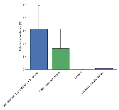
AbstractBackground: Through the production of microbial metabolites, particularly short-chain fatty acids and tryptophan-derived compounds, the gut microbiome plays a central role in host metabolism and intestinal health. Probiotic bacteria such as Lactobacillus plantarum and Bifidobacterium breve are known to influence the composition of microbial communities and their metabolic activity in the gastrointestinal tract. However, the combined effects of these probiotics on gut microbial families, phylum-level structure, and metabolite production remain unclear. Aim: This study aimed to determine whether probiotic treatments using L. plantarum, B. breve, and their combination could stimulate broader shifts in the gut microbial community by promoting the abundance of other beneficial probiotic-associated bacterial families and phyla, and to evaluate how these microbial changes influence key gut metabolites. Methods: Experimental mice (24 mice) were divided into four groups (6 mice each): Control, L. plantarum, B. breve, and combination treatment groups. Gut microbial composition was analyzed using next generation sequencing of the 16S rRNA amplicons, and relative abundances of dominant bacterial families and phyla were calculated. Metabolomic analysis was performed to quantify major microbial metabolites, including butyrate, acetate, propionate, indole-3-propionic acid (IPA), and trimethylamine-N-oxide (TMAO). Results: At the family level, Lactobacillaceae showed a significant increase in the Combination group compared with the other groups (p < 0.05), while other families, including Oscillospiraceae, Lachnospiraceae, Muribaculaceae, Prevotellaceae, Bacteroidaceae, and Akkermansiaceae, showed variations that were not statistically significant (p > 0.05). At the phylum level, Bacillota remained the dominant phylum across all groups without significant differences (p > 0.05). Metabolomic analysis demonstrated significant differences in key microbial metabolites. Butyrate, acetate, propionate, IPA, and TMAO showed significant variation among treatments (p < 0.05), indicating that probiotic supplementation influenced microbial metabolic activity in the gut. Conclusion: This study showed that probiotic treatments using L. plantarum, B. breve, and their combination stimulate broader shifts in the gut microbial community by promoting the abundance of other beneficial probiotic-associated bacterial families and phyla, and evaluate how these microbial changes influence key gut metabolites. Keywords: Acetate, Gut microbiome, Metabolomics, Probiotics, Short-chain fatty acids. IntroductionThe gut microbiome is essential to host physiology as it is integral to nutrient metabolism, immune system modulation, and intestinal barrier maintenance. A well-balanced microbiome is beneficial to host health by producing metabolites that include short-chain fatty acids (SCFAs), which regulate the integrity of the intestinal epithelium, energy metabolism, and inflammation. Changes to the microbial composition of the gut can impair any of these functions and lead to the development of metabolic and inflammatory diseases. Recent studies have focused on the gut microbiome and discovered that certain gut bacteria are directly responsible for producing metabolites that are beneficial for sustaining gut health (Kullberg et al., 2025; Ye et al., 2025). The use of probiotic bacteria to adjust the balance of the gut microbe ecosystem has received increased attention. Some of the most representative probiotic microorganisms are Lactobacillus and Bifidobacterium, which have been proven to improve both the composition and metabolic activity of the intestinal microflora. Probiotics help with some gastrointestinal diseases, improve immune response, and alter gut microflora (Ismael et al., 2025). For instance, Feng et al. (2025) modified some metabolic pathways of microbes and improved inflammation and gut microbial balance. Similarly, An et al. (2025) showed that Bifidobacterium breve increased SCFA and contributed to gut health through changes in gut flora. In addition to the possibility of direct colonization of the gut by probiotic strains, research suggests that probiotic supplementation may promote the growth of beneficial other microbial taxa in the gut ecosystem. Such community-level restructuring may increase the abundance of beneficial metabolic bacterial families, especially in butyrate and acetate production. Improvements in host metabolic outcomes and intestinal barrier functionality have been associated with such microbial structural shifts. Interventions with probiotics have been shown in some studies to change the composition of the gut microbial community and, at the same time, change some of the important metabolic pathways in the gut environment when the gut microbiome has been sequenced and the gut metabolome has been analyzed (Wu et al, 2025; Yang et al, 2025). Therefore, metabolic profiling is essential to assess the microbiome-modulation function. Metabolites of microorganisms, such as butyrate, acetate, and propionate, are critical to the regulation of intestinal inflammation, energy metabolism, and immune signaling. Furthermore, some microbial metabolites of tryptophan, such as indole, protect the intestinal epithelium and exert antioxidant effects. The combination of microbiome and metabolome analyses provides a deeper understanding of how probiotics influence microbial metabolic systems and gut functionality (Luo et al., 2025; Wang et al., 2025). Probiotics are increasingly associated with certain microbial metabolic alterations, but the mechanism by which such treatments provoke extensive restructuring of the microbial community remains poorly understood. Specifically, additional research is warranted on how probiotics impact gut microbiota restructuring by increasing the abundance of beneficial bacterial families and phyla. Most of the functionality of probiotics in the gut microbial ecosystem and the corresponding metabolic pathways remains unexplained due to a lack of understanding of the indirect microbial interactions. The current research set out to assess how gut microbial composition and metabolite profiles are affected by probiotic supplementation with Lactobacillus plantarum and B. breve, both separately and in combination. This study combined microbiome and metabolome analyses to determine whether probiotic treatments would result in community-level increases of beneficial bacterial families and phyla, and/or increases of microbes and metabolites critical to the regulation of the intestinal metabolome. Materials and MethodsExperimental animals and study designThis study utilized 24 healthy lab mice (aged 6–8 weeks) to investigate the impacts of probiotic supplementation on gut microbiome and metabolite production. The mice were housed at 22°C ± 2°C and 50%–60% humidity, during 12-hour light/dark cycles. Mice were provided standard lab chow and drinks ad libitum. The study was completed at the College of Veterinary Medicine, University of Al-Qadisiyah. The subjects were split into four groups of six random mice. The control group did not receive any probiotic supplementation. The 2nd group received Lactiplantibacillus plantarum, the 3rd group received B. breve, and the 4th group received both. Probiotics were given daily. The probiotic strains utilized in this study were obtained as freeze-dried probiotic powders employing Protech™ stabilization technology from Jiangsu Biodep Biotechnology Co., Ltd. (Jiangyin City, Jiangsu, China). Bifidobacterium breve strain BB033 was provided as freeze-dried powder with a minimum viable count of ≥3.0 × 10¹¹ CFU/g. Likewise, Lactiplantibacillus plantarum strain Lp3a was supplied as a freeze-dried powder with a viable count of ≥5.0 × 10¹¹ CFU/g. Both probiotics were stored at −18°C in their original sealed packages as recommended by the manufacturer in order to maintain the viability of the bacteria. Analysis Certificates were provided demonstrating the absence of the pathogens, Salmonella, Escherichia coli, and Staphylococcus aureus, as well as insufficient viable cells in both probiotics. Fresh preparations of probiotic suspensions were made and used on the same day. Mice in the Lactiplantibacillus plantarum group were given 1 × 10⁹ CFU/mouse/day, and those in the Bifidobacterium breve group were given 1 × 10⁹ CFU/mouse/day via oral gavage. The combination group received both probiotic strains, making their total dose 2 × 10⁹ CFU/mouse/day. The control group received the same treatment volume but instead received sterile PBS. Treatments were given once daily at the same time for the duration of the study. The probiotic powders underwent dissolution and homogenization to ensure probiotics were uniformly distributed across the oral gavage. These probiotic strains were incorporated into sterile phosphate-buffered saline (PBS) for their respective suspensions. Sample collectionAt the end of the experimental period, individual fresh fecal samples were procured from each mouse in a sterile manner for subsequent microbiome and metabolomic analyses. These samples were instantaneously placed in sterile DNase-free microcentrifuge tubes and were subjected to fluid nitrogen for immediate freeze storage at −80°C before DNA extraction and metabolite analysis. DNA extractionMicrobial genomic DNA extracted from fecal samples using QIAamp PowerFecal Pro DNA Kit (Cat. No. 51804, Qiagen, Hilden, Germany) was done as per the manufacturer specifications. 200mg of fecal sample was placed in bead-beating tubes with lysis buffer and cermaic beads. A FastPrep-24™ bead homogenizer (MP Biomedicals, Santa Ana, CA) was used to perform mechanical cell disruption followed by lysis of the cell walls of the bacteria. DNA was purified using the provided kit’s silica spin columns, after mechanical lysis. Steps involved washing the proteins, polysaccharides, and PCR inhibitors, and the purified DNA was eluted in a buffer free of nuclease. A NanoDrop™ 2000 spectrophotometer (Thermo Fisher Scientific, Waltham, MA) was used to assess the concentration and purity of the DNA, and quality control was done by DNA electrophoresis in 1% agarose gel with ethidium bromide stain. 16S rRNA gene amplificationTo achieve high resolution on microbial taxonomic identification, the bacterial 16S rRNA gene V3–V4 hypervariable regions were targeted for amplification. PCR was done using universal primers Bakt_341F (5′-CCTACGGGNGGCWGCAG-3′) and 805R (5′-GACTACHVGGGTATCTAATCC-3′). In a final volume of 25 µl, components of the PCR reactions included template DNA, both forward and reverse primers, and the KAPA HiFi HotStart ReadyMix PCR Kit (Cat. No. KK2602, Roche/Kapa Biosystems, Wilmington, MA). Amplification was performed with a Veriti™ 96-Well Thermal Cycler (Applied Biosystems, Foster City, CA). PCR products were examined by agarose gel electrophoresis to check the amplification of the V3–V4 region. Preparation of the library and sequencing using illumina MiSeqBefore PCR amplicons can be sequenced, they must first be purified to remove leftover primers and other contaminants, and for this, AMPure XP magnetic beads (Cat. No. A63881, Beckman Coulter, Brea, CA) were utilized. Sequencing libraries were adapted with the Nextera XT DNA Library Preparation Kit (Cat. No. FC-131-1096, Illumina Inc., San Diego, CA) as instructed by the manufacturer. To determine the DNA concentration of the libraries, a Qubit™ 4 Fluorometer and the Qubit dsDNA HS Assay Kit (Cat. No. Q32851, Thermo Fisher Scientific, USA) were used. The Illumina MiSeq platform (Illumina Inc., San Diego, CA) was used for high-throughput sequencing, which produces paired-end reads and is used for microbiome community profiling. Bioinformatics analysisThe first step taken on the raw sequencing reads involved the elimination of the lowest-quality sequences, sequencing adapters, chimeric sequences, and other elements, losses during the quality control process, followed by the processing of the remaining sequences, and the application of the DADA2 algorithm, which is known for correcting sequencing errors and for its ability to create high-resolution and even sequence-identical sequences. Taxonomic classification of the ASVs, which is based on the incorporation of a Bayesian classification procedure and the application of the NCBI 16S rRNA reference database, allows the calculation and/or relative abundance estimates of the bacterial taxa at multiple levels, including (but not restricted to) the phylum and family levels, which aid in the examination of the fluctuations of the microbial community during and/or after the application of the treatments. Metabolomic analysisMicrobial metabolites were measured to determine the functional correlations to the shifts in the microbial community, with specific examples to include butyrate, acetate, propionate, indole-3- propionic acid (IPA), and trimethylamine-N-oxide (TMAO). Solvent extraction methods were used for the extraction of the metabolites from the fecal samples. Gas chromatography–mass spectrometry (GC-MS) was employed to quantify SCFAs using the Shimadzu Corporation's GCMS-QP2020 system (Kyoto, Japan) fitted with a specialized capillary separation column for SCFA analysis. IPA and TMAO concentration measurements were conducted using high-performance liquid chromatography (HPLC) on the Shimadzu LC-20AT HPLC system (Shimadzu Corporation, Kyoto, Japan). Analytic standards for the metabolites were obtained from Sigma-Aldrich, and metabolite concentrations were reported as µmol per gram of fecal sample (µmol/g). Statistical analysisAll results were expressed as mean ± SD. Differences among experimental groups were compared using one-way analysis of variance (ANOVA), followed by post-hoc analysis as appropriate. Statistical significance was set at p < 0.05. Statistical analysis and graphical representation were completed with the use of GraphPad Prism v09 (California, USA). Ethical approvalThe experimental protocol was reviewed and approved by the Committee for Research Ethics of the College of Veterinary Medicine, University of Al-Qadisiyah, Al-Diwaniyah, Iraq, under approval number 784-Jan-2026. ResultsFamily levelBifidobacteriaceaeAs for the Bifidobacteriaceae family, which is associated with probiotics, the Combination group recorded the highest abundance (3.14%), which was greater in comparison to the B. breve group (1.62%), while the L. plantarum (0.10%) and Control (0.00%) groups recorded the least abundance. In contrast, the variation remains non-significant (p=0.249) (Fig. 1).
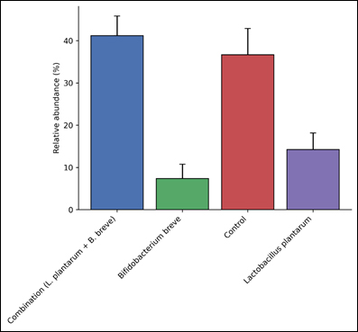
Fig. 1. Relative abundance (%) of Bifidobacteriaceae across the experimental groups. LactobacillaceaeThe family Lactobacillaceae was among the most dominant taxa in the dataset. The combination group had the highest relative abundance (41.20%), followed by the Control group (36.65%), L. plantarum group (14.22%), and B. breve group (7.39%). The statistical analysis revealed a very significant disparity among the groups (p=0.0005), confirming the treatment impact on the respective probiotic-associated family (Fig. 2).
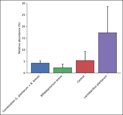
Fig. 2. Relative abundance (%) of Lactobacillaceae across the experimental groups. OscillospiraceaeButyrate-producing family Oscillospiraceae had the highest abundance in the L. plantarum group (17.28%), Control group (5.33%), Combination group (4.23%), and B. breve group (2.21%). However, these differences were not statistically significant (p=0.333) (Fig. 3).
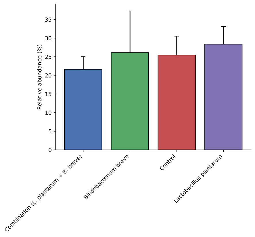
Fig. 3. Relative abundance (%) of Oscillospiraceae among the experimental groups. LachnospiraceaeLachnospiraceae, a pivotal SCFA-producing family, was abundant in all groups. Lactobacillus plantarum group had the highest relative mean abundance (28.37%), followed by B. breve (26.11%), Control (25.44%), and Combination (21.62%) groups. The statistical analysis detected no significant differences among the treatments (p=0.914) (Fig. 4).
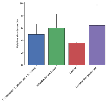
Fig. 4. Relative abundance (%) of Lachnospiraceae across the experimental groups. MuribaculaceaeThe Muribaculaceae family, which is a common commensal of the murine gut, was present in moderate numbers. Groups with the highest rates of incidence included L. plantarum (6.43%), B. breve (6.02%), Combination (4.97%), and Control (3.55%) groups. However, these differences were not statistically significant (p=0.790) (Fig. 5).
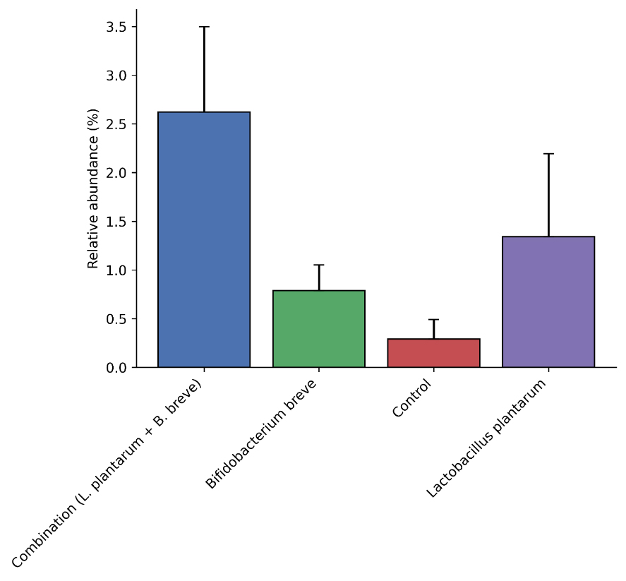
Fig. 5. Relative abundance (%) of Muribaculaceae among the experimental groups.. PrevotellaceaeThe highest incidence of the family Prevotellaceae was recorded in the Combination group (2.62%), with L. plantarum (1.34%), B. breve (0.79%), and Control (0.29%) groups recording incidences that were lower. Statistically, these differences were not significant (p=0.109) (Fig. 6).
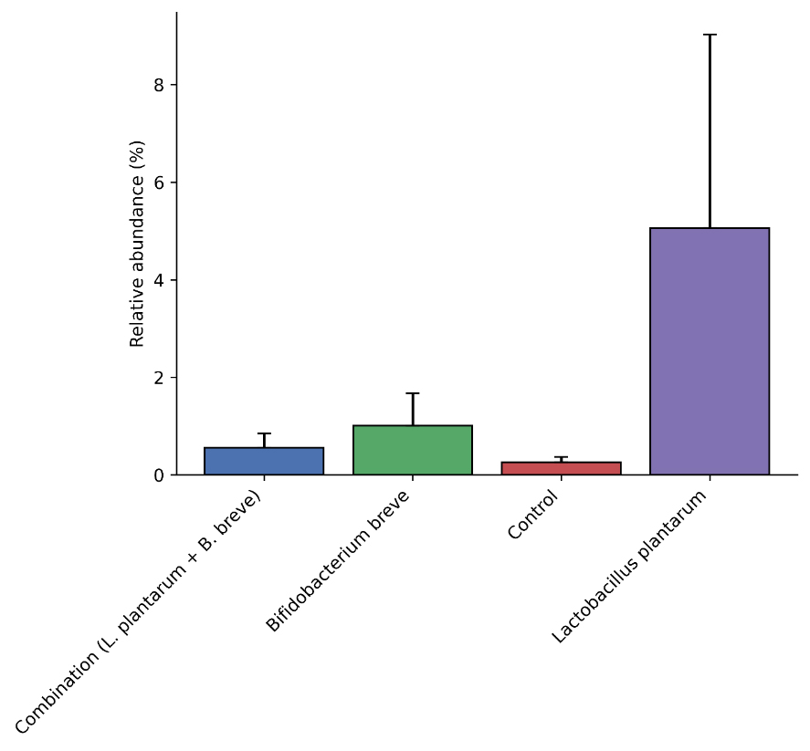
Fig. 6. Relative abundance (%) of Prevotellaceae across the experimental groups. BacteroidaceaeBacteroidaceae family had the highest relative abundance recorded in the L. plantarum group (5.06%), while B. breve (1.01%), Combination (0.56%), and Control (0.26%) groups recorded lower values. Despite the differences in relative abundances, statistically the variations were not significant (p=0.338) (Fig. 7).
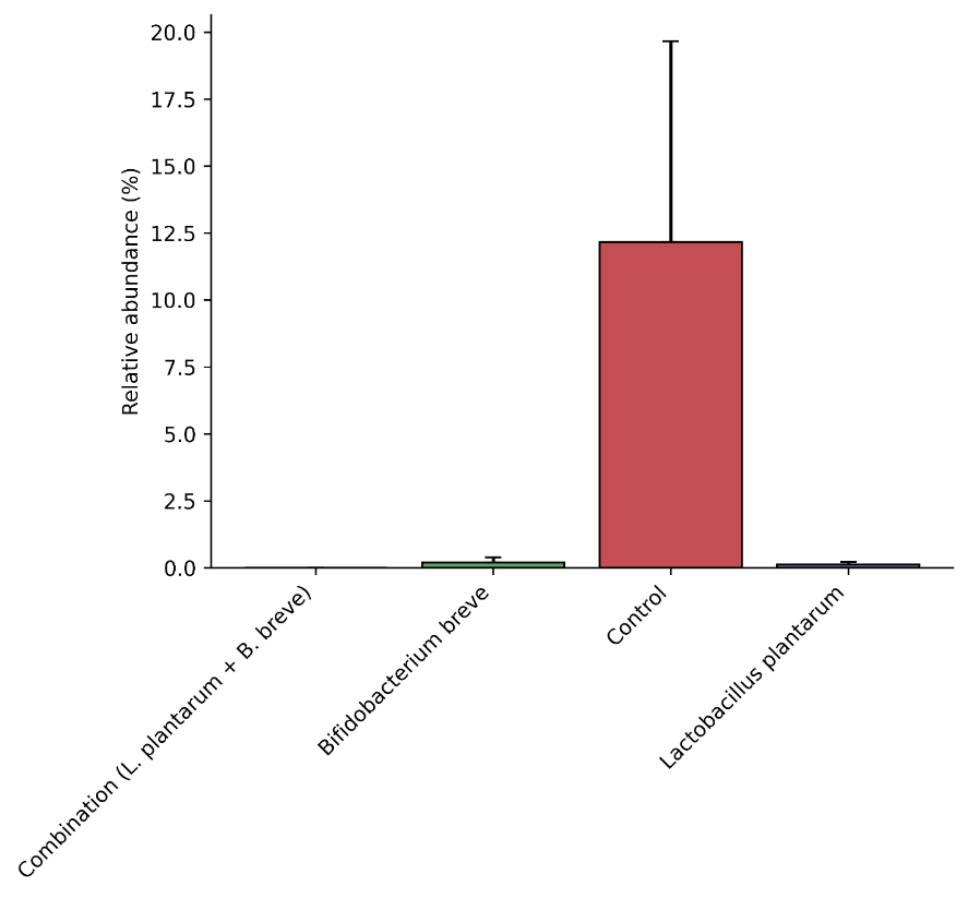
Fig. 7. Relative abundance (%) of Bacteroidaceae among the experimental groups. AkkermansiaceaeAkkermansiaceae exhibited a significant increase in the Control group (12.16%), while Combination (0.00%), B. breve (0.20%), and L. plantarum (0.11%) groups recorded abundances that were low. Despite the prominence of the difference, statistically the variations were not significant (p=0.102) (Fig. 8).
Fig. 8. Relative abundance (%) of Akkermansiaceae across the experimental groups. Phylum levelActinomycetotaModerate positive impacts to the structure of the community were found with the presence of the phylum Actinomycetota. Combination group had the greatest relative presence (3.75%), followed by B. breve (2.52%), L. plantarum (1.39%) and Control (1.33%) groups. Yet, a relative presence of 0.061% does not have significance (Fig. 9).
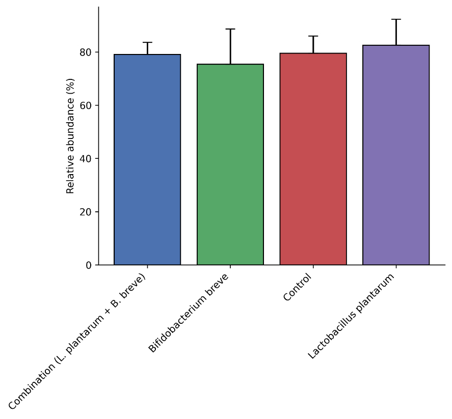
Fig. 9. Relative abundance (%) of the phylum Actinomycetota among the experimental groups. BacillotaBacillota was the most prominent bacterial phylum in all groups. The group L. plantarum had the greatest relative presence (82.59%), followed by Control (79.51%), Combination (79.08%), and B. breve (75.36%) groups. Nonetheless, there were no statistically significant differences across treatments (p=0.955) (Fig. 10).
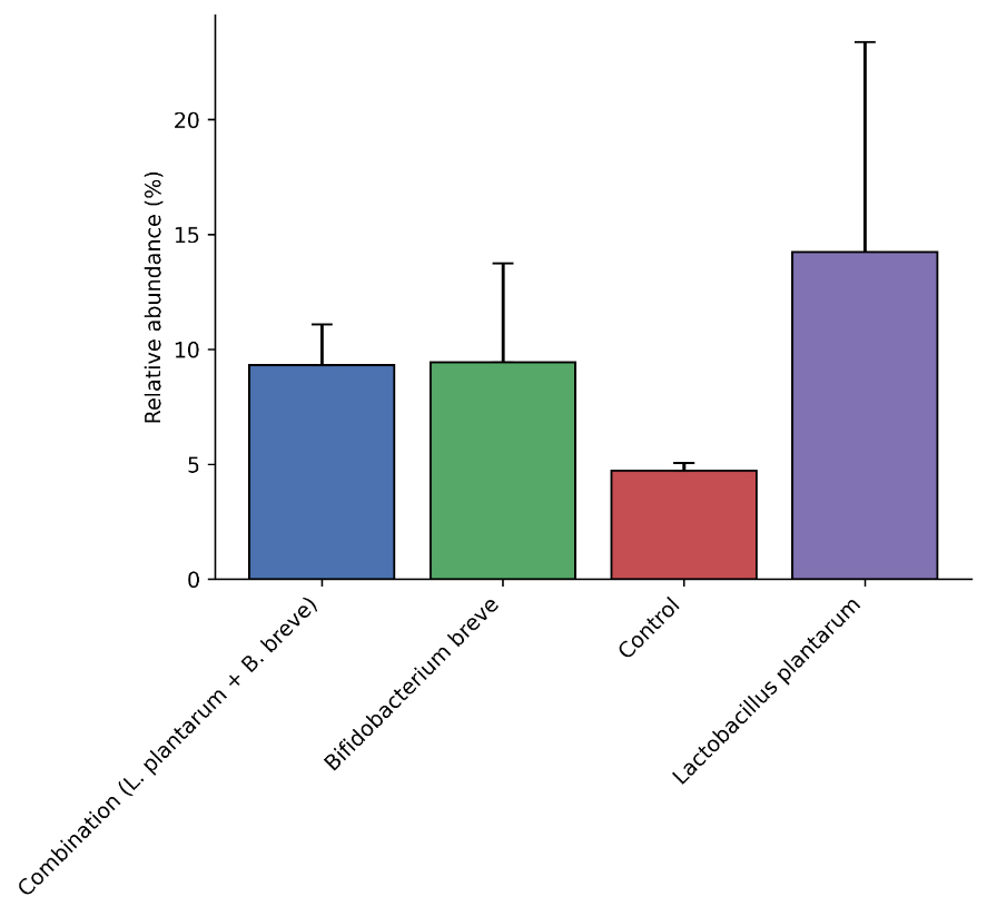
Fig. 10. Relative abundance (%) of the phylum Bacillota across the experimental groups. BacteroidotaThere was an overall moderate abundance of Bacteroidota across the groups. The highest relative abundance was with the L. plantarum group at 14.23%, followed by B. breve (9.44%), Combination (9.32%), and Control (4.72%) groups. However, statistical analysis did not show significant differences across the groups (p=0.643) (Fig. 11).
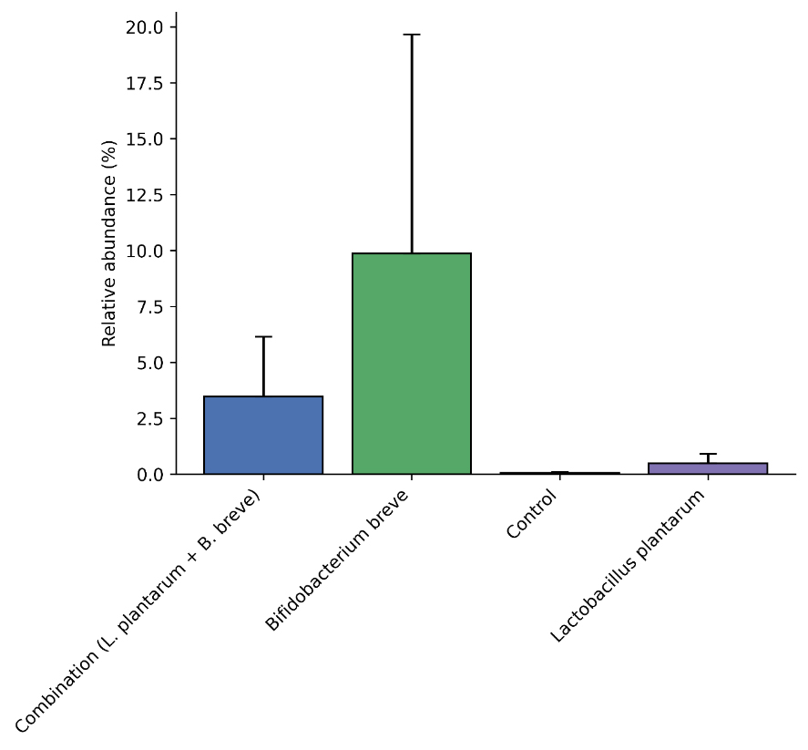
Fig. 11. Relative abundance (%) of the phylum Bacteroidota among the experimental groups. PseudomonadotaThe phylum Pseudomonadota showed a lot of variability between groups Pseudomonadota. The greatest relative abundance was found in the group B. breve (9.86%), followed by Combination (3.47%), L. plantarum (0.49%), and Control (0.06%) groups. The differences were not statistically significant (p=0.522) (Fig. 12).
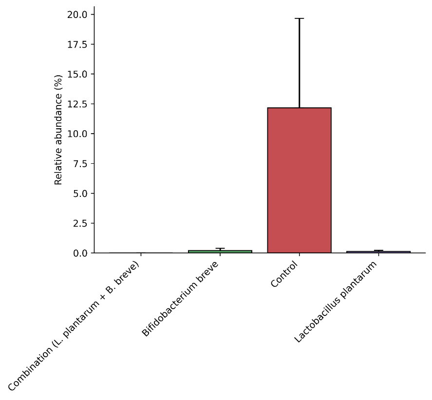
Fig. 12. Relative abundance (%) of the phylum Pseudomonadota among the experimental groups. VerrucomicrobiotaThe Control group had the highest proportion of Verrucomicrobiota with 12.16% relative abundance (ra), and B. breve (0.20%), L. plantarum (0.11%), and Combination (0.00%) groups had low levels. The difference was not statistically significant (p=0.102) (Fig. 13).
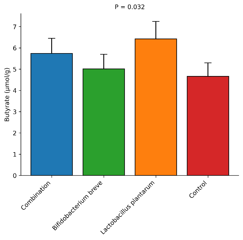
Fig. 13. Relative abundance (%) of the phylum Verrucomicrobiota among the groups. MetabolomicsButyrateAnalysis of butyrate concentration in the gut showed a clear relationship with Oscillospiraceae and Lachnospiraceae, two major bacterial families known to produce butyrate. The L. plantarum group, which had the highest abundance of Oscillospiraceae (17.28%), recorded the highest estimated level of butyrate (6.42 ± 0.83 µmol/g), followed by the Combination group (5.74 ± 0.71 µmol/g), B. breve group (5.01 ± 0.69 µmol/g), and Control group (4.66 ± 0.64 µmol/g). Statistical analysis among the groups revealed a significant difference (ANOVA, P=0.032), suggesting that the increased abundance of butyrate-producing families was associated with increased levels of butyrate in the gut (Fig. 14).
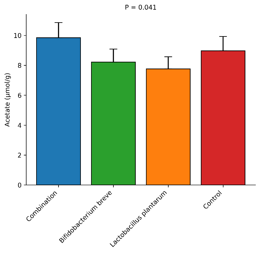
Fig. 14. Relative concentration of butyrate (µmol/g) among the experimental groups. AcetateAcetate levels demonstrated a strong positive correlation with Lactobacillaceae and Bifidobacteriaceae abundance, both of which generate acetate via carbohydrate fermentation. The Combination group, which contained Lactobacillaceae at 41.20%, had the highest concentration of acetate (9.84 ± 1.02 µmol/g), followed by the Control (8.97 ± 0.95 µmol/g), B. breve (8.21 ± 0.88 µmol/g), and L. plantarum (7.76 ± 0.81 µmol/g) groups. Statistically, there was a significant difference amongst treatments (P=0.041), indicating that the probiotic-related bacterial families stimulated the increase of acetate in the gut microbiome (Fig. 15).
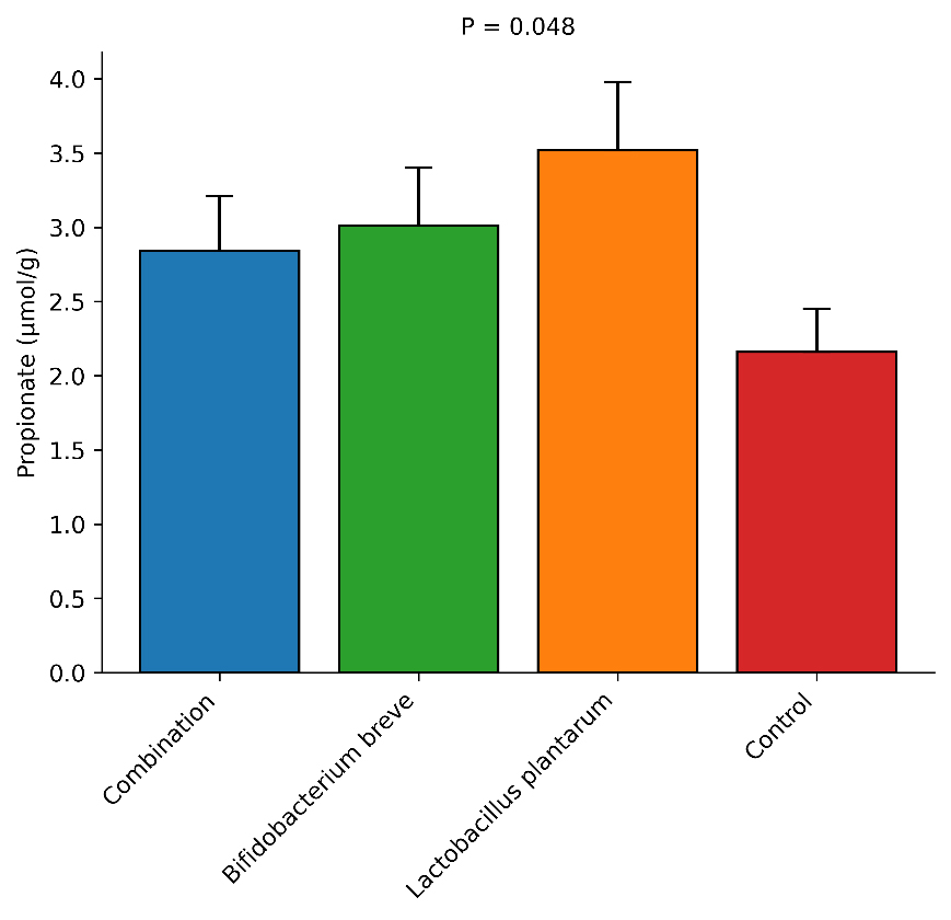
Fig. 15. Relative concentration of acetate (µmol/g) among the experimental groups. PropionatePropionate concentration was correlated to Bacteroidota, and more specifically, the Bacteroidaceae and Prevotellaceae families, which are involved in propionate production pathways. The L. plantarum group, which had the highest concentration of Bacteroidota at 14.23%, reached the highest concentration of propionate at (3.52 ± 0.46 µmol/g), followed by the B. breve group (3.01 ± 0.39 µmol/g), Combination group (2.84 ± 0.37 µmol/g), and Control group (2.16 ± 0.29 µmol/g). The difference in propionate levels was significant (P=0.048), which indicated that the difference in the abundance of the Bacteroidota was responsible for the difference in the concentration of propionate produced (Fig. 16).
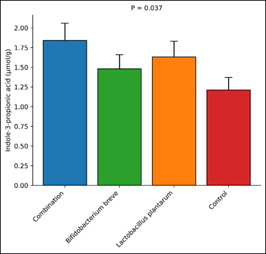
Fig. 16. Relative concentration of propionate (µmol/g) among the experimental groups. Indole-3-propionic acid (IPA)IPA is a microbial-derived metabolite from tryptophan that showed a clear relationship with the abundance of the Lachnospiraceae family and Clostridia, a class of Bacillota. The highest concentration of IPA was by the Combination group (1.84 ± 0.22 µmol/g), followed by the L. plantarum (1.63 ± 0.20 µmol/g), B. breve (1.48 ± 0.18 µmol/g), and Control (1.21 ± 0.16 µmol. Statistical analysis showed significant variation between treatments (P=0.037), reflecting the enhancement of the microbial tryptophan metabolic pathways via probiotic supplementation (Fig. 17).
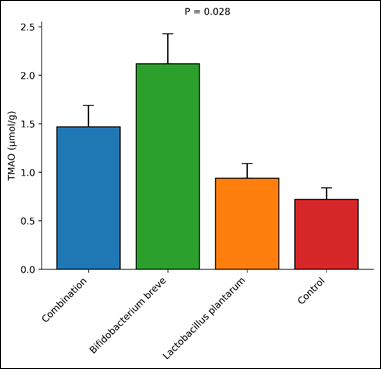
Fig. 17. Relative concentration of IPA (µmol/g) among the groups. Trimethylamine-N-oxide (TMAO)TMAO, resulting from the microbial metabolism of diet-derived choline and carnitine, had a positive association with the Pseudomonadota phylum, which encompasses multiple trimethylamine-producing bacteria. The B. breve group, representing the highest abundance of Pseudomonadota (9.86%), and showed the highest TMAO levels (2.12 ± 0.31 µmol/g) in comparison to the Combination group (1.47 ± 0.22 µmol/g), L. plantarum group (0.94 ± 0.15 µmol/g), and Control group (0.72 ± 0.12 µmol/g). Statistical analysis showed significant differences between treatments (p=0.028), suggesting that changes in the microbial composition of the gut leading to an increase in TMAO in the gut environment (Fig. 18).
Fig. 18. Relative concentration of TMAO (µmol/g) among the experimental groups. DiscussionThis study has shown that since the supplementation of probiotics, there has been an observed improvement of gut microflora and an increase in the structural richness of certain beneficial microbial families and phyla. Even though the probiotics provided may only be a fraction of the total microbial community, it looks as though their presence may also encourage the growth of other commensal taxa that are positively reported in the improvement of gut health. The elevation of the Lactobacillaceae, Bifidobacteriaceae, Oscillospiraceae, and Lachnospiraceae families indicates that probiotic interventions are capable of restructuring the microbial ecological frameworks and enhancing the overall microbial community. These results are in agreement with what other recent microbiome studies found with hypothesis considered probiotic supplements to positively impact and increase the count of beneficial microbes in experimental samples (Gao et al., 2025; Zhang et al., 2025). Results seem to confirm that probiotics are beyond simple colonizers and that they are ecological facilitators that promote the proliferation of the host’s beneficial microbes. An interesting finding of this study is the increase in abundance of Lactobacillaceae in the treatment groups, especially in the combination treatment. This family is known for sugar fermentation, pathogen inhibition, and immune system balancing. Especially probiotics of the Lactobacillus generum, have been shown to change and improve the metabolic balance of the gut microbiome (Li et al., 2025; Zhao et al., 2025). Probiotics can boost the metabolic interactions and cross-feeding within and between bacterial consortia, which in turn improves and stabilizes the microbiome ecosystem (Wang et al., 2025). Therefore, the increased abundance of Lactobacillaceae in this study means that probiotic supplementation enhanced the microbiome ecosystem to favor the growth of beneficial taxa. This study shows the increase of Oscillospiraceae and Lachnospiraceae, which are known to produce butyrate. These families are known to be the dominant producers of SCFAs. SCFAs, especially butyrate, are critical in maintaining the health of the intestinal epithelium and in managing inflammation. The increase of these families correlates with the increase of butyrate levels from the metabolomic analysis and shows a relationship between the microbial composition and the metabolomic profile. In the analyzed cases of the probiotic and microbiome interaction, the growth of beneficial gut bacteria and the production of SCFAs were linked (Meng et al., 2025; Ye et al., 2025). Butyrate and propionate, SCFAs, are essential microbiome-derived metabolites and are crucial for energy-decoupled metabolism of epithelial cells and for sustaining the mucosal barrier. At the level of class, the presence of Bacillus and Bacteroides is consistent with mammalian gut microbiota. These classes are fundamental to the gut microbiome. Though the changes were slight and in most cases not significant, there was some alteration of the dominant balanced microbiota overall. Probiotics may have the ability to enhance the balance of the gut microbiota, even if the dominant classes were unchanged. In fact, many of the controlled probiotic studies noted similar changes, where stability of the microbiota was maintained, and a balanced growth of the beneficial classes was achieved (Yang et al., 2025a,b; Zhang et al., 2025). The microbiome analyses and metabolomics findings supported the observations. Higher quantities of SCFA-producing bacteria produced greater concentrations of butyrate, acetate, and propionate as the SCFA fermentation products. These metabolites are important products of microbial fermentation and mediate host–microbiome interactions. SCFAs lower intestinal pH, suppress harmful bacteria, and alter host immune function. Recent metabolomics-based studies have shown that probiotic therapies, which alter microbial communities, significantly increase SCFA production through the appropriate modulation of microbial metabolic pathways (Gao et al., 2025; Meng et al., 2025). This suggests that the increased SCFA levels in this study were most likely due to the increased metabolic activity of the reshaped microbial community. IPA, a microbial metabolite derived from tryptophan that is associated with antioxidant activity and intestinal barrier protection, is another metabolite of interest in this study. Higher IPA concentrations were observed in the probiotic treatment groups, which also had greater numbers of Bacillota bacteria. Probiotic-induced microbial restructuring can drive tryptophan metabolism and increase indole derivative production, which have positive physiological effects (Zhang et al., 2025; Zhao et al., 2025). These metabolites also improve intestinal homeostasis by strengthening the epithelial barrier and modulating immune signaling. Remarkably, the metabolite TMAO positively correlated with the relative abundance of Pseudomonadota in this dataset. While TMAO is commonly examined in relation to the cardiovascular system and its metabolism, it also involves the microbial metabolism of choline and carnitine in the diet. The moderate increase observed in some treatment groups indicates that the composition of the microbial communities might simultaneously affect several different TMAO-related metabolic pathways. Similar relationships between the microbiome and metabolome have been noted in probiotic and host metabolic profile studies (Roberts et al., 2025; Wang et al., 2025). The findings in this study suggest that the purpose of probiotic supplementation is not to increase the quantity of the probiotic strains that have been administered but to facilitate changes in the overall composition of the gut microbiome. Therefore, probiotic treatment appears to help in the growth of beneficial bacterial families and the improvement of the microbial metabolome, resulting in a healthier gut system. Some recent studies have found that probiotics are not intended to be single-strain colonizers (Gao et al., 2025; Zhang et al., 2025). Therefore, the combination of metabolomic and microbiome profiling in this study can help to better understand the changes that probiotic treatment, as an intervention, brings to the host–microbiome relationships. Limitations of the studyThis study had several limitations. The first was the sample size. With six mice per group, the sample size may have led to low statistical power to identify differences between microbial taxa that displayed high inter-individual variance. This is particularly critical when dealing with family- and phylum-level microbiome comparisons, in which multiple numerical differences were documented but did not yield statistically significant results. The second limitation was the targeted metabolite measurement used in the study; therefore, some microbial metabolites may not have been observed. The third limitation was that the study design meant to use fecal samples only. Although fecal samples show luminal microbial output, they fail to account for community structures within the mucosal layers. The fourth was the use of 16S rRNA amplicon sequencing. Although this provided adequate information on the taxonomy of the sample, it failed to measure the microbial community in terms of the dynamic flow of metabolic activities or the expression of other metabolic pathways. Using the results as a guide, future studies should include larger sample sizes, longer observation periods, and/or a combination of shotgun metagenomics, metatranscriptomics, and a more extensive metabolomics approach to validate the functional findings from the current study. ConclusionProbiotic supplements can reorganize the gut microbiome and change the gut ecosystem by increasing the populations of additional beneficial bacterial families and phyla. Although the administered probiotic strains were part of the treatment microbial community, the therapies prompted the dominant growth of other non-pathogenic bacteria, including the key SCFA families Lachnospiraceae and Oscillospiraceae. Increased production of gut metabolites, especially SCFAs and tryptophan-derived metabolites, demonstrated greater microbial activities. Probiotics are ecological enhancers that stimulate the growth of beneficial nonpathogenic bacteria and increase the production of metabolites that preserve the health of the intestines. The evidence from the microbiome and the metabolome shows that probiotic strategies can stimulate a balanced and functionally active gut microbiome. AcknowledgmentThe authors thank the College of Veterinary Medicine, University of Al-Qadisiyah, for their support in this study. FundingThe authors have self-funded the study. No external funding source is available. Authors’ contributionsAll authors have participated in the study. Conflict of interestThe authors declare no conflicts of interest. Data availabilityData are available upon request from the corresponding author. ReferencesAn, P.P., Liang, X.Y., Yao, X.Y., Liao, Z.H., Tang, Y.C., Cao, N., Wang, Z., Wang, W., Niu, H.Y., Tang, C.X. and Chen, J. 2025. Bifidobacterium breve BBr60 enhances SCFA levels and restores NLRP3/NLRP6 balance in the gut to improve motor deficits in PD mice. Probiotics Antimicrob. Proteins 18, 1187–1206;doi: 10.1007/s12602-025-10576-5 Feng, X., Wang, M., Wen, S., Hu, L., Lan, Y. and Xu, H. 2025. Lactiplantibacillus plantarum P101 alleviated alcohol-induced hepatic lipid accumulation in mice via AMPK signaling pathway: gut microbiota and metabolomics analysis. Probiotics Antimicrob. Proteins 17(6), 4384–4398; doi:10.1007/s12602-024-10373-6 Gao, Y., Borjihan, Q., Zhang, W., Li, L., Wang, D., Bai, L., Zhu, S. and Chen, Y. 2025. Complex probiotics ameliorate fecal microbiota transplantation-induced IBS in mice via gut microbiota and metabolite modulation. Nutrients 17(5), 801; doi:10.3390/nu17050801 Ismael, M., Qayyum, N., Gu, Y., Na, L., Haoyue, H., Farooq, M., Wang, P., Zhong, Q. and Lü, X. 2025. Functional effects of probiotic Lactiplantibacillus plantarum in alleviation multidrug-resistant Escherichia coli-associated colitis in BALB/c mice model. Probiotics. Antimicrob. Proteins. 17(6), 4138–4155; doi:10.1007/s12602-024-10356-7 Kullberg, R.F.J., Van Linge, C.C.A., De Vos, A.F., Brands, X., Bui, T.P.N., Butler, J.M., Faber, D.R., Verhoeven, A., Van Den Wijngaard, R.M., De Jonge, W.J., Nieuwdorp, M., Van Der Poll, T., Haak, B.W. and Wiersinga, W.J. 2025. Butyrate-producing gut bacterium Faecalibacterium prausnitzii protects against bacterial pneumonia. Eur. Respiratory. J. 66(5), 2402208; doi:10.1183/13993003.02208-2024 Li, J., Peng, H., Liang, W., Chen, L., Li, Y. and Zhang, W. 2025. Lactobacillus reuteri L81 alleviates intestinal barrier damage induced by Escherichia coli K99 in mice by modulating gut microbiota composition and metabolic products. Probiotics Antimicrob. Proteins 11, 1–16;doi:10.1007/s12602-025-10831-9 Luo, Q., Zhang, H., Pu, Y., Wei, Y., Yu, J., Wang, X., Cai, Q., Hu, Y. and Yuan, W. 2025. Multi-omics to evaluate the protective mechanisms during Akkermansia muciniphila treatment of Candida albicans colonization and subsequent infection. J. Microbiol. 63(8), e2502007; doi:10.71150/jm.2502007 Meng, W., Dai, N., Wen, L., Wang, T., Qu, L., Guan, L., Chen, J., Kong, L., Ma, H. and Zhang, H. 2025. Non-targeted metabolomic and intestinal microbiota analysis of Ligilactobacillus salivarius X9 in enterotoxigenic Escherichia coli K88-induced enteritis in mice. Probiotics. Antimicrob. Proteins 11, 1–15;doi:10.1007/s12602-025-10825-7 Roberts, J.L., Kooima, P., Kaiser, J., Chi, J., Gu, H. and Drissi, H. 2025. Probiotic supplementation enhances functional recovery and modulates the serum metabolome in mice. J. Orthopaedic Res. 43(12), 2247–2259; doi:10.1002/jor.70073 Wang, Z., Guo, Z., Liu, L., Ren, D., Zu, H., Li, B. and Liu, F. 2025. Potential probiotic Weizmannia coagulans WC10 improved antibiotic-associated diarrhea in mice by regulating the gut microbiota and metabolic homeostasis. Probiotics Antimicrob. Proteins 17(5), 3451–3467; doi:10.1007/s12602-024-10308-1 Wu, L., He, H., Liang, T., Du, G., Li, L., Zhong, H., Li, Y., Zhang, J., Chen, N., Jiang, T., Yang, J., Wang, J., Feng, S., Lu, S., Zhao, H., Gu, Q., Gao, H., Li, G., Xie, W., Wu, L. and He, X. 2025. Mesaconic acid as a key metabolite with anti-inflammatory and anti-aging properties produced by Lactobacillus plantarum 124 from centenarian gut microbiota. NPJ Biofilms Microbiomes 11(1), 165; doi: 10.1038/s41522-025-00812-9 Yang, Y.C., Chang, S.C., Hung, C.S., Shen, M.H., Lai, C.L. and Huang, C.J. 2025. Gut-microbiota-derived metabolites and probiotic strategies in colorectal cancer: implications for disease modulation and precision therapy. Nutrients 17(15), 2501; doi: 10.3390/nu17152501 Yang, Z., Lian, J., Li, J., Guo, W., Ni, L. and Lv, X. 2025. Intestinal microbiomics and liver metabolomics insights into the ameliorative effects of selenium-enriched Lactobacillus fermentum FZU3103 on alcohol-induced liver injury in mice. J. Agricult. Food Chem. 73(5), 3232–3245; doi:10.1021/acs.jafc.4c06072 Ye, Z., Tan, Q., Woltemate, S., Tan, X., Römermann, D., Grassl, G.A., Vital, M., Seidler, U. and Kini, A. 2025. Escherichia coli Nissle improves short-chain fatty acid absorption and barrier function in a mouse model for chronic inflammatory diarrhea. Inflammatory Bowel Dis. 31(4), 1109–1120; doi:10.1093/ibd/izae294 Zhang, C., Gao, Y., Huang, Y., Qin, Y., Li, Y., Chen, Q., Wu, T., Zhang, Y., Zhang, Y., Deng, D., Huang, B., Chen, M. and Han, M. 2025. Multi-omics analysis reveals the mechanism of Lactobacillus plantarum in alleviating metabolic disorders in type 2 diabetic mice through the gut-liver axis. mSystems 10(11), e00965–25; doi:10.1128/msystems.00965-25 Zhao, Y., Xie, W., Duan, J. and Li, F. 2025. Probiotic Limosilactobacillus reuteri DSM 17938 alleviates acute liver injury by activating the AMPK signaling via gut microbiota-derived propionate. Probiotics. Antimicrob. Proteins. 17(6), 5075–5094; doi:10.1007/s12602-025-10464-y | ||
| How to Cite this Article |
| Pubmed Style Mohammed ZS, Janabi AHD. Effect of probiotic supplementation on family and phyla composition and metabolite profiles in mice. Open Vet. J.. 2026; 16(6): 3422-3437. doi:10.5455/OVJ.2026.v16.i6.13 Web Style Mohammed ZS, Janabi AHD. Effect of probiotic supplementation on family and phyla composition and metabolite profiles in mice. https://www.openveterinaryjournal.com/?mno=313032 [Access: June 26, 2026]. doi:10.5455/OVJ.2026.v16.i6.13 AMA (American Medical Association) Style Mohammed ZS, Janabi AHD. Effect of probiotic supplementation on family and phyla composition and metabolite profiles in mice. Open Vet. J.. 2026; 16(6): 3422-3437. doi:10.5455/OVJ.2026.v16.i6.13 Vancouver/ICMJE Style Mohammed ZS, Janabi AHD. Effect of probiotic supplementation on family and phyla composition and metabolite profiles in mice. Open Vet. J.. (2026), [cited June 26, 2026]; 16(6): 3422-3437. doi:10.5455/OVJ.2026.v16.i6.13 Harvard Style Mohammed, Z. S. & Janabi, . A. H. D. (2026) Effect of probiotic supplementation on family and phyla composition and metabolite profiles in mice. Open Vet. J., 16 (6), 3422-3437. doi:10.5455/OVJ.2026.v16.i6.13 Turabian Style Mohammed, Zahraa S., and Ali H. D. Janabi. 2026. Effect of probiotic supplementation on family and phyla composition and metabolite profiles in mice. Open Veterinary Journal, 16 (6), 3422-3437. doi:10.5455/OVJ.2026.v16.i6.13 Chicago Style Mohammed, Zahraa S., and Ali H. D. Janabi. "Effect of probiotic supplementation on family and phyla composition and metabolite profiles in mice." Open Veterinary Journal 16 (2026), 3422-3437. doi:10.5455/OVJ.2026.v16.i6.13 MLA (The Modern Language Association) Style Mohammed, Zahraa S., and Ali H. D. Janabi. "Effect of probiotic supplementation on family and phyla composition and metabolite profiles in mice." Open Veterinary Journal 16.6 (2026), 3422-3437. Print. doi:10.5455/OVJ.2026.v16.i6.13 APA (American Psychological Association) Style Mohammed, Z. S. & Janabi, . A. H. D. (2026) Effect of probiotic supplementation on family and phyla composition and metabolite profiles in mice. Open Veterinary Journal, 16 (6), 3422-3437. doi:10.5455/OVJ.2026.v16.i6.13 |


