Open Veterinary Journal, (2026), Vol. 16(6): 3966-3985
Research Article
10.5455/OVJ.2026.v16.i6.65
Efficacy of biological products in mitigating clinical signs, T-2 mycotoxicosis, and vibriosis in gilthead seabream (Sparus aurata L.)
Ahmed Said Al-Souti1, Mahmoud A. Elnakeeb2*, Ahmed A. El-bahlol2, Mohamed M. Elnawsany2,
Saad M. Alsaiad2, Mohamed E. Abou El Atta3 and Tamer Mohammed Monir Abdelrahiem3
1Department of Marine Science and Fisheries, College of Agricultural and Marine Sciences, Sultan Qaboos University, Al-Khoud, Sultanate of Oman
2Fish Production Department, Faculty of Agriculture, Al-Azhar University, Nasr City, Egypt
3Department of Fish Health and Management, Central Laboratory for Aquaculture Research, Agricultural Research Center, Abbassa, Egypt
*Corresponding Author: Mahmoud A. Elnakeeb. Assistant Professor, Fish Production Department, Faculty of Agriculture, Al-Azhar University, Cairo, Egypt. Email: mahmoud.biotech [at] azhar.edu.eg; mahmoud.biotech [at] gmail.com
Submitted: 16/03/2026 Revised: 25/05/2026 Accepted: 07/06/2026 Published: 30/06/2026
© 2025 Open Veterinary Journal
This is an Open Access article distributed under the terms of the Creative Commons Attribution-Non Commercial-No Derivatives License (http://creativecommons.org/licenses/by-nc-nd/4.0/), which permits non-commercial re-use, distribution, and reproduction in any medium, provided the original work is properly cited, and is not altered, transformed, or built upon in any way.
Abstract
Background: T-2 toxin, a type A trichothecene, exhibits hepatotoxic, immunosuppressive, and oxidative effects in fish.
Aim: This study examines the protective effects of Mycofix Plus and Mycosorb A+ commercial mycotoxin binders (MBs) against T-2-induced toxicity in gilthead seabream.
Methods: In a 6-week feeding trial, 180 fingerlings (initial weight 100 g) were allocated into 6 groups in triplicate. The dietary groups consisted of a control, binder-only diets, a T-2 toxin-challenged group, and contaminated diets supplemented with either binder (1 g kg⁻¹). Measurements included clinical manifestations, innate immunity, oxidative status, enzymatic activity, histological changes, and survival following a V. harveyi challenge.
Results: T-2 toxin-exposed fish exhibited severe clinical signs, organ damage, and immunosuppression of the innate immune response compared with control fish (p < 0.05). These changes were accompanied by a significant increase in cortisol and oxidative stress indicators (glutathione reductase and lipid peroxidase). In the disease-resistance trial, the T-2 group showed the highest susceptibility to V. harveyi (mortality: 90%). However, the inclusion of Mycofix Plus and Mycosorb A+ in the diets significantly counteracted the impact of the toxin, improving the antioxidant status and reducing clinical symptoms. Notably, these treatments restored survival rates to 70%–80% and provided substantial relative protection (57.14%–71.43%) compared to fish receiving the T-2 toxin alone.
Conclusion: Mycofix® Plus and Mycosorb® A+ are MBs used as functional feed additives in gilthead seabream to reduce T-2 toxin toxicity and enhance disease resistance to V. harveyi.
Keywords: Gilthead seabream, Immunosuppression, Mycotoxin binders, T-2 toxin, Vibrio harveyi.
Introduction
Mariculture in the northern part of Egypt, especially in the Mediterranean area and around the Suez Canal, is increasingly contributing to fish production in the country (Paruğ et al., 2024). Among the marine species cultivated, the gilthead sea bream (Sparus aurata) is one of the most valuable species (Eissa et al., 2025) in terms of economic benefits due to its high market demand and ability to thrive in intensive culture systems (Mhalhel et al., 2023). Although mariculture in Egypt remains less developed than freshwater aquaculture, it has shown steady expansion in response to the scarcity of freshwater and the need to sustainably exploit marine resources (Shaalan et al., 2018).
Despite this growth, the intensification of sea bream farming is constrained by several environmental and nutritional stressors that adversely affect fish health, growth performance, and survival (Rennie et al., 2019; Dara et al., 2023; Taylor et al., 2025). Contamination of aquafeeds with mycotoxins represents a major and often underestimated risk (Pulvirenti et al., 2024; Gruber-Dorninger et al., 2025; Lorusso et al., 2025). Trichothecenes, a large group of mycotoxins produced by Fusarium species, are of concern in aquaculture due to their known toxicity and common occurrence in plant-based ingredients of aquafeeds (Vulić et al., 2025).
T-2 toxin is one of the most toxic mycotoxins to animals, including fish (Meneely et al., 2023). It is described as a highly lipophilic molecule that dissolves in nonpolar solvents, thereby enhancing its bioavailability (Zhang et al., 2022). There has been a surge in the formulation of fish feeds with plant-based compounds, leading to T-2 toxin contamination in aquafeeds due to their susceptibility to fungal contamination during cultivation, storage, and processing (Lorusso et al., 2025). Previous studies have shown that aquaculture feeds are prone to contamination with mycotoxins, thereby reducing feed efficiency, nutrient utilization, and growth performance, as well as affecting vital organs, especially the liver and kidneys (Gruber-Dorninger et al., 2025).
T-2 toxin has been well documented at a concentration of 500 μg kg⁻¹ as a hepatotoxin and hepatocarcinogen in a variety of animal species, including fish, with the liver being the principal target organ (Qiu et al., 2016; Yin et al., 2020; Kihal et al., 2022). Toxins trigger pathological changes, impair liver function, and cause significant changes in liver enzyme activity, indicating impaired metabolic function (Abdelrahiem et al., 2025). T-2 toxin has also been reported to induce immunosuppression and oxidative stress and to increase the risk of disease in fish, posing a major threat to fish welfare and mariculture sustainability (Kihal et al., 2022; Goda et al., 2025). In light of the emerging issues of mycotoxin contamination of aquafeeds in marine aquaculture, there is a pressing need to develop strategies to mitigate the effects of mycotoxins in fish, which have become a major concern for both researchers and the aquafeed industry (Agriopoulou et al., 2020).
Mycotoxin binders (MBs) reduce toxicity by adsorbing mycotoxins with sufficient affinity to prevent their interaction with animal tissues and to limit their absorption across the gastrointestinal tract (Puvača et al., 2023). Numerous adsorbent materials have been tested, including activated carbon, aluminosilicates, which cover a wide variety of materials from clay and bentonite to zeolite and other phyllosilicates, complex indigestible carbohydrates, which cover cellulose and polysaccharides derived from yeast and bacterial cell walls, and cholestyramine, polyvinylpyrrolidone, and their derivatives (Arafa et al., 2025). Several commercial mycotoxin-binding products, such as Mycofix Plus, Mycosorb A+, Agresol, and CAP T2, have been investigated for their efficacy in aquafeeds (Puvača et al., 2024; Anandaraj et al., 2025; Zahran et al., 2026). However, the binding ability of these products varies widely depending on the type of binder used, its source, level of inclusion, and the target mycotoxin’s chemical structure (Kihal et al., 2022). Some of these products may also negatively influence growth performance due to a lack of specificity in binding mycotoxins because they can also bind other nutrients such as vitamins and minerals (Hussain et al., 2024).
The broad efficacy of commercial mycotoxin-binding agents in aquaculture feeds has been demonstrated. For instance, Mycofix, a commercial formulation based on clay minerals and specific enzymatic components, reduced mycotoxin bioavailability by more than 70% when included at a low dietary level of 1 g kg⁻¹ (Chefchaou et al., 2019). Several studies have confirmed the effectiveness of Mycofix and similar binders across aquaculture species, indicating their general applicability rather than species-specific action (Koletsi et al., 2021). In parallel, Mycosorb A+ has shown considerable promise as an alternative adsorbent; dietary inclusion at 0.2%–0.4% in aflatoxin T2-contaminated diets resulted in improved hepatic function, immune responses, toxin residues, and intestinal microbial profiles comparable to those of uncontaminated controls (Kihal et al., 2022). These outcomes support the effectiveness of both mineral-based binders and biological adsorbents in mitigating mycotoxin-induced toxicity in aquafeed formulations (Agriopoulou et al., 2020).
Disease outbreaks, particularly vibriosis induced by Gram-negative Vibrio spp., represent a primary impediment to the advancement of global mariculture (Mohamad et al., 2019). Vibrio harveyi is a prominent pathogen that affects numerous teleost species, including S. aurata, often resulting in systemic septicemia and mass mortality (Triga et al., 2024). Consequently, V. harveyi challenge trials are widely employed to evaluate disease resistance and immunological competence in fish subjected to various physiological stressors (Serna-Duque et al., 2023). Despite extensive research on MBs in terrestrial livestock and, to a lesser extent, freshwater fish species, their functional efficacy under concurrent toxicological and infectious stressors in marine aquaculture systems remains poorly understood. Studies integrating chronic dietary exposure to T-2 toxin with a subsequent pathogenic challenge are extremely limited, thereby constraining our understanding of the interaction between mycotoxicosis, immune suppression, and disease susceptibility. Furthermore, comparative evaluations of distinct commercial MBs under such dual-stress conditions are scarce, especially in economically important marine species, such as S. aurata. Thus, the current study combines dietary toxin exposure, physiological and histological analysis, and a controlled V. harveyi challenge to provide a complete picture of the host’s resilience. This study is based on the idea that supplementing the diet with Mycofix Plus and Mycosorb A+ may influence immune function and oxidative stress in different ways. These treatments could help reduce the negative effects of toxin exposure and improve the resistance of S. aurata to V. harveyi infection.
Materials and Methods
Experimental site and the fish
The current study was conducted in a controlled indoor setup that simulates mariculture conditions using seawater sourced from a commercial fish farm located in Alexandria, Egypt. A cohort of 180 healthy S. aurata fingerlings, with an initial mean body weight of 100 ± 0.16 g, was used in this study. Fish were obtained from a single hatchery batch to ensure uniformity and minimize biological variability. Fingerlings were acclimated under laboratory conditions for 2 weeks before the start of the experiment. During the acclimation period, the fish were fed a basal diet at a rate of 3% body weight per day.
After acclimation, the fish were randomly distributed in glass aquaria measuring 70 × 35 × 50 cm, with 10 fish per aquarium. Continuous aeration was maintained using an electric air compressor. Partial water exchange (approximately one-third of the volume) was conducted daily to maintain the water quality within optimal levels. Water salinity was maintained at 32 ± 0.02 ppt under continuous aeration and partial daily water exchange, while water temperature ranged from 26 °C to 28 °C; dissolved oxygen remained above 5 mg l⁻¹.
Diets and incorporation of T-2 toxin
A basal diet was designed to meet the specific nutrient requirements of seabream fingerlings (NRC, 2011). The diet comprised 42% crude protein, 18% ether extract, 7% crude fiber, 6.5% ash, and 26.5% nitrogen-free extract. The main ingredients included fish meal, soybean meal, yellow maize, wheat bran, sunflower oil, and a vitamin premix. All ingredients were ground into a fine powder, thoroughly homogenized, and extruded into 1-mm diameter pellets using a feed manufacturing machine (Okolie et al., 2019). After extrusion, the pellets were air-dried at room temperature until a constant weight was achieved. The dried diets were stored in airtight containers under cool, dry conditions to prevent moisture uptake and minimize the risk of fungal growth or unintended mycotoxin contamination prior to feeding (Ilesanmi et al., 2024). T-2 toxin, at a concentration of 500 μg kg⁻¹, was incorporated into the toxin-contaminated diets according to the experimental protocols (Matejova et al., 2017). The toxin was made soluble in an appropriate solvent for its even distribution before being incorporated into the diets.
Two commercial MBs were used in this study: (1) Mycofix Plus and (2) Mycosorb A+. Both binders were incorporated into the experimental diets at an inclusion level of 1 g kg⁻¹ feed, either alone or in combination with T-2 toxin, as specified in the experimental design.
Experimental design
The study employed a randomized design with 6 dietary treatments, each in triplicate (Table 1, Fig. 1). Gilthead seabream were stocked at a rate of 10 fish per aquarium, with 3 aquariums per treatment. The fish were fed twice daily at 3% of their body weight for 6 weeks. The neutralizing capacity of Mycofix Plus and Mycosorb A+ in the presence of T-2 toxin was assessed. Feed rations were adjusted weekly according to weight measurements. Water quality parameters were rigorously monitored and maintained within the optimal ranges required for seabream cultivation throughout the experimental period.
Table 1. Experimental groups and S. aurata fingerling dietary treatments for 42 days.
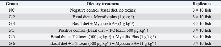
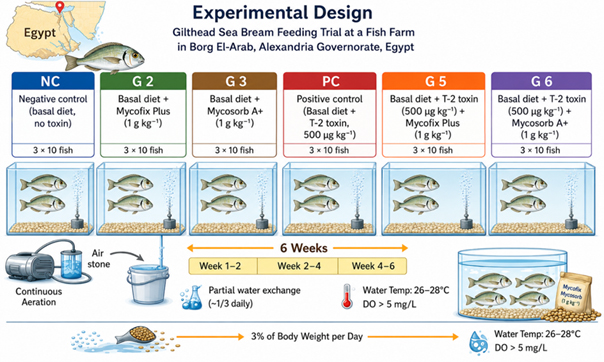
Fig. 1. Experimental groups and dietary treatments of S. aurata fingerlings for 42 days.
Sampling
At the termination of the trial, all fish were sampled using a standard procedure following a 24-hours fasting period. The number of fish was estimated, their weight was measured, and any growth variation, clinical symptoms, and morphological changes caused by the diets were evaluated. Three fish per replication aquarium (n=9 fish per treatment) were randomly chosen, sacrificed, and dissected for the inspection of internal organs and lesion sites to conduct a postmortem examination and histopathological investigation. After this, liver and kidney samples were harvested for histopathological examination. Five fish per replication (n=15 fish per treatment) were randomly selected, anesthetized, and blood samples were collected individually to evaluate the levels of nonspecific humoral immunity (NBT, lysozyme, and bactericidal activities), stress response markers (plasma cortisol), oxidative stress markers (glutathione reductase [GR] and lipid peroxidation), antioxidant enzymes (catalase [CAT], superoxide dismutase [SOD], and Glutathione S-transferase [GST]), and metallothionein according to the methodology outlined by Ashry et al. (2024).
Clinical and postmortem examination
Throughout the 6-week trial period, the fish were observed daily for signs of toxicity, behavior, swimming patterns, and appearance (Mello et al., 2025). At the end of the second, fourth, and sixth weeks, fish from each test group were randomly selected and autopsied for examination. The internal organs of the fish, such as the liver, gills, and kidneys, were examined in situ for any gross pathological changes (Shrivastava et al., 2025).
Nonspecific humoral immune assays
Nonspecific humoral immune parameters were analyzed to measure the innate immune potential. Respiratory burst of circulating phagocytes was measured by assessing nitroblue tetrazolium (NBT) reduction. The NBT reduction assay was performed following the method described by Biller-Takahashi et al. (2013). In brief, 15 μl of blood was mixed with NBT solution under specific conditions, and ROS formation was quantified by measuring intracellular formazan granules. Plasma lysozyme activity, based on the Sitós-Bobadilla method (Saera-Vila et al., 2009), was measured by turbidimetry using the lysis of Micrococcus lysodeikticus. After incubation for 0.5 and 4.5 minutes, turbidity was measured at 450 nm using a spectrophotometer. The unit of measurement for plasma lysozyme activity was IU/ml. Bactericidal activity of sera was measured as described by Mizaeva et al. (2023), in which plasma samples were mixed with pathogenic bacterial suspensions, and activity was determined by counting CFU relative to the control.
Analysis of stress and oxidative biomarkers
Physiological stress and oxidative status were evaluated to assess the biological response to treatment. Cortisol levels were determined according to the guidelines provided in the handbook for a commercial enzyme-linked immunosorbent assay kit (Colás-Ruiz et al., 2022). GR activity was determined as the NADPH oxidation rate at 340 nm in the presence of GSSG (Gisbert et al., 2015). The malondialdehyde (MDA) content was determined using the TBARS test, a marker of lipid peroxidation as a symptom of membrane oxidative damage (Janik et al., 2021).
Evaluation of the antioxidant capacity
Key enzyme activities were measured to assess the antioxidant status of the system. The rate of hydrogen peroxide (H₂O₂) breakdown at 240 nm was used to gauge CAT activity (Kim et al., 2017). The ability of SOD to prevent the photochemical reduction of NBT was used to measure SOD activity (Elnakeeb et al., 2025). GST was assessed by measuring the conjugation rate of 1-chloro-2,4-dinitrobenzene (CDNB) and reduced glutathione (GSH) at 340 nm, as described by Gisbert et al. (2015).
Metallothionein profiling
Total, oxidized, and reduced metallothionein (MT) concentrations in whole blood were quantified using a modified spectrophotometric method based on the reaction of thiol groups with Ellman’s reagent (DTNB), as described by Bejaoui et al. (2024). MT levels were expressed as μmol thiol per milligram of protein.
Histopathological examination
At the end of the trial, a set of fish from each treatment group was euthanized, and samples of the target organs, particularly the liver and kidney, were collected. Following fixation in 10% neutral buffered formalin for 24–48 hours, the tissues were rinsed under running water, dehydrated in ethanol, clarified in xylene, and then embedded in paraffin wax (Bisai et al., 2025). Sections of the paraffin block were placed on glass slides and stained with hematoxylin and eosin (H&E) after being cut to a thickness of 5–6 µm. Prepared sections were then examined microscopically for any pathological changes following standard histological procedures (Sivakumar et al., 2022).
Resistance to Bacterial Infection
Vibrio harveyi strain was obtained from naturally affected marine fish exhibiting classical signs of vibriosis and was pathogenically characterized using standard microbiological, biochemical, and molecular biology techniques, including 16S rRNA sequences (Aly et al., 2025). Cultured cells of the selected strain were stored in the microbiology laboratory and periodically subcultured on tryptic soy agar (TSA) supplemented with 2% NaCl. Bacteria were grown in tryptic soy broth containing 2% NaCl at 28 °C for 12–18 hours. The culture was then centrifuged at 3000 × g for 10 minutes at 25 °C, after which the bacteria were suspended in phosphate-buffered saline (PBS) and washed twice. Bacterial concentration was standardized to 1 × 10⁸ CFU ml⁻¹. To prepare the final challenge dose, a 10-fold serial dilution in sterile PBS was performed to obtain a working concentration of 1 × 10⁶ CFU ml⁻¹ per fish. This dose was selected based on a previously determined LD₅₀ value reported by El-Waziry et al. (2025).
At the end of the sixth week of the experiment, a challenge test was conducted using 10 fish per experimental group. The fish were anesthetized with clove oil (Sigma-Aldrich, USA; 50 mg l⁻¹) and intraperitoneally injected with the prepared bacterial suspension (Aly et al., 2024). The infected fish were monitored for 2 weeks for any signs of disease, and the mortality rate was recorded to calculate the relative percentage survival (RPS) using the following equation described by Amend (1981):

Where: SR=survival rate; MR=mortality rate; RPS=relative percent survival.
The RPS values indicate the treatment’s efficacy in improving disease resistance, with higher values signifying greater protection against the pathogen relative to the control.
Data analysis
Statistical analysis was performed using SAS software version 9.1 (Littell WW Stroup and Freund, 2002), and all experimental data were expressed as the arithmetic mean ± standard deviation (M ± SD). To validate the assumptions of normality and homoscedasticity, datasets were evaluated using the Shapiro–Wilk test before further processing. The aquarium (n=3 per treatment) served as the experimental unit for all statistical tests to ensure statistical independence and avoid pseudoreplication. Multiple fish were sampled from each aquarium to assess the immunological, biochemical, and antioxidant properties of the specimens. However, data from individual fish were not treated as separate experimental units; only the average of these data was considered. The treatment effect was separately analyzed at each sampling interval (weeks 2, 4, and 6) using one-way analysis of variance after confirming the assumption of normality and homogeneity of variances. LSD was used in situations where there were differences at p ≤ 0.05 to determine whether there was any separation of means (Gomez and Gomez, 1984). This post hoc approach was selected because of the balanced experimental design and the hypothesis-driven nature of the comparisons, which required maintaining statistical sensitivity to detect biologically relevant differences.
Ethical approval
Specimens were obtained from the Central Laboratory for Aquaculture Research (CLAR), Egypt. They were obtained in accordance with the ethical standards established for conducting biological research. The research design and protocols were ratified officially by the Ethics and Research Committee of the Fish Production Department, Faculty of Agriculture, Al-Azhar University, Cairo, Egypt. They were conducted under the official authorization code (APR-111-fish. Dep. Issue No. 25/202, Dec., 2025). All research protocols were conducted following the established animal welfare regulations. The investigation design and protocols were conducted as per the ARRIVE guidelines (https://arriveguidelines.org; accessed on 20 April 2025).
Results
Clinical examination of seabream fingerlings exposed to T-2 toxin
External clinical signs
Throughout the experimental period, fingerlings of gilthead sea bream exposed to dietary T-2 toxin exhibited pronounced clinical signs compared to the control and binder-treated groups, as is well documented regarding the impact of dietary mycotoxin contamination on fish health and welfare under intensive aquaculture conditions (Lorusso et al., 2025). After two weeks of T-2 toxin dietary exposure (500 μg kg⁻¹), fingerlings expressed evident abdominal distension due to ascites, which is related to the initial signs of systemic toxicity (Fig. 2A). T-2 toxin has already been shown to cause multi-organ damage, oxidative stress, and immunosuppression in aquatic and terrestrial animals (Matejova et al., 2017; Wang et al., 2020). With prolonged exposure for 4 weeks, severe external hemorrhages were observed, including fin erosion and fin rot, with varying degrees of sloughing observed on dorsal, caudal, and pectoral fins (Fig. 2B), which is consistent with reports regarding the progressive worsening of animal condition, tissue integrity, and external signs of mycotoxicosis in fish and livestock (Diab et al., 2018; Barany et al., 2021).
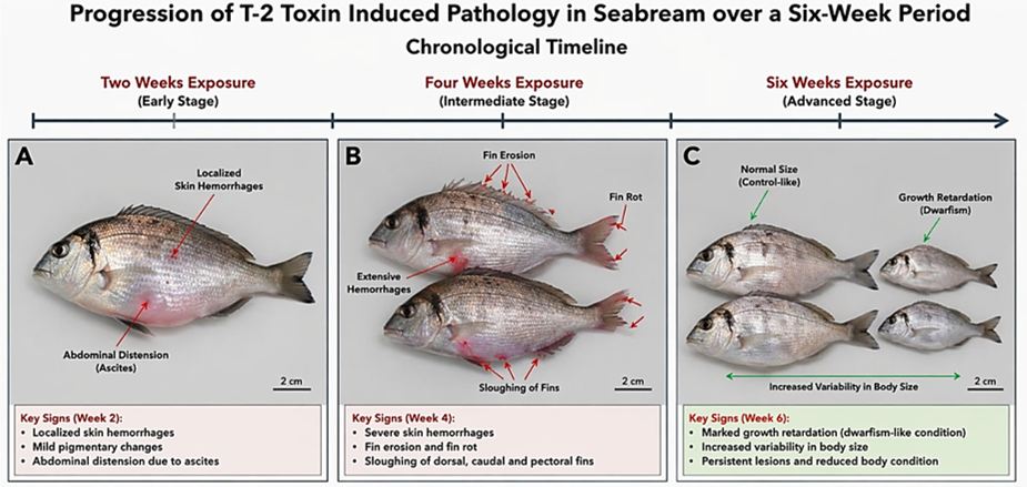
Fig. 2. Progression of T-2 toxin-induced external pathologies in S. aurata during a 6-week trial (A) Early-stage (week 2) localized skin hemorrhages and pigmentary changes (red arrows). (B) The mid-stage (week 4) manifestation of extensive hemorrhaging (red arrows). (C) Late-stage (week 6) consequences, including marked growth retardation and increased body size variability, are observed in fish (green arrows).
By the end of the 6-week experimental period, fish in the T-2 toxin group displayed marked growth retardation, as reflected by noticeable differences in size among the experimental groups (Fig. 2C). This dwarfism-like condition is consistent with previous reports of impaired growth and development following trichothecene exposure in fish species, including carp and Nile tilapia, in which chronic dietary mycotoxins reduce weight gain and growth efficiency (Matejova et al., 2017; Diab et al., 2018; Barany et al., 2021). On the contrary, seabream fingerlings receiving diets supplemented with Mycofix Plus or Mycosorb A+, either alone or in combination with T-2 toxin, showed a clear attenuation of these external clinical signs, maintaining normal body conformation and fin integrity relative to the toxin-only group. This pattern is consistent with evidence that commercial MBs such as Mycofix and Mycosorb mitigate T-2 and aflatoxin toxicity, improve performance, and alleviate clinical and pathological alterations in different animal species (Jaćević et al., 2020; Kihal et al., 2022; Koynarski et al., 2025.
Postmortem signs
Postmortem examination revealed progressive internal pathological alterations in seabream fingerlings exposed to T-2 toxin, including hepatic congestion, gill damage, and renal abnormalities observed over the experimental period. During the second week of exposure, the fish exhibited hepatic congestion, gill congestion, and exophthalmia, reflecting acute circulatory and inflammatory disturbances (Fig. 3A). These findings are consistent with previous reports of vascular congestion, hemorrhage, and early degenerative changes in the gills and liver following exposure to toxicants and mycotoxins (Shah and Parveen, 2022; Wang et al., 2022).
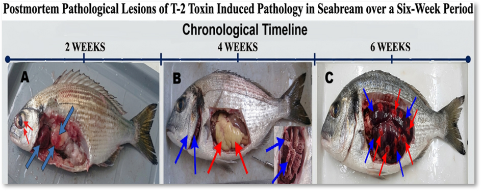
Fig. 3. Postmortem alterations and tissue lesions in gilthead seabream specimens. (A) Early necropsy results showing severe visceral lesions and abdominal tissue degeneration (blue arrows) together with exophthalmia and eye hemorrhages (red arrow). (B) Hepatic degeneration and accumulation of visceral fat within the abdominal cavity are evident (red arrows), together with severe gill degeneration (blue arrows) after 4 weeks of exposure. (C) Extensive hemorrhage and severe internal organ damage are observed within the abdominal cavity (red arrows) and marked vascular congestion (blue arrows) after 6 weeks of exposure.
More pronounced lesions were evident after 4 weeks of intoxication, including hepatomegaly, characterized by an enlarged liver with a pale whitish appearance, as well as severe gill degeneration marked by sloughing and destruction of the secondary lamellae (Fig. 3B). Similar hepatomegaly, hepatic degeneration, and lamellar sloughing or fusion of gill secondary lamellae have been described as typical responses to chronic toxic insult in fish, including exposure to T-2 toxin and other environmental contaminants (Shah and Parveen, 2022; Wang et al., 2024). At the sixth week, chronic pathological changes were observed, particularly in the kidneys, which appeared enlarged, congested, and mottled with sporadic hemorrhagic patches distributed across the organ surface (Fig. 3C), consistent with previous descriptions of nephrotoxic effects of T-2 and related mycotoxins, including congestion, tubular degeneration, and hemorrhages in renal tissues. These findings are consistent with those of earlier studies reporting severe edema, visceral organ damage, and intestinal or mucosal loss in animals exposed to T-2 toxin and other potent toxicants (Shah and Parveen, 2022; Wang et al., 2024).
Notably, postmortem lesions were markedly less severe or absent in fish receiving dietary supplementation with Mycofix Plus or Mycosorb A+, indicating their protective role in mitigating T-2 toxin-induced organ damage, which is supported by evidence that Mycosorb, Mycofix, and other mycotoxin-binding feed additives significantly reduce T-2-associated histopathological lesions and improve organ integrity in experimentally intoxicated animals (Rodrigues et al., 2017; Singh, 2021).
Temporal effects of T-2 toxin and MBs on nonspecific humoral immune responses
Table 2 summarizes the effects of dietary treatments on nonspecific humoral immune responses. A clear time-dependent pattern was observed, in which fish exposed to T-2 toxin alone exhibited a progressive and significant decline (p < 0.05) in all immune parameters, including NBT activity, lysozyme activity, and bactericidal capacity, with the most pronounced suppression recorded at the 6th week. This represents a reduction of approximately 30%–40% compared with the control group, indicating severe impairment of innate immune defenses.
Table 2. Immunological responses in S. aurata fingerlings fed with T-2 toxin and MBs for 42 days.
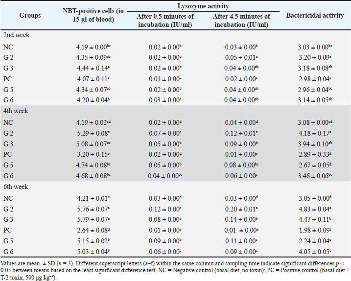
On the contrary, fish fed Mycofix Plus (G2) or Mycosorb A+ (G3) showed a consistent and significant enhancement of immune parameters across all sampling periods, with the highest values recorded at week 6. Notably, G2 and G3 exhibited up to 35%–60% higher lysozyme and bactericidal activity than the control, demonstrating a strong immunostimulatory effect.
Immune parameters were partially restored under combined stress conditions (G5 and G6), with values significantly higher than the T-2 group but still lower than binder-only groups. This indicates that while both binders mitigate T-2-induced immunosuppression, their protective efficacy is incomplete under continuous exposure to toxins.
Effects of T-2 toxin and MBs on stress and oxidative biomarkers
Table 3 presents the effects of dietary treatments on stress and oxidative biomarkers. A pronounced, time-dependent increase in plasma cortisol, GR activity, and MDA levels was observed in fish in the T-2 toxin alone (PC) group. Increased MDA is a hallmark of oxidative damage, and elevated GR activity reflects a compensatory enzymatic response to neutralize toxin-induced free radicals. These effects intensified over time, reaching peak levels at the 6th week, where cortisol and MDA levels were 40%–70% higher than those of the NC group. These findings, specifically the elevated cortisol levels and a sustained stress response, indicate the activation of the hypothalamic–pituitary–interrenal (HPI) axis in T-2 toxin-exposed fish.
Table 3. Stress and antioxidant biomarkers in S. aurata fingerlings fed with T-2 toxin and MBs for 42 days.
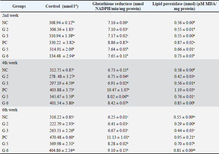
Conversely, fish fed diets supplemented with Mycofix Plus (G2) or Mycosorb A+ (G3) maintained significantly lower levels of cortisol and MDA throughout the experiment. The reduction in these biomarkers, particularly with Mycofix Plus during the later stages, demonstrates a high degree of protection against toxin-related physiological stress and oxidative imbalance.
The results for the combined groups (G5 and G6) indicated intermediate values of the measured parameters. These were significantly lower in the T-2 toxin-exposed group than in the control and binder groups; however, they were higher than in the control and binder groups. Therefore, partial protection from the stress caused by toxin action can be assumed.
Effects of T-2 toxin and MBs on antioxidant enzyme activities
The findings regarding the antioxidant enzyme activity are summarized in Table 4. The activities of CAT, SOD, and GST were significantly decreased throughout the experiment. In particular, the most pronounced decreases in activity were registered during week 6 for all studied enzymes in the T-2 toxin group PC. The constant ROS production resulting from T-2 toxin exposure led to a continuous endogenous antioxidant defense system weakening.
Table 4. Antioxidant enzyme activities in S. aurata fingerlings fed with T-2 toxin and MBs for 42 days.
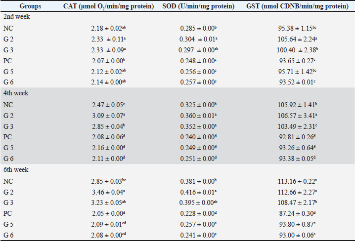
On the contrary, Mycofix Plus (G2) and Mycosorb A+ (G3) treatments led to a greater increase in enzyme activity than the control and T-2 toxin groups. A significant increase in antioxidant enzyme activity was observed at weeks 4 and 6. During these periods, CAT and SOD activities increased by 20%–40%. In other words, the use of Mycofix Plus and Mycosorb A+ protects fish from ROS damage.
Fish in the combined-treatment groups (G5 and G6) exhibited partial restoration compared to the T-2 toxin group but remained significantly lower than those observed in the binder-only groups. Although the binders alleviate oxidative stress, the presence of T-2 toxin continues to suppress antioxidant systems.
Effects of T-2 toxin and MBs on metallothionein profiles in whole blood
The MT content in whole blood for each experimental group is presented in Table 5. Fish exposed to T-2 toxin only PC displayed statistically significant reductions (p < 0.05) in overall, oxidized, and reduced MTs, especially at the 6th week, when a decrease in total MTs exceeded 40% compared with the control group. It may be assumed that T-2 toxin causes serious disturbances in the functioning of thiol-based antioxidant and metal scavenging systems.
Table 5. Metallothionein profiles in S. aurata fingerlings fed with T-2 toxin and MBs for 42 days.
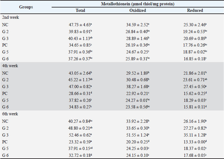
On the contrary, fish from groups G2 and G3, which were given Mycofix Plus and Mycosorb A+, respectively, had significantly higher MT content than other groups. Simultaneously, fish from G3 had the maximum MT content at the 6th week of the experiment. This result may indicate that metallothioneins are more effective in maintaining the redox state and protecting cells.
Fish from the combined treatment groups (G5 and G6) showed partial restoration of MT levels compared with the PC group, but these levels were still significantly lower compared with the groups receiving the binders only. This means that MT synthesis is partially inhibited in the presence of T-2 toxin even after adding binders to feed.
Taken together, these findings confirm that T-2 toxin severely impairs MT-mediated defense systems, whereas MBs restore thiol-dependent antioxidant capacity and enhance resilience against oxidative and toxic stress.
Histopathological findings in seabream fingerlings exposed to T-2 toxin
Histopathological findings indicated significant alterations in liver and kidney tissues of T-2 toxin-exposed seabream fingerlings, while dietary inclusion of MBs reduced the adverse effects of T-2 toxin, consistent with previous findings indicating T-2 toxin and other trichothecene derivatives cause damage to liver and kidney tissues (Fig. 4), although such adverse effects can be minimized using adsorbent products (Koletsi et al., 2021; Gruber-Dorninger et al., 2025).
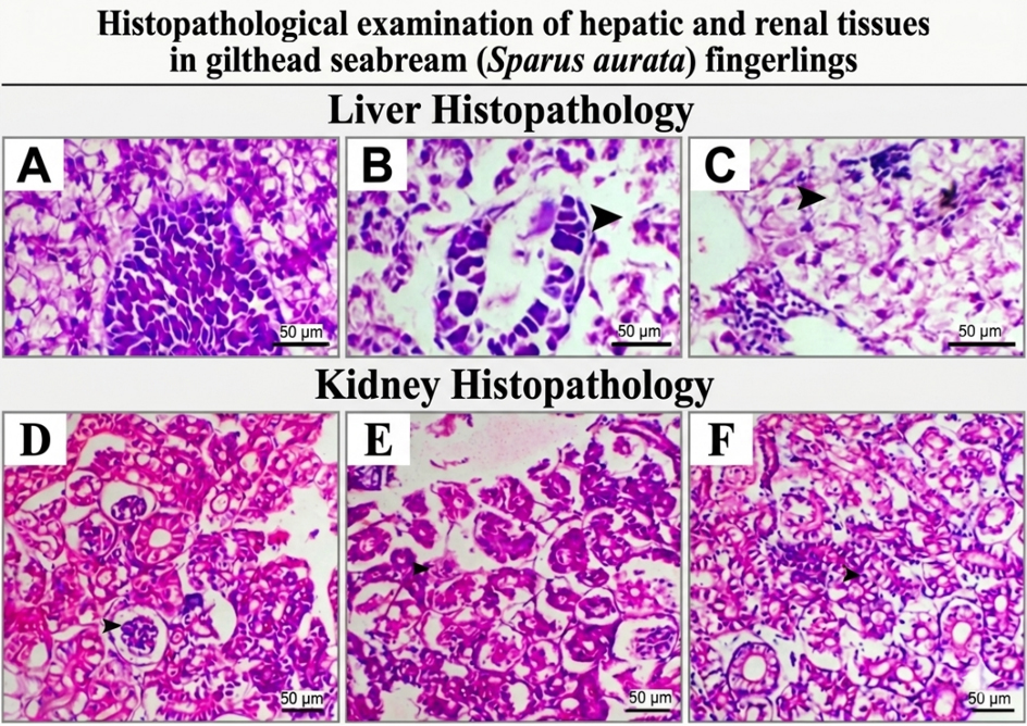
Fig. 4. Histopathological alterations in the hepatic and renal tissues of S. aurata fingerlings after exposure to dietary T-2 toxin and supplementation with MB. The upper part (A–C) represents histological sections of the liver. In the control group (A), normal histological features are observed with typical cytoplasmic vacuolation. In the T-2 toxin group (B), marked hepatic and pancreatic tissue damage is observed with extensive tissue disintegration (arrowhead). In the MB group (C), extensive vacuolation is observed in the hepatic tissue (arrowhead), indicating protection against tissue necrosis. The lower part (D–F) represents histological sections of the kidney; normal glomerular and tubular structures are observed in the control group (D, arrowhead); tubular tissue damage is observed in the T-2 toxin group (E, arrowhead); and marked improvement in kidney tissue with normal histology is observed in the MB group (F, arrowhead), indicating protection against T-2 toxin-induced tissue damage. (H&E stain; scale bars=50 µm).
The liver of control seabream fingerlings had normally structured liver tissue, consisting of well-organized hepatocytes, whereas those of T-2 toxin-treated fingerlings showed severe damage, including hepatocyte and pancreatic cell disintegration, consistent with previous findings of T-2 toxin hepatotoxicity and hepatocarcinogenicity in fish and other vertebrates (Taroncher et al., 2025). Moreover, there was evident vacuolation of the liver tissue of fingerlings exposed to T-2 toxin and Mycofix Plus, indicating some degree of protection against T-2 toxin-induced damage, although this damage was not completely abolished, aligning with previous findings indicating Mycofix to reduce but not completely abolish mycotoxin-induced liver damage (Chefchaou et al., 2019; Koletsi et al., 2021).
Histological assessment of kidney tissue from fingerlings in the control group showed normal kidney tissue with normally structured glomeruli and renal tubules. On the other hand, histological assessment of kidney tissue from seabream fingerlings exposed to T-2 toxin alone showed focal renal tubular degeneration, indicating severe nephrotoxic effects consistent with reported T-2 toxin-induced vacuolar degeneration, tubular necrosis, and congestion in mammalian kidney tissue (Janik et al., 2021). However, seabream fingerlings exposed to T-2 toxin in combination with Mycofix Plus showed improvement in kidney histology, with normally structured renal tubules, indicating the protective effects of T-2 toxin binder against T-2 toxin-induced kidney damage, consistent with the reported effects of T-2 toxin binder on mycotoxin bioavailability and kidney tissue damage (Agriopoulou et al., 2020).
Histopathological findings corroborate the biochemical and immunological results, confirming that T-2 toxin induces severe hepatic and renal damage, whereas Mycofix Plus and Mycosorb A+ markedly attenuate tissue injury and support organ integrity in seabream fingerlings, supporting the broader evidence that dietary MBs are an effective strategy to mitigate T-2-associated organ toxicity in farmed animals (Nácher-Mestre et al., 2015; Chefchaou et al., 2019; Agriopoulou et al., 2020).
Resistance to V. harveyi infection
The survival and RPS of gilthead seabream fingerlings following experimental challenge with V. harveyi are presented in Table 6. Among fish exposed to dietary T-2 toxin PC, the highest mortality rate (90%) and lowest survival rate (10%) were observed, confirming the immunosuppressive effects of T-2 toxin observed in the immunological assays. Fish receiving diets supplemented with MBs (G2 and G3) showed significantly improved survival, with rates of 70% and 80%, respectively, following bacterial infection.
Table 6. Survival and relative protection levels of S. aurata fingerlings challenged against V. harveyi following dietary exposure to T-2 toxin and MBs.
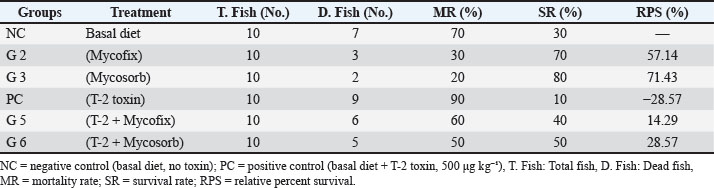
The calculated RPS values reached 57.14% in G2 and 71.43% in G3, indicating a strong protective effect of both binders against V. harveyi infection. Fish receiving combined treatments of T-2 toxin with Mycofix Plus (G5) or Mycosorb A+ (G6) showed partial disease resistance recovery, with survival rates of 40% and 50% corresponding to RPS values of 14.29% and 28.57%, respectively. However, these values were lower than those recorded in the binder-only groups. These findings demonstrate that dietary exposure to T-2 toxin significantly increases the susceptibility of seabream to bacterial infection, whereas supplementation with MBs enhances disease resistance and improves survival following pathogenic challenge.
Discussion
This study shows that dietary exposure to T-2 toxin causes severe adverse effects in S. aurata, including clinical manifestations, immunological parameters, oxidative status, and histopathological alterations, while dietary supplementation with Mycofix Plus and Mycosorb A+ MBs significantly reduced these effects. These data underscore the potential risks associated with trichothecenes in fish diets and confirm the efficacy of supplementation with MB in mariculture systems (Gruber-Dorninger et al., 2025; Lorusso et al., 2025).
Clinical and postmortem manifestations of T-2 toxin exposure
Adverse clinical signs, including abdominal distension, fin erosion, and stunted growth, were observed in fish exposed to T-2 toxin, consistent with its cytotoxic and protein synthesis-inhibiting activity (Matejova et al., 2017). The observed dwarfism and body size variability likely result from the negative effects of T-2 toxin on nutrient utilization and growth efficiency, as previously reported in species such as common carp and Nile tilapia (Diab et al., 2018; Barany et al., 2021). These clinical signs indicate systemic toxicity and disruptions in the metabolic and physiological processes of the animals caused by T-2 toxin.
T-2 toxin binds to the 60S ribosomal subunit at the cellular level (Zhang et al., 2022), inhibiting peptidyl transferase activity and consequently blocking protein synthesis (Janik et al., 2021). This leads to impaired tissue regeneration, reduced enzyme production, and metabolic pathway disruption, which collectively explain growth retardation and external deformities (Vörösházi et al., 2024; Abdelrahiem et al., 2025).
Postmortem examination revealed severe organ damage, including enlarged and discolored livers, congested kidneys, and gill lamellae degeneration. These changes indicate cumulative T-2 toxicity in metabolically active organs involved in detoxification, osmoregulation, and immune defense (Shah and Parveen, 2022; Wang et al., 2024). Similar vascular congestion, hemorrhages, and degenerative changes in internal organs have been observed in other aquatic and terrestrial animals after exposure to trichothecenes (Shah and Parveen, 2022).
Such organ-specific damage is closely linked to T-2 toxin-induced mitochondrial dysfunction and cellular apoptosis, particularly in highly metabolically active tissues such as the liver and kidney, where toxin accumulation and oxidative stress are most pronounced (Janik et al., 2021).
Immunosuppressive effects and protective role of MBs
In the current study, non-specific humoral defense mechanisms were found to be severely suppressed in fish exposed to T-2 toxin, as evidenced by decreased respiratory burst activity, plasma lysozyme levels, and bactericidal capacity. A notable outcome of this study is the differential efficacy observed between Mycofix Plus and Mycosorb A+, particularly in terms of immune modulation. Fish receiving Mycosorb A+ consistently exhibited slightly higher lysozyme activity, bactericidal capacity, and respiratory burst responses than those receiving Mycofix Plus. This difference may be attributed to the glucomannan nature of Mycosorb A+ obtained from yeast cell walls, its high binding capacity for trichothecene mycotoxins through hydrogen bond formation, and its complementary structural features (Agriopoulou et al., 2020; Kihal et al., 2022). However, although the efficacy of Mycofix Plus is proven, it depends on its multicomponent composition, including detoxification and adsorption, which may exhibit differential efficiency at varying toxin levels.
These results are consistent with those of earlier studies on the negative impact of T-2 toxin on leukocyte activity, cytokine production, and phagocytosis (Rocha et al., 2005; Matejova et al., 2017; Modra et al., 2018; Chen et al., 2025). These observations are also supported by the results of Rocha et al. (2005), who reported a significant suppression of cell and humoral immunity caused by T-2 toxin, and by other studies revealing the immunotoxicity of T-2 toxin induced through oxidation, inflammasome activation, and apoptosis in immune organs (Rehberger et al., 2017; Kooij et al., 2025). Immunosuppression is achieved by interfering with the biosynthesis of proteins that contribute to immune processes (Matejova et al., 2017) and by inducing the apoptosis of immune cells, including macrophages and lymphocytes. Dysregulation of cellular signaling pathways, such as MAPK and NF-κB, also contributes to this condition by disrupting cytokine secretion (Bai and Guo, 2025). Consequently, T-2-intoxicated seabream displayed the highest mortality rate following V. harveyi challenge, confirming functional immunosuppression.
On the other hand, the immune systems of fish supplemented with an MB were restored to a greater extent than in the control group. This recovery can be attributed primarily to the adsorption of T-2 toxin within the gastrointestinal tract, thereby reducing systemic exposure and restoring immune cell function (Koynarski et al., 2025; Tayyab et al., 2025). Furthermore, Mycosorb A+ may contribute to immunomodulation by enhancing gut-associated immune responses (Jaćević et al., 2020; Mesgar et al., 2022).
Endocrine stress response and oxidative damage
The elevated cortisol levels of T-2-exposed fish indicate the activation of the hypothalamic–pituitary–interrenal axis and a pronounced stress response (Modra et al., 2018; Kulcsár et al., 2023). This endocrine response is linked to cellular stress signaling due to the adverse effects of toxins, which cause oxidative stress and metabolic disturbances. Increased GSH reductase activity and MDA levels indicate oxidative stress and cell membrane damage (Modra et al., 2020; Kulcsár et al., 2023). On the molecular level, the T-2 toxin causes ROS overproduction due to disturbances in mitochondrial transport, lipid oxidation, proteins, and DNA (Kihal et al., 2022; Papatsiros et al., 2023; Goda et al., 2025). The addition of dietary binders helped alleviate these effects (Cuccato et al., 2025; Raz et al., 2025). Differences in the modulation of the stress and oxidative markers between the two binders were also evident. Mycosorb A+ exhibited a significantly higher decrease in cortisol concentrations and lipid peroxidase during the latter sampling times, implying that it better inhibited stress and oxidative injury. This might be attributed to its superior ability to block toxin uptake, thus avoiding the subsequent triggering of stress pathways and ROS production. The slower or incomplete detoxification process, involving both toxin binding and enzyme-mediated conversion, might explain the relatively mild effect observed with Mycofix Plus.
Antioxidant enzyme modulation
The observed suppression of CAT, SOD, and GST activities in fish treated with T-2 toxin indicates a definite disturbance of the antioxidant mechanism and increased sensitivity to ROS-mediated injury. These effects have been previously reported for rainbow trout as well as in other vertebrates, where feeding on T-2 toxin caused decreased CAT activity and deterioration of antioxidant properties, even with some transient activation of other GSH -dependent enzymes, thereby revealing oxidative stress and exhaustion of enzymatic defense (Deng et al., 2017; Modra et al., 2020; Taroncher et al., 2025). Moreover, higher T-2 levels could lead to even more suppressed SOD activity after a preliminary adaptive response, indicating a breakdown of the antioxidant system when faced with constant ROS generation.
Feeding fish on diets containing Mycofix Plus and Mycosorb A+, on the other hand, helped to improve the activities of antioxidant enzymes and decrease lipid peroxidation, providing considerable protection against ROS-induced oxidative damage. The observed results are due to reduced systemic toxins resulting from digestive fluid adsorption and do not reflect any direct antioxidant properties of the tested mycotoxin deactivators, which help preserve redox balance and prevent excessive ROS formation. Similar increases in CAT and SOD activities, as well as decreases in oxidative damage, were observed in carp, poultry, and other animals supplemented with Mycosorb or other mycotoxin sorbents, supporting their protective effects (Puvača et al., 2024; Koynarski et al., 2025).
The partial improvement observed with the combination of treatments demonstrates a specific, dose- and formulation-dependent effect of MBs. Mycosorb A+ consistently showed higher CAT, SOD, and GST activities than Mycofix Plus, indicating a more effective preservation of redox homeostasis. This confirms that rapid toxin sequestration in the gastrointestinal tract plays a critical role in preventing oxidative damage, whereas delayed detoxification mechanisms may allow transient oxidative stress to occur before it is mitigated. These results are also in agreement with previous reports suggesting that the protective efficiency of the studied adsorbents with respect to T-2 depends on the degree of its binding ability, composition, and inclusion in feed (Deng et al., 2017; Modra et al., 2020). Overall, these data are consistent with the established knowledge of the effects of mycotoxins, including T-2, on antioxidant enzyme systems, such as CAT, SOD, and GST, across various animals.
Metallothionein (MT) response
Metallothioneins (MTs) are widely recognized as sensitive biomarkers of oxidative stress and toxic exposures due to their high content of sulfur-containing amino acids and thiol-mediated metal-binding properties, and they represent crucial components of cellular redox systems in aquatic organisms (Roesijadi, 1992; Abdelghany et al., 2023). In this study, fingerlings of seabream exposed to T-2 toxin showed a persistent decrease in total, oxidized, and reduced MT fractions across all treatment periods, indicating the inhibition of thiol-mediated antioxidant mechanisms. The known effects of trichothecene mycotoxins increase oxidative stress and interfere with sulfhydryl-containing defenses, leading to decreased antioxidant protection (Modra et al., 2018; Puvača et al., 2024). These changes imply that the detoxifying abilities of cells are lost due to MT reduction.
In contrast to the toxin-exposed group, diet supplementation with mycotoxin adsorbents increased MT concentration, indicating the ability of these additives to protect the cell redox status. The mechanism is based on reduced toxin uptake, thereby limiting its effects on cellular metabolism and oxidative stress. In particular, fingerlings consuming Mycosorb A+ had maximum MT concentrations after 6 weeks, implying better efficiency in protecting and restoring redox homeostasis. This is in line with previous research confirming the higher effectiveness of yeast-based organic adsorbents in reducing the impact of T-2 toxin (Jaćević et al., 2020; Wang et al., 2023).
In addition, fish receiving combinations of additives showed only a slight increase in the MT fraction compared with the toxin group, with levels still significantly lower than those in groups with adsorbers. This finding shows that detoxifying activity depends on the dose and type of additives, similar to data from other studies performed with various animal species (Dai et al., 2019; Jaćević et al., 2020). Altogether, these results indicate a strong modulation of MT concentration in fish in accordance with contaminant exposure and oxidative stress. This confirms the applicability of MTs as biomarkers of diet-induced mycotoxin stress and the efficiency of additives in aquaculture systems (Roesijadi, 1992; Bejaoui et al., 2024).
Histopathological alterations
The histopathological alterations observed in this study provide clear evidence of the physiological and biochemical disruptions caused by the exposure of the sea bream fingerlings to T-2 toxin. Extreme hepatocellular vacuolation, hepatocyte disruption, and pancreatic tissue damage, in addition to severe degenerative renal tubular changes and congestion, indicate the toxic effects of the T-2 toxin on the liver and kidneys. The findings are in line with the already established ability of trichothecene mycotoxins to cause oxidative stress-based cytotoxicity resulting in progressive degenerative changes of liver and kidney tissues in vertebrates (Koletsi et al., 2021; Taroncher et al., 2025; Gruber-Dorninger et al., 2025). The described histopathological changes, such as vacuolation and tubular necrosis, form the structural background for the functional disorders identified in the current biochemical and immunological analyses.
On the contrary, the consumption of MBs reduced the degree of histopathological impairment and maintained tissue integrity in both the liver and kidney in most tested cases. In particular, the hepatocytes of the treated fish presented organized cell structure without any visible vacuolation, thus reflecting partial protection against toxin-induced degeneration. The tissues of the kidneys in fish receiving Mycofix Plus and Mycosorb A+ supplements had almost intact glomerular and tubular structures compared with the extremely degenerated structures observed following exposure to T-2 toxin. Thus, the dietary intake of such substances appears to be effective in preventing the cell uptake of toxins and, as a result, their subsequent harmful influence on organ structure.
Despite the presence of residual alterations in the livers of some experimental fish after using binder supplements and exposure to multiple treatments, the overall improvement in tissue integrity indicates a clear protective effect of the tested supplements. Previous research has shown that the use of mycotoxin adsorbent additives leads to a decrease in adverse outcomes for organs in the case of their exposure but does not fully prevent damage to tissue structure (Chefchaou et al., 2019; Agriopoulou et al., 2020). Thus, the improvement in tissue structure is clearly correlated with the positive changes in biochemical and immunological characteristics described above, indicating that oxidative stress mitigation and immune suppression also affect tissue morphology.
Resistance to infection
Long-term feeding of T-2 toxin had significant effects on the innate immune system of gilthead seabream, characterized by decreased NBT capacity, plasma lysozyme concentration, and bactericidal activity. The immunosuppressive effect made gilthead seabream extremely vulnerable to V. harveyi infection (SR=10%; RPS=−28.57%). Cortisol and MDA levels were also elevated in gilthead seabream, indicating increased stress and oxidative damage in this species, which may lead to immunosuppression (Modra et al., 2018; Kulcsár et al., 2023). Histopathology revealed hepatocyte and renal tubular cell disintegration, which is consistent with previous findings of T-2-induced organ toxicity in teleosts (Shah and Parveen, 2022).
Dietary supplementation with Mycofix Plus (G2) or Mycosorb A+ (G3) successfully restored immune function, antioxidant enzyme activities (CAT, SOD, and GST), and MT levels. This physiological recovery led to significantly improved survival during the bacterial challenge (SR=70%–80%; RPS=57%–71%). On the contrary, the combined treatment groups (G5 and G6) exhibited intermediate protection (SR=40%–50%; RPS=14%–29%), indicating only partial mitigation of T-2 toxicity during periods of ongoing exposure (Dai et al., 2019; Kihal et al., 2022).
The differences in survival rate and RPS further indicate that Mycosorb A+ is more effective than Mycofix Plus under these experimental conditions. The survival rates and RPS were higher in the fish treated with Mycosorb A+ than Mycofix Plus. Therefore, it can be inferred that better immunity and antioxidant capacity lead to increased disease resistance. This study shows the significance of the composition and working of binders in their efficacy against mycotoxins’ immunosuppressive effects.
Moreover, the strong correlation between immune biomarkers and survival rates highlights the predictive value of NBT, lysozyme, and bactericidal capacity for assessing disease resistance under mycotoxin-induced stress (Rocha et al., 2005; Matejova et al., 2017). These findings demonstrate that commercial MBs effectively counteract T-2-induced immunosuppression, preserve organ integrity, and enhance resistance to opportunistic bacterial infections. These results support the implementation of these binders as a robust preventive strategy for sustainable aquaculture management.
Limitations
The present study has several limitations that warrant consideration. The evaluation of a single T-2 concentration level precludes the observation of dose-dependent effects, and reliance on a controlled laboratory environment may not fully capture the complex ecological and management dynamics of commercial mariculture facilities. The absence of toxin residue analysis in fish tissues (e.g., liver and muscle) prevents a definitive quantification of reduced T-2 bioavailability; consequently, the observed recovery in physiological and antioxidant parameters infers the adsorptive efficacy of the biological binders. Furthermore, the histopathological assessments presented here are descriptive; the absence of a standardized quantitative scoring system for tissue lesions limits the precise indexing of the degree of pathological mitigation. Accordingly, these findings were interpreted as primary experimental observations requiring further validation through multi-dose designs, long-term trials, and advanced molecular approaches. Further investigation into gut microbiome dynamics and validation at the field scale under commercial farming conditions are required to enhance the practical applicability of these results.
Conclusion
Dietary exposure of gilthead seabream fingerlings (S. aurata) to T-2 toxin induced pronounced immunosuppression, oxidative stress, and tissue damage, thereby increasing their vulnerability to V. harveyi infection. This study clearly demonstrates the significant risks posed by trichothecene contamination in fish feed. Partial protection against the harmful effects of dietary T-2 was observed with Mycofix Plus and Mycosorb A+ administration, resulting in improvements in immunity, antioxidant status, and tissue structure, as well as increased survival after bacterial challenge. This protective effect is probably due to reduced toxin availability and stimulation of physiological defense processes. Nevertheless, the protection provided by the tested binders remained insufficient under continuous exposure of fish to the toxin, and further investigation is required. Furthermore, the use of a single toxin concentration and the absence of molecular-level analyses limit this study. Future studies should involve dose–response relationship assessment, long-term exposure experiments, and underlying mechanism investigation.
Acknowledgments
The authors would like to extend their deepest gratitude to the members of the participating research groups for their outstanding support. We thank the Aquaculture Laboratory at the Department of Fish Production, Faculty of Agriculture, Al-Azhar University.
Funding
This study received no external funding.
Authors' contributions
All authors contributed equally.
Conflict of interest
The authors declare no conflicts of interest.
Data availability
The data could be provided to the corresponding author upon request.
References
Abdelghany, F.M., Abdel-Warith, A.W.A., Younis, E.M., Davies, S.J., Elnakeeb, M.A., El-Nawasany, M.M., El-Bahlol, A.A., Bauomi, M.A., Abu-Almaaty, A.H. and Omer, M.Y. 2023. Genetic differentiation of five sea cucumber species from the Red Sea, Hurghada, Egypt. Braz. J. Biol. 83, 83; doi:10.1590/1519-6984.271983
Abdelrahiem, T.M.M., Hanbal, M.M., Salem, M.F. and Elnakeeb, M.A. 2025. Effect of some heavy metals (Pb, Hg, Cd and Cu) on physiological and immunological parameters in the Nile Tilapia (Oreochromis niloticus). Egypt. J. Aquat. Biol. Fish. 29(6), 849–866; doi:10.21608/ejabf.2025.439975.6947
Agriopoulou, S., Stamatelopoulou, E. and Varzakas, T. 2020. Advances in occurrence, importance, and mycotoxin control strategies: prevention and detoxification in foods. Foods 9(2), 137; doi:10.3390/foods9020137
Aly, S.M., Elbanna, N.I., Elatta, M.A., Hegazy, M. and Fathi, M. 2024. Effects of natural and synthetic immunostimulants on growth, feed utilization, immune status and disease resistance against vibriosis in sea bream (Sparus aurata). Aquac. Int. 32(3), 2739–2756; doi:10.1007/s10499-023-01294-2
Aly, S.M., ElBanna, N.I., Elatta, M.A., Hegazy, M. and Fathi, M. 2025. Prevalence, molecular typing, antibiogram and histopathological changes of Vibrio harveyi and Vibrio parahaemolyticus isolated from gilthead seabream (Sparus aurata). Aquac. Fish. 10(2), 281–288; doi:10.1016/j.aaf.2023.11.002
Amend, D.F. 1981. Potency testing of fish vaccines. Dev. Biol. Stand. 49, 447–454.
Anandaraj, A., Cleatus, B., Thirukumaran, V., Solayappan, M. and Thirunavukkarasu, R. 2025. Aflatoxigenic fungi in fish and fish feed. In: Aflatoxigenic fungi. Eds., Swamy, M., Somasundaram, S. and Deivasigamani, B. Boca Raton, FL: CRC Press, pp. 172-182; doi: 10.1201/9781003487043-22
Arafa, A., Eissa, A.E., Khallaf, M.A., Ghetas, H.A., Ragab, R.H., Mansour, S.M., Younis, E.M., Ali, S.F., Ismail, E.M., Rashwan, E.H., Diab, H.M., Hamouda, A.H., Abuelkasem, S.S., Elnakeeb, M.A., Abd-Elghany, M., Ismail, M.M., Abdel Hady, H.A., Korany, R. and Younis, N.A. 2025. Photobacterium damselae subsp. piscicida Infection, a Persistent Cause of Mortalities in Old Hatchery-reared Gilthead Seabream (Sparus aurata) Broodstocks: molecular-pathological Evidences and Control Strategy. Egypt. J. Vet. Sci. 0(0), 1–13; doi:10.21608/ejvs.2025.339899.2526
Ashry, A.M., Habiba, M.M., Abdel-Warith, A.W.A., Younis, E.M., Davies, S.J., Elnakeeb, M.A., Abdelghany, M.F., El-Zayat, A.M. and El-Sebaey, A.M. 2024. Dietary effect of powdered herbal seeds on zootechnical performance, hemato-biochemical indices, immunological status, and intestinal microbiota of European sea bass (Dicentrarchus labrax). Aquac. Rep. 36, 102074; doi:10.1016/j.aqrep.2024.102074
Bai, R. and Guo, J. 2025. SPI1 upregulated LILRB2 to enhance the immunosuppressive phenotype of LPS-tolerant macrophages by inhibiting TLR8-mediated MyD88/NF-κB signaling. Biol. Direct 20(1), 73; doi: 10.1186/s13062-025-00669-0
Barany, A., Guilloto, M., Cosano, J., De Boevre, M., Oliva, M., De Saeger, S., Fuentes, J., Martínez-Rodriguez, G. and Mancera, J.M. 2021. Dietary aflatoxin B1 (AFB1) reduces growth performance, impacting growth axis, metabolism, and tissue integrity in juvenile gilthead sea bream (Sparus aurata). Aquaculture 533, 736189; doi:10.1016/j.aquaculture.2020.736189
Bejaoui, S., Chetoui, I., Ghribi, F., Belhassen, D., Abdallah, B.B., Fayala, C.B., Boubaker, S., Mili, S. and Soudani, N. 2024. Exposure to different cobalt chloride levels produces oxidative stress and lipidomic changes and affects the liver structure of Cyprinus carpio juveniles. Environ. Sci. Pollut. Res. 31(39), 51658–51672; doi:10.1007/s11356-024-34578-y
Biller-Takahashi, J., Takahashi, L., Saita, M., Gimbo, R. and Urbinati, E. 2013. Leukocytes respiratory burst activity as indicator of innate immunity of pacu Piaractus mesopotamicus. Braz. J. Biol. 73(2), 425–429; doi:10.1590/s1519-69842013000200026
Bisai, K., Roy, A., Pati, M.K. and Das, B.K. 2025. Histological techniques in fish disease diagnosis. In: Laboratory techniques for fish disease diagnosis. Eds., Das, B.K., Roy, A. and Pati, M.K. Springer, Singapore, pp. 359–374; doi: 10.1007/978-981-96-4620-3_16
Chefchaou, H., Mzabi, A., Tanghort, M., Moussa, H., Chami, N., Chami, F. and Remmal, A. 2019. A comparative study of different mycotoxin adsorbents against DON, T2 toxin, aflatoxins and fumonisins production in maize flour. Livest. Res. Rural. Dev, 31, 35. https://doi.org/10.3923/ijps.2019.187.194
Chen, M., Zhang, Y., Zhao, Y., Cao, K., Niu, R., Guo, D. and Sun, Z. 2025. Complex immunotoxic effects of T-2 Toxin on the murine spleen and thymus: oxidative damage, inflammasomes, apoptosis, and immunosuppression. Ecotoxicol. Environ. Saf. 289, 117476; doi:10.1016/j.ecoenv.2024.117476
Colás-Ruiz, N.R., Ramirez, G., Courant, F., Gomez, E., Hampel, M. and Lara-Martín, P.A. 2022. Multi-omic approach to evaluate the response of gilt-head sea bream (Sparus aurata) exposed to the UV filter sulisobenzone. Sci. Total. Environ. 803, 150080; doi:10.1016/j.scitotenv.2021.150080
Cuccato, M., Amminikutty, N., Spalenza, V., Conte, V., Bagatella, S., Greco, D., D’Ascanio, V., Gai, F., Schiavone, A., Avantaggiato, G., Nebbia, C. and Girolami, F. 2025. Innovative mycotoxin detoxifying agents decrease the absorption rate of aflatoxin B1 and counteract the oxidative stress in broiler chickens exposed to low dietary levels of the mycotoxin. Toxins 17(2), 82; doi:10.3390/toxins17020082
Dai, C., Xiao, X., Sun, F., Zhang, Y., Hoyer, D., Shen, J., Tang, S. and Velkov, T. 2019. T-2 toxin neurotoxicity: role of oxidative stress and mitochondrial dysfunction. Arch. Toxicol. 93(11), 3041–3056; doi:10.1007/s00204-019-02577-5
Dara, M., Carbonara, P., La Corte, C., Parrinello, D., Cammarata, M. and Parisi, M.G. 2023. Fish welfare in aquaculture: physiological and immunological activities for diets, social and spatial stress on Mediterranean aquacultured species. Fishes 8(8), 414; doi:10.3390/fishes8080414
Deng, Y., Wang, Y., Zhang, X., Sun, L., Wu, C., Shi, Q., Wang, R., Sun, X., Bi, S. and Gooneratne, R. 2017. Effects of T‐2 toxin on Pacific white shrimp Litopenaeus vannamei: growth, and antioxidant defenses and capacity and histopathology in the Hepatopancreas. J. Aquat. Anim. Health. 29(1), 15–25; doi:10.1038/s41598-019-49004-4
Diab, A.M., R.M., S., Abeer, E.K.M.S., Ali, G.I.E. and El-Habashi, N. 2018. Experimental ochratoxicosis A in Nile tilapia and its amelioration by some feed additives. Int. J. Vet. Sci. Med. 6(2), 149–158; doi:10.1016/j.ijvsm.2018.09.004
Eissa, A.E., Ziltne, R.E., Edrees, A., El-Ramlawy, A.O., Sharaf, M.S., Korany, R., Abdel-Latif, H.M.R., Ibrahim, I.H., Mahmoud, A.E., Qorany, R., Mansour, S., Ibrahim, S.A.A., Younis, E.M.M., Abdelrahman, M.A., Abulelkasem, S.S., Afiffi, E. and Ragab, R.H. 2025. Epidemiological tracking and control strategies of early mortalities in hatchery-reared gilthead seabream (Sparus aurata) larvae. Aquac. Int. 33(6), 468; doi:10.1007/s10499-025-02148-9
Elnakeeb, M.A., Ashry, A.M., El-Zayat, A.M., Abdel-Warith, A.W.A.M., Habiba, M.M., Younis, E.M.I., Davies, S.J., Ibrahim, I.M., Elzhraa, F. and El-Sebaey, A.M. 2025. Impact of effective probiotic microorganisms (EPMs) on growth performance, hematobiochemical panel, immuno-antioxidant status, and gut cultivable microbiota in striped catfish (Pangasianodon hypophthalmus). Fishes 10(11), 573; doi:10.3390/fishes10110573
El-Waziry, I.A.K., Eissa, A.E., El-Tarabili, R.M., Abouelhassan, E.M., Ghetas, H.A., Ismail, E.M., Elnakeeb, M.A., Abdel Hady, H.A., Aboelnaga, H.S. and Dessouki, A.A. 2025. Parasitic crustaceans as a potential vector of MDR Vibrio alginolyticus infection among farmed marine fish in some earthen-pond based Egyptian aquaculture facilities: molecular, epidemiological and pathological evidences. Parasitol. Int. 108, 103066; doi:10.1016/j.parint.2025.103066
Gisbert, E., Skalli, A., Campbell, J., Solovyev, M.M., Rodríguez, C., Dias, J. and Polo, J. 2015. Spray-dried plasma promotes growth, modulates the activity of antioxidant defenses, and enhances the immune status of gilthead sea bream (Sparus aurata) fingerlings. J. Anim. Sci. 93(1), 278–286; doi:10.2527/jas.2014-7491
Goda, A.A., Shi, J., Xu, J., Liu, X., Zhou, Y., Xiao, L., Abdel-Galil, M., Salem, S.H., Ayad, E.G., Deabes, M., Pooe, O., Donia, M.A.A., Abou-Arab, A.A.K. and Ramzy, S. 2025. Global health and economic impacts of mycotoxins: a comprehensive review. Environ. Sci. Eur. 37(1), 122; doi:10.1186/s12302-025-01166-x
Gomez, K.A. and Gomez, A.A. 1984. Statistical procedures for agricultural research. New York, Ny: John wiley & sons.
Gruber-Dorninger, C., Müller, A. and Rosen, R. 2025. Multi-mycotoxin contamination of aquaculture feed: a global survey. Toxins 17(3), 116; doi:10.3390/toxins17030116
Hussain, S.M., Bano, A.A., Ali, S., Rizwan, M., Adrees, M., Zahoor, A.F., Sarker, P.K., Hussain, M., Arsalan, M.Z.U.H. and Yong, J.W.H. 2024. Substitution of fishmeal: Highlights of potential plant protein sources for aquaculture sustainability. Heliyon 10(4), e26573 ;doi: 10.1016/j.heliyon.2024.e26573
Ilesanmi, A.I., Adeoba, M.I., Bubu-Davies, O.A., Osemwegie, O.O. and Dan-Ologe, I.A. 2024. Possible methods of suppressing mycotoxin in aquatic organisms feed. In: Marine bioprospecting for sustainable blue-bioeconomy. Eds., Shanthi, S., Prakash, S. and Santhanam, P. Cham, Switzerland: Springer Nature Switzerland, pp. 227–250; doi: 10.1007/978-3-031-68911-6_12
Jaćević, V., Dumanović, J., Lazarević, M., Nepovimova, E., Resanović, R., Milovanović, Z., Wu, Q. and Kuča, K. 2020. Antidotal potency of the novel, structurally different adsorbents in rats acutely intoxicated with the T-2 toxin. Toxins 12(10), 643; doi:10.3390/toxins12100643
Janik, E., Niemcewicz, M., Podogrocki, M., Ceremuga, M., Stela, M. and Bijak, M. 2021. T-2 Toxin—the most toxic trichothecene mycotoxin: metabolism, toxicity, and decontamination strategies. Molecules 26(22), 6868; doi: 10.3390/molecules26226868
Kihal, A., Rodríguez-Prado, M. and Calsamiglia, S. 2022. The efficacy of mycotoxin binders to control mycotoxins in feeds and the potential risk of interactions with nutrient: a review. J. Anim. Sci. 100(11), 328; doi:10.1093/jas/skac328
Kim, J.H., Park, H.J., Kim, K.W. and Kang, J.C. 2017. Oxidative stress and non-specific immune responses in juvenile black sea bream, Acanthopagrus schlegelii, exposed to waterborne zinc. Fisheries Aquatic Sci. 20(1), 11; doi:10.1186/s41240-017-0056-x
Koletsi, P., Schrama, J.W., Graat, E.A.M., Wiegertjes, G.F., Lyons, P. and Pietsch, C. 2021. The occurrence of mycotoxins in raw materials and fish feeds in Europe and the potential effects of deoxynivalenol (DON) on the health and growth of farmed fish species—a review. Toxins 13(6), 403; doi:10.3390/toxins13060403
Kooij, N., Fowle, A., Lyons, S., Yu, A.M. and King-Heiden, T.C. 2025. Comparative immunomodulatory effects of chronic exposure to imidacloprid and thiamethoxam on the respiratory burst response in zebrafish and fathead minnow larvae. Fish Shellfish Immunol. (165), 110556; doi: 10.2139/ssrn.5158437
Koynarski, T., Nedelcheva, D., Valchev, I., Hristov, T., Nikolov, N., Kostadinov, N. and Binev, R. 2025. Mitigating T-2 toxin-induced immunosuppression in ducks: effects of Mycofix® Plus 5.0 and silymarin on innate immunity. Pak. Vet. J. 45(3), 1419–1422; doi:10.29261/pakvetj/2025.245
Kulcsár, S., Kövesi, B., Balogh, K., Zándoki, E., Ancsin, Z., Erdélyi, M. and Mézes, M. 2023. The co-occurrence of T-2 toxin, deoxynivalenol, and fumonisin B1 activated the glutathione redox system in the EU-limiting doses in laying hens. Toxins 15(5), 305; doi:10.3390/toxins15050305
Littell, W.W. and Freund, S. 2002. SAS for linear models. Cary, NC: John Wiley & Sons.
Lorusso, P., Rusco, G., Manfredi, A., Iaffaldano, N., Di Pinto, A. and Bonerba, E. 2025. Emerging mycotoxins in aquaculture: current insights on toxicity, biocontrol strategies, and occurrence in aquafeed and fish. Toxins 17(7), 356; doi:10.3390/toxins17070356
Matejova, I., Faldyna, M., Modra, H., Blahova, J., Palikova, M., Markova, Z., Franc, A., Vicenova, M., Vojtek, L., Bartonkova, J., Sehonova, P., Hostovsky, M. and Svobodova, Z. 2017. Effect of T-2 toxin-contaminated diet on common carp (Cyprinus carpio L.). Fish. Shellfish. Immunol. 60, 458–465; doi:10.1016/j.fsi.2016.11.032
Mello, F.V., Pereira, C., Özkan, B., Maulvault, A.L., Soares, F., Pousão-Ferreira, P., Fernandes, J.O., Cunha, S.C., Marques, A. and Anacleto, P. 2025. Toxicological responses of juvenile gilthead seabream to enniatin B and fumonisin B1. Int. J. Mol. Sci. 26(12), 5676; doi:10.2139/ssrn.4912891
Meneely, J., Greer, B., Kolawole, O. and Elliott, C. 2023. T-2 and HT-2 toxins: toxicity, occurrence and analysis: a review. Toxins 15(8), 481; doi:10.3390/toxins15080481
Mesgar, A., Aghdam Shahryar, H., Bailey, C.A., Ebrahimnezhad, Y. and Mohan, A. 2022. Effect of dietary L-threonine and toxin binder on performance, blood parameters, and immune response of broilers exposed to aflatoxin B1. Toxins 14(3), 192; doi:10.3390/toxins14030192
Mhalhel, K., Levanti, M., Abbate, F., Laurà, R., Guerrera, M.C., Aragona, M., Porcino, C., Briglia, M., Germanà, A. and Montalbano, G. 2023. Review on Gilthead seabream (Sparus aurata) aquaculture: life cycle, growth, aquaculture practices and challenges. J. Mar. Sci. Eng. 11(10), 2008; doi:10.3390/jmse11102008
Mizaeva, T., Alieva, K., Zulkarneev, E., Kurpe, S., Isakova, K., Matrosova, S., Borvinskaya, E. and Sukhovskaya, I. 2023. Antibacterial activity of rainbow trout plasma: in vitro assays and proteomic analysis. Animals 13(22), 3565; doi:10.3390/ani13223565
Modra, H., Palikova, M., Hyrsl, P., Bartonkova, J., Papezikova, I., Svobodova, Z., Blahova, J. and Mares, J. 2020. Effects of trichothecene mycotoxin T-2 toxin on haematological and immunological parameters of rainbow trout (Oncorhynchus mykiss). Mycotoxin Res. 36(3), 319–326; doi:10.1007/s12550-020-00396-7
Modra, H., Sisperova, E., Blahova, J., Enevova, V., Fictum, P., Franc, A., Mares, J. and Svobodova, Z. 2018. Elevated concentrations of T‐2 toxin cause oxidative stress in the rainbow trout (Oncorhynchus mykiss). Aquac. Nutr. 24(2), 842–849; doi:10.1111/anu.12613
Mohamad, N., Amal, M., Yasin, I.S.M., Saad, M., Nasruddin, N.S., Al-Saari, N., Mino, S. and Sawabe, T. 2019. Vibriosis in cultured marine fishes: a review. Aquaculture 512, 734289; doi: 10.1016/j.aquaculture.2019.734289
Nácher-Mestre, J., Serrano, R., Beltrán, E., Pérez-Sánchez, J., Silva, J., Karalazos, V., Hernández, F. and Berntssen, M.H. 2015. Occurrence and potential transfer of mycotoxins in gilthead sea bream and Atlantic salmon by use of novel alternative feed ingredients. Chemosphere 128, 314–320; doi:10.1016/j.chemosphere.2015.02.021
NRC. 2011. Nutrient requirements of fish and shrimp. The National Academies Press. https://doi.org/10.1007/s10499-011-9480-6
Okolie, P.C., Chukwujike, I.C., Chukwuneke, J.L. and Dara, J.E. 2019. Design and production of a fish feed pelletizing machine. Heliyon 5(6), e02001; doi: 10.1016/j.heliyon.2019.e02001
Papatsiros, V.G., Eliopoulos, C., Voulgarakis, N., Arapoglou, D., Riahi, I., Sadurní, M. and Papakonstantinou, G.I. 2023. Effects of a multi-component mycotoxin-detoxifying agent on oxidative stress, health and performance of sows. Toxins 15(9), 580; doi:10.3390/toxins15090580
Paruğ, Ş., Jabbr, A. and Lazrag, F.A. 2024. Sustainable development of fisheries and aquaculture in North Africa: current status and future prospects. Menba. J. Fish. Fac. 10(1), 90–105; doi:10.58626/ menba.1455763
Pulvirenti, E., Favara, C., Rapisarda, P., Castrogiovanni, M., Sica, C., Aquilia, E., Cristaldi, A., Conti, G.O. and Ferrante, M. 2024. Use of chemical–physical and natural ingredients of food factors modulation for freshwater aquaculture systems fish wellness: a systematic review. Aquac. Res. 2024(1), 1284780; doi:10.1155/are/1284780
Puvača, N., Ljubojević Pelić, D. and Tufarelli, V. 2023. Mycotoxins adsorbents in food animal production. https://doi.org/10.55817/gyic7602
Puvača, N., Shtylla Kika, T., Brkić, I., Ljubojević Pelić, D., Tufarelli, V., Nikolova, N., Kostić, B., Radojičić, I., Brkanlić, S. and Bursić, V. 2024. Influence of mycostop mycotoxin adsorbents on production, oxidative stress, and economic cost of laying hens intoxicated with T-2 toxin. J. Hell. Vet. Med. Soc. 75(4), 8445–8456; doi:10.12681/jhvms.37778
Qiu, M., Wang, Y., Wang, X., Sun, L., Ye, R., Xu, D. and Gooneratne, R. 2016. Effects of T-2 toxin on growth, immune function and hepatopancreas microstructure of shrimp (Litopenaeus vannamei). Aquaculture 462, 35–39; doi:10.1016/j.aquaculture.2016.04.032
Raz, M., Bagherzadeh-Kasmani, F., Karimi-Torshizi, M.A., Ghazaghi, M., Mokhtarpour, A. and Mehri, M. 2025. Boosting antioxidant defense and enhancing product quality by biochar and probiotics under chronic aflatoxicosis in quails. Poult. Sci. 104(7), 105183; doi:10.1016/j.psj.2025.105183
Rehberger, K., Werner, I., Hitzfeld, B., Segner, H. and Baumann, L. 2017. 20 Years of fish immunotoxicology–what we know and where we are. Crit. Rev. Toxicol. 47(6), 516–542; doi:10.1080/10408444.2017.1288024
Rennie, M.D., Kennedy, P.J., Mills, K.H., Rodgers, C.M.C., Charles, C., Hrenchuk, L.E., Chalanchuk, S., Blanchfield, P.J., Paterson, M.J. and Podemski, C.L. 2019. Impacts of freshwater aquaculture on fish communities: a whole-ecosystem experimental approach. Freshw. Biol. 64(5), 870–885; doi:10.1111/fwb.13269
Rocha, O., Ansari, K. and Doohan, F.M. 2005. Effects of trichothecene mycotoxins on eukaryotic cells: a review. Food. Addit. Contam. 22(4), 369–378; doi:10.1080/02652030500058403
Rodrigues, S., Antunes, S., Nunes, B. and Correia, A. 2017. Histological alterations in gills and liver of rainbow trout (Oncorhynchus mykiss) after exposure to the antibiotic oxytetracycline. Environ. Toxicol. Pharmacol. 53, 164–176; doi:10.1016/j.etap.2017.05.012
Roesijadi, G. 1992. Metallothioneins in metal regulation and toxicity in aquatic animals. Aquatic. Toxicol. 22(2), 81–113; doi:10.1016/0166-445x(92)90026-j
Saera-Vila, A., Benedito-Palos, L., Sitjà-Bobadilla, A., Nácher-Mestre, J., Serrano, R., Kaushik, S. and Pérez-Sánchez, J. 2009. Assessment of the health and antioxidant trade-off in gilthead sea bream (Sparus aurata L.) fed alternative diets with low levels of contaminants. Aquaculture 296(1-2), 87–95; doi:10.1016/j.aquaculture.2009.07.028
Serna-Duque, J., Espinosa-Ruíz, C. and Esteban, M. 2023. Hepcidin and piscidin modulation and antibacterial response in gilthead seabream (Sparus aurata) infected with Vibrio harveyi. Fish Shellfish Immunol. (139), 108899; doi: 10.1016/j.fsi.2023.108899
Shaalan, M., El-Mahdy, M., Saleh, M. and El-Matbouli, M. 2018. Aquaculture in Egypt: insights on the current trends and future perspectives for sustainable development. Rev. Fish. Sci. Aquac. 26(1), 99–110; doi:10.1080/23308249.2017.1358696
Shah, Z.U. and Parveen, S. 2022. Oxidative, biochemical and histopathological alterations in fishes from pesticide contaminated river Ganga, India. Sci. Rep. 12(1), 3628; doi:10.1038/s41598-022-07506-8
Shrivastava, Y., Nayak, S.K. and Mogalekar, H. 2025. Aquatic animal health management. In: Management of fish diseases. Eds., Das, B.K., Mogalekar, H.S. and Nayak, S.K. Springer, Singapore, pp. 189–211; doi: 10.1007/978-981-96-0270-4_8
Singh, D.B.M. 2021. Histopathological study of Channa gachua exposed to sublethal concentration of malathion. Int. J. Fish. Aquat. Stud. 9(3), 174–179; doi:10.22271/fish.2021.v9.i3c.2472
Sivakumar, J., Richardson, P., Sammaddar, C., Babu, S.R., Bhuvaneswari, S. and Venu, S. 2022. A study on anatomical and histological characterization of Juvenile Asian Seabass, Lates calcarifer (Bloch, 1790). Int. J. Res. Anal. Rev. 9(4), 411–412.
Taroncher, M., Franco-Campos, F., Rodríguez-Carrasco, Y. and Ruiz, M.J. 2025. T-2 toxin-induced hepatotoxicity in HepG2 cells involves the inflammatory and Nrf2/HO-1 pathways. Toxins 17(8), 397; doi:10.3390/toxins17080397
Taylor, B., Ofori, K.F., Parsaeimehr, A., Akdemir Evrendilek, G., Attarwala, T. and Ozbay, G. 2025. Exploring the complexities of seafood: from benefits to contaminants. Foods 14(9), 1461; doi:10.3390/foods14091461
Tayyab, M., Islam, W., Waqas, W. and Zhang, Y. 2025. Probiotic–vaccine synergy in fish aquaculture: exploring microbiome-immune interactions for enhanced vaccine efficacy. Biology 14(6), 629; doi:10.3390/biology14060629
Triga, A., Issa, Z., Smyrli, M., Fenske, L. and Katharios, P. 2024. Virulence and pangenome analysis of Vibrio harveyi strains from Greek and Red Sea marine aquaculture. Aquaculture (587), 740839; doi: 10.1016/j.aquaculture.2024.740839
Vörösházi, J., Neogrády, Z., Mátis, G. and Mackei, M. 2024. Pathological consequences, metabolism and toxic effects of trichothecene T-2 toxin in poultry. Poult. Sci. 103(3), 103471; doi:10.1016/j.psj.2024.103471
Vulić, A., Kudumija, N., Šegvić-Bubić, T. and Lešić, T. 2025. Mycotoxins in fish aquaculture—occurrence and future perspective. Foods 14(24), 4301; doi:10.3390/foods14244301
Wang, C., He, J., Jin, H., Xiao, H., Peng, S., Xie, J., Zhang, L. and Guo, J. 2023. T-2 toxin induces cardiotoxicity by activating ferroptosis and inhibiting heme oxygenase-1. Chemosphere 341, 140087; doi:10.1016/j.chemosphere.2023.140087
Wang, C., Wang, X., Huang, Y., Bu, X., Xiao, S., Qin, C., Qiao, F., Qin, J.G. and Chen, L. 2020. Effects of dietary T-2 toxin on gut health and gut microbiota composition of the juvenile Chinese mitten crab (Eriocheir sinensis). Fish. Shellfish. Immunol. 106, 574–582; doi:10.1016/j.fsi.2020.08.019
Wang, J.Q., Hussain, R., Ghaffar, A., Afzal, G., Saad, A.Q., Ahmad, N., Nazir, U., Ahmad, H.I., Hussain, T. and Khan, A. 2022. Clinicohematological, mutagenic, and oxidative stress induced by pendimethalin in freshwater fish bighead carp (Hypophthalmichthys nobilis). Oxid. Med. Cell. Longev. 1(1), 2093822; doi:10.1155/2022/2093822
Wang, P., Sun, L. -H., Wang, X., Wu, Q. and Liu, A. 2024. Effective protective agents against the organ toxicity of T-2 toxin and corresponding detoxification mechanisms: a narrative review. Anim. Nutr. 16, 251–266; doi:10.1016/j.aninu.2023.12.001
Yin, H., Han, S., Chen, Y., Wang, Y., Li, D. and Zhu, Q. 2020. T-2 toxin induces oxidative stress, apoptosis and cytoprotective autophagy in chicken hepatocytes. Toxins 12(2), 90; doi:10.3390/toxins12020090
Zahran, E., Elbahnaswy, S., Abdelhamid, F., Abdel., Fattah, S.M., Mansour, A.I., Ibrahim, I., Ahmed, F., El., Houseiny, W., León, E., Codina, R. and Castro. 2026. Evaluation of a novel anti‐mycotoxin agent specific for aquatic species in mitigating aflatoxin B1‐induced changes in Nile tilapia: a comprehensive study. JWAS 57(1), e70075; doi:10.1111/jwas.70075
Zhang, J., Liu, X., Su, Y. and Li, T. 2022. An update on T2-toxins: metabolism, immunotoxicity mechanism and human assessment exposure of intestinal microbiota. Heliyon 8(8), e10012; doi: 10.1016/j.heliyon.2022.e10012