





| Research Article | ||
Open Vet. J.. 2026; 16(6): 3731-3748 Open Veterinary Journal, (2026), Vol. 16(6): 3731-3748 Research Article Circulating Babesia sp. and Ehrlichia sp. in Egyptian dogs: Molecular detection, risk assessment, and hematobiochemical parametersShahenaz M. H. Hassan1, Rasha Diabb2, Neveen S. Satour3 and Emad Beshir Ata4*1Department of Clinical Pathology, Alexandria Regional Laboratory, Animal Health Research Institute (AHRI), Agriculture Research Center (ARC), Alexandria, Egypt 2Department of Immunity, Alexandria Regional Laboratory, Animal Health Research Institute (AHRI), Agriculture Research Center (ARC), Alexandria, Egypt 3Department of Parasitology, Alexandria Regional Laboratory, Animal Health Research Institute, Agriculture Research Center, Alexandria, Egypt 4Department of Parasitology and Animal Diseases, Veterinary Research Institute, National Research Centre, Cairo, Egypt *Corresponding Author: Emad Beshir Ata. Department of Parasitology and Animal Diseases, Veterinary Research Institute, National Research Center, Cairo, Egypt. Email: emadvet2003 [at] yahoo.com Submitted: 07/04/2026 Revised: 17/05/2026 Accepted: 24/05/2026 Published: 16/06/2026 © 2025 Open Veterinary Journal
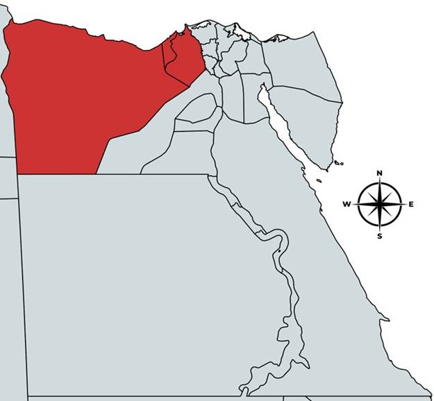
AbstractBackground: Canine babesiosis and ehrlichiosis are common diseases that significantly affect the health of infected dogs, particularly in tropical areas. Aim: This study aimed to identify the circulating Babesia sp. and Ehrlichia sp. in Egypt and to determine the related risk factors, with a particular focus on the hematobiochemical parameters. Methods: A total of 157 blood samples were used for nucleic acid extraction for molecular identification, phylogenetic analysis, and evaluating the expression level of tumor necrosis factor-α (TNF-α) and interferon-γ (IFN-γ) by qPCR, respectively. Hematobiochemical parameters were also measured. Animal data were obtained during clinical investigation and used for risk assessment. Multiple clinical signs were recorded with varying degrees of intensity. Results: The results confirmed the identification of Babesia canis subspecies vogeli. and E. canis with a rate of 17.83% and 12.73%, respectively, which was higher than the microscopic examination. The obtained Babesia sp. were uploaded to the National Center for Biotechnology Information under the accession numbers (PQ821446.1, PQ821447.1, PQ821448.1). For Ehrlichia sp., were (PQ821442.1, PQ821443.1, PQ821444.1). IFN-γ and TNF-α genes were significantly expressed in the infected cases compared with the healthy ones. The presence of stray dogs and external ticks was the most significant associated risk factor. Hemolytic anemia, thrombocytopenia, lymphocytosis, and hyperbilirubinemia are the principal hemato-biochemical alterations. Conclusion: B. canis vogeli, Ehrlichia canis is the most common circulating type, accompanied by hyperexpression of IFN-γ and TNF-α genes, hemolytic anemia, and external parasitic infestation. This study not only highlighted the prevalence of canine haematogones, related signs, and risk factors but also recommended the application of effective control strategies, especially for stray dogs. Keywords: Babesiosis, Ehrlichiosis, Prevalence, Risk factors, Hematobiochemical parameters. IntroductionVector-borne diseases were categorized as emerging infections causing health concerns for different mammals. They resulted in severe economic losses due to high mortality rates and diminished profits in the global livestock industry. The importance of such diseases has increased globally, especially with the recorded climatic changes and increased international trade, which influenced their epidemiology and distribution (Abdoli et al., 2024; Ata et al., 2024). Canine babesiosis is a tick-borne disease caused by an intraerythrocytic parasite of the genus Babesia. Morphologically, Babesia sp. are categorized into large and small species. Although canine babesiosis could be caused by several types, Babesia canis, including the subspecies B. canis canis, B. canis vogeli, and B. canis rossi, are the most common (Zygner et al., 2023). Canine hemorrhagic fever or ehrlichiosis might be fatal, but it is not a contagious disease. It is caused by the obligatory intracellular Gram-negative bacterium E. canis (family Anaplasmataceae, order Rickettsiales). It is transmitted by the brown dog tick (Rhipicephalus sanguineus) (Ferrolho et al., 2025). Babesia infection can lead to a variety of clinical signs, ranging from anorexia, pallor, lethargy, pigmenturia, fever, splenomegaly, and circulatory shock to dysfunction of different organs, which may be complicated (Chan et al., 2025). The variance in virulence between the species and isolates could be the main factor related to the difference in manifestation between the animals (Matjila et al., 2009). However, many dogs could be sub-clinically infected and serve as reservoirs (Salim et al., 2019). Elevated liver enzymes and hyperbilirubinemia were recorded in severe cases, and additional abnormalities, such as hypokalemia, hyperglobulinemia, and azotemia, could be determined (Bilwal et al., 2017). In addition, ehrlichiosis signs vary from asymptomatic infection to severe and even life-threatening disease, with clinical manifestations such as depression, fever, leukopenia, and thrombocytopenia (Ferrolho et al., 2025). Both pathogens have wide epidemiological patterns, mainly in animals in tropical and subtropical areas. Temperature and humidity are potent factors related to disease occurrence, as high levels favor the multiplication of vectors and parasites (Abdoli et al., 2024). These pathogens are usually determined through the microscopical investigation of stained blood smears of peripheral blood (Mahmoud et al., 2024). The development of molecular techniques and sequencing approaches has provided sensitive tools for identification, the study of the evolutionary epidemiology of many pathogens (Ata et al., 2023a, 2023b), and a better understanding of new dog-infecting species (Mahmoud et al., 2024). Different biological parameters, including hematological ones or the cytokine profile, were found to be correlated with blood parasite infections and were incorporated in the pathogenesis, mainly the host defense mechanism against the pathogen (Zygner et al., 2014). The response in infected dogs was characterized by increased levels of inflammatory cytokines, such as TNF-α and INF-γ, especially in the acute phase, which helps in early disease detection (Khalifa et al., 2025). Establishing a control strategy for tick-borne diseases requires studying the true prevalence and related risk factors. (Gboeloh et al., 2023). The role of stray dogs is always underestimated, although they have a great role as reservoirs for the persistence of these diseases, especially as they suffer from external parasites (Paladsing et al., 2024). At the national level, there was a gap in the real situation of these diseases, especially in that stray dogs were less surveyed and controlled (Mahmoud et al., 2024). Therefore, the aim of this research was to identify and phylogeny the circulating Babesia sp. and Ehrlichia sp. in some Egyptian governorates. Determination of cytokine expression levels by quantitative polymerase chain reaction and study of related risk factors with special concern to the accompanying hematobiochemical and serological parameters. Materials and MethodsSampling areaA total of 157 dogs from 3 Egyptian governorates, including Alex (31°11′51′′N 29°53′33′′E), Beheira (30.61°N 30.43°E.), and Marsa Matrouh (31°20′N 27°13′E) (Fig. 1), located in the north of Egypt, were used in this study. The animals were classified as either stray dogs from multiple dog shelters or indoors with their owners, who were able to afford regular vaccination programs, mainly against viral diseases and general antiparasitic treatment. Animals were presented to private clinics for health checks. The animals were physically examined (Mehra et al., 2024). The history of cases and other related risk factors, including age, sex, breed, season, management, and location, were recorded.
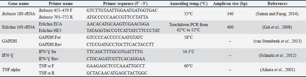
Fig. 1. Egypt map shows the location of the governorates (red) used for sample collection. Samples’ collectionA total of 157 blood samples were obtained through cephalic vein puncture. The samples were kept on ice and rapidly delivered to the laboratory, and divided into 3 parts. The first one was used for measuring the hematological parameters, while the second part was used for Ribonucleic acid (RNA) extraction and preserved at −80°C until use. The third part was used for Deoxy ribonucleic acid (DNA) extraction, which was kept at −20°C. Serum samples were used for biochemical analysis. Microscopical examinationFreshly collected blood samples were used to prepare blood smears, followed by Gimsa staining, and subsequently examined microscopically using an oil immersion lens. The was examined by inspecting 20 microscopic fields per stained slide. Inspection was performed by 3 independent analysts for the presence of any blood parasite infection (Khalifa et al., 2025). Molecular identificationDNA extractionPreserved whole blood samples were used for whole genomic DNA extraction. The instructions of the DNeasy Blood and Tissue Kits for DNA Isolation (Qiagen, Germany)® were followed. The purity and concentration of the extracted nucleic acid were assessed using a Nanodrop Microvolume Spectrophotometer. Molecular detectionConventional polymerase chain reaction (PCR) was used for detecting Babesia sp. and Ehrlichia sp. based on 18S and 16S rRNA, respectively. For each tested sample, the PCR mix consisted of 12.5 µl 2X Emerald Amp Max PCR Master Mix (Takara, Japan), 20 pmol of each primer, 50–100 ng of DNA template, RNase-free water for a total of 25 µl reaction. The same reaction was used for detecting Ehrlichia sp., but with the related primers (Table 1). Table 1. Primer’s nucleotide sequences, target genes, and amplicon sizes used in this study.
For the detection of Babesia sp. and Ehrlichia sp., the thermal programs were performed as previously described (Salem and Farag, 2014) and (Gal et al., 2008), respectively. The obtained amplicons were electrophoresed using 1.5% agarose gel (El-gbily et al., 2025; Hassan et al., 2025). Sequencing and phylogenetic analysisThe amplified PCR products were purified from the gel using the QIAquick Gel Extraction Kit–Gel Purification Kit (Qiagen, Germany)®, and their concentrations were measured. Representative samples were sent for direct sequencing. The sequences were assembled and edited using the free Bioedit 7.2 tool (https://bioedit.software.informer.com/7.2/). The edited sequences of Babesia sp. were uploaded to the National Center for Biotechnology Information (NCBI) database under the accession numbers PQ821446.1, PQ821447.1, and PQ821448.1. For the Ehrlichia sp., the sequences PQ821442.1, PQ821443.1, and PQ821444.1 were uploaded. The Basic Local Alignment Search Tool of the NCBI was used for comparison with similar sequences. The phylogenetic analysis was performed using the minimum evolution method (Rzhetsky and Nei, 1992). The evolutionary distances were calculated using the maximum composite likelihood method (Tamura et al., 2004) and in units of the number of base substitutions per site. The initial tree was generated using the neighbor-joining algorithm (Saitou and Nei, 1987). Evolutionary analyses were conducted using MEGA 7 (Kumar et al., 2016). The analysis involved 28 nucleotide sequences. All gaps and missing data positions were eliminated. There were 248 positions in the final dataset. The confidence level of the Neighbor joining tree was assessed by bootstrapping with 1,000 replicates. Cytokines expressionRNA extractionBlood sampling was conducted from the blood samples using the QIAamp RNeasy Mini kit (Qiagen, Germany, GmbH) according to the instruction manual, taking into consideration all precautions for RNA processing and DNase digestion to remove the residual genomic DNA. The purity and concentration of the extracted nucleic acid were assessed using a Nanodrop Microvolume Spectrophotometer. Gene expression as determined by quantitative real-time PCRReal-time PCR was used to detect the expression level of TNF-α and IFN-γ. The housekeeping gene Glyceraldehyde 3-phosphate dehydrogenase (GAPDH) was used as a reference gene for normalization of the samples. For each tested sample, the PCR mix consisted of a 25 µl reaction containing 12.5 µl of the 2x Quanti Tect SYBR Green PCR Master Mix (Qiagen, Germany, GmbH), 0.25 µl of RevertAid Reverse Transcriptase (200 U/µl) (Thermo Fisher), 0.5 µl of each primer at 20 pmol concentration, 3 µl of RNA template, and 8.25 µl of water. The annealing temperatures are shown in Table 1. For analysis, amplification curves and cycle threshold (Ct) values were determined using the Stratagene MX3005P software. To estimate gene expression variation. The Ct of each sample was compared with that of the positive control group according to the "ΔΔCt” method (Yuan et al., 2006), using the following ratio: (2-∆∆ct), whereas ΔΔCt=ΔCt reference—ΔCt target, where ΔCt target=Ct control Ct treatment, and ΔCt reference=Ct control Ct treatment. Haemato-biochemical analysisFreshly obtained whole blood samples were used to assess hematological parameters, including hemoglobin, packed cell volume, total erythrocyte count, total leucocyte count, differential leucocyte count, and platelet count, using an automatic Vet CBC counter (Sysmex XT 2000 iV Corporation, KOBE, Japan). Thin blood smears were prepared and stained with Gimsa. The slides were examined to evaluate the morphological alterations in the red and white blood cells. Screening for intraerythrocytic parasites, as described in Davis and Garcia (2023). Serum samples were used to estimate the levels of different biochemical parameters in the confirmed infected and non-infected cases. Blood urea nitrogen (BUN), creatinine, alanine aminotransferase (ALT), aspartate aminotransferase (AST), alkaline phosphatase (ALP), and total bilirubin were measured using specific kits purchased from Biodiagnostic©. Serum total protein and albumin levels were determined using the Diamond Diagnostics) © kit, according to the manufacturers’ guidelines, while serum globulin levels and albumin/globulin ratio (A/G ratio) were determined. Statistical analysisThe analysis of the study results was conducted using IBM SPSS Statistics 30.0 (https://www.ibm.com/support/pages/downloading-ibm-spss-statistics-30). Data were presented as mean ± SD. Qualitative data comparisons were carried out utilizing the chi-square and Fisher’s exact tests to determine any significant differences across various parameters, with a threshold of (p-value ≤ 0.05) set for statistical significance. Additionally, an unpaired Student’s t-test was employed to compare the blood parasite-infected and non-infected groups for immune response gene expression, hematological, and serum biochemical parameters. Ethical approvalAll animal experiments, including examination and sample collection, were conducted in accordance with the Animal Welfare Directives. The Ethics Committee of the Animal Health Research Institute approved this study (approval numbers ARC/AHRI/6425). The ethical approval date is 18 August 2025. ResultsMicroscopy and prevalence rateThe stained blood smears of the samples collected were subjected to microscopy to determine the prevalence of the disease. Large merozoites and trophozoites of Babesia sp., along with Ehrlichia sp. morulae, were detected within the red blood cells (RBCs). 12.1% (19/157) was determined against Babesia sp., and 7% (11/157) was determined against Ehrlichia sp. Table 2 Table 2. Total prevalence of blood parasites in the dogs examined by blood film and polymerase chain.
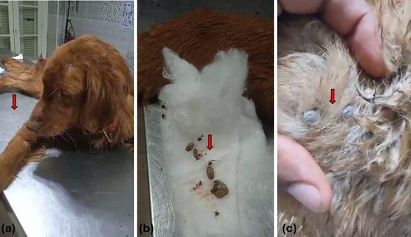
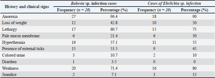
Physical examination of the animalsClinical examinati on of the infected dogs revealed multiple signs with variable degrees of intensity in the confirmed cases by PCR. The most prominent features were anorexia (96.4%), weakness (71.4%), and lethargy (60.7%) in Babesia sp.-infected patients. Meanwhile, the percentages in Ehrlichia sp.-infected cases were (90%), (80%), and (75%), respectively. In addition, hyperthermia, the presence of ticks (Fig. 2), and weight loss were determined in 57.1%, 53.5%, and 42.8% of Babesia sp. confirmed cases, respectively. Similar percentages of (55%), (45%), and (50%) were determined in the Ehrlichia sp.-infected animals.
Fig. 2. Different dog breeds infected with external ticks during physical examinations at veterinary clinics. The relatively less recorded signs were the presence of pale mucus membrane, colored urine, jaundice, and diarrhea. There was no clinical significance in the determined clinical features in animals infected with either Babesia sp. or Ehrlichia sp. (Table 3). Table 3. Frequency and percentage of the most prominent case history and clinical findings of infected dogs (n=28).
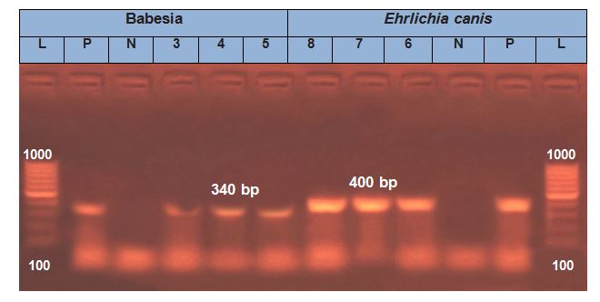
Molecular identification of blood parasiteConventional PCR was used to determine the Babesia sp. infection rate. Specific bands of 340 bp were successfully amplified using the primers. A rate of 17.83 % (28/157) was recorded. Similarly, 400-bp bands were successfully detected using the Ehrlichia 16S rRNA primers, but the infection rate was 12.73% (20/157) (Fig. 3). The examined animals showed no mixed infection.
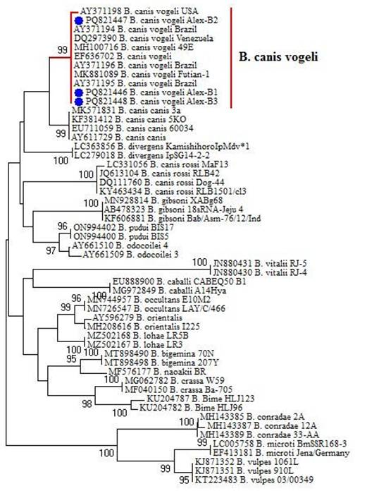
Fig. 3. Molecular detection of the tested blood parasites, including Babesia sp. and Ehrlichia sp., using polymerase chain reaction (PCR) depending on specific primers. PhylogenySequencing Babesia 18S rRNA amplicon samples revealed that the detected Babesia sp. was of the B. canis subspecies vogeli. The genetic relationship with the previously detected reference strains uploaded to the gene bank was determined through phylogenetic analysis. The final cladogram based on the Babesia 18S rRNA gene included 53 nucleotide sequences. The obtained Egyptian strains were closely related to those recorded in Brazil (AY371194, AY371195, and AY371196) with an identity of 100%. The (PQ821447) was more closely related to (AY371198). Although all of them were present in the same clade (Fig. 4).
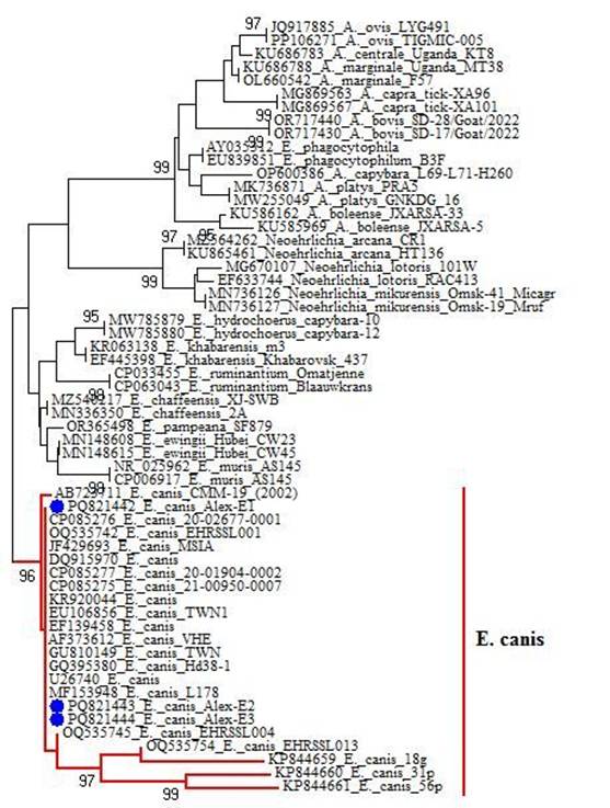
Fig. 4. Phylogenetic analysis of the obtained B. canis vogeli (accession numbers: PQ821446, PQ821447, and PQ821448) according to the 18s rRNA sequence. Blue circles refer to the isolates of this study. Sequencing of amplified Erlichia 16S rRNA amplicons revealed that the detected Ehrlichia sp. was of E. canis only. The final tree based on the Ehrlichia 16S rRNA primer set included 58 nucleotide sequences. The obtained Egyptian strains were closely related to KR920044 in Malaysia, CP085275 in Australia, and EF139458 in Thailand, with an identity of 100%. Although PQ821442.1 was more closely related to CP085276 in Australia. All strains were present in the same clade (Fig. 5).
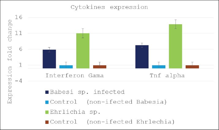
Fig. 5. Phylogenetic analysis of Ehrlichia canis (accession numbers: PQ821442, PQ821443, and PQ821444) according to Erlichia 16S rRNA. Blue circles refer to the isolates of this study. Cytokine expressionThe expression level of IFN-γ and TNF-α genes was assessed using qPCR in both blood parasite-infected and noninfected dogs. The results showed that in Babesia spp.-positive dogs, IFN-γ was expressed 5.8 (4–8.1) times compared with that in healthy dogs. Ehrlichia-infected animals had a higher expression level of 11.0 (9.7–12.5) times with an average of 11 times compared to Ehrlichia-non–infected animals (Fig. 6).
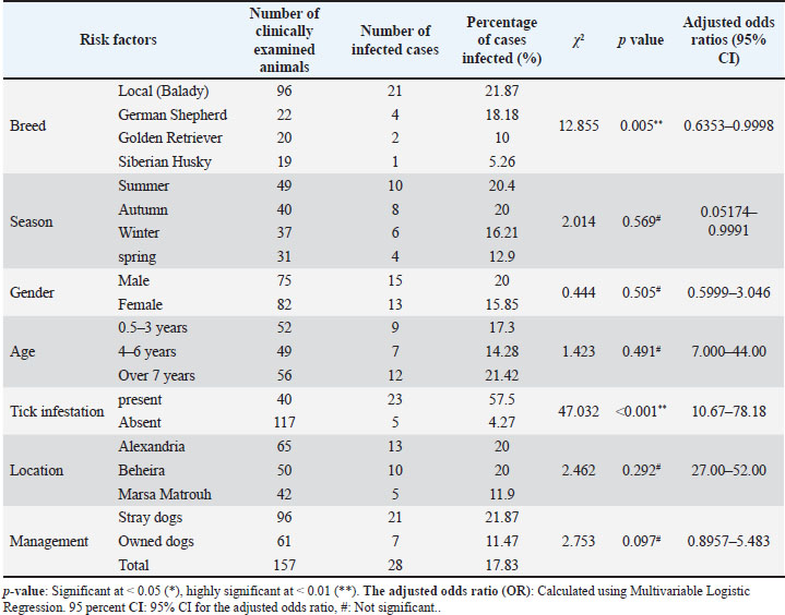
Fig. 6. Expression profiles of the proinflammatory cytokines (IFN-γ, and TNF-α) in noninfected and infected animals. Data are presented as mean ± SD. Significant increases in the cytokine expression of both genes were observed. Similarly, the TNF-α gene expression level had an average of 7.3- fold (5–9.2) in the confirmed babesia-infected animals compared to the healthy ones. In addition, the Ehrlichia-infected animals showed a greater expression of 13.9 (12.5–16.1) times (6). Risk factors related to Babesia sp. infectionMultiple risk factors correlated with Babesia sp. infection were assessed and statistically analyzed, including breed, sex, age, tick infestation, season, management, and location. The statistical analysis revealed the presence of significant differences related only to the animal breed and ticks. Local dogs (Balady) were found to have the highest significant (p=0.005, 95% CI=0.6353–0.9998) infection rate (21.87%) compared with German Shepherd (18.18%), Golden Retriever (10%), and Siberian Husky (5.26%). The presence of tick infestation was found to be highly correlated with Babesia sp. infection, with 57.5% of the infected cases having ticks. While 4.27% of the animals were infected in the absence of ticks (p < 0.001, 95% CI=10.67–78.18). Regarding season, the highest infection rate was in the summer (20.4%) compared to the lowest in the spring (p=0.56), with no significant difference (p=0.569, 95% CI=0.05174–0.9991). Male animals (20%) were more infected than females (15.85%), with no statistically significant difference (p=0.505, 95% CI=0.5999–3.046). Old age animals (over 7 years) were highly infected (21.42%) compared to those of (4-6 years) (14.28%), with no significant difference (p= 0.491, 95% CI=7.00–44.00). In addition, the lowest infection rate was determined in animals located at Marsa Matrouh Governorate (11.9%) compared to Alexandria and Behira (20%) (p=0.292, 95% CI=27.00–52.00). Stray dogs were found to be highly infected (21.87%) compared with those owned (11.47%) (p=0.097, 95% CI= 0.8957–5.483) (Table 4). Table 4. Risk factors associated with Babesia sp. infection in dogs.
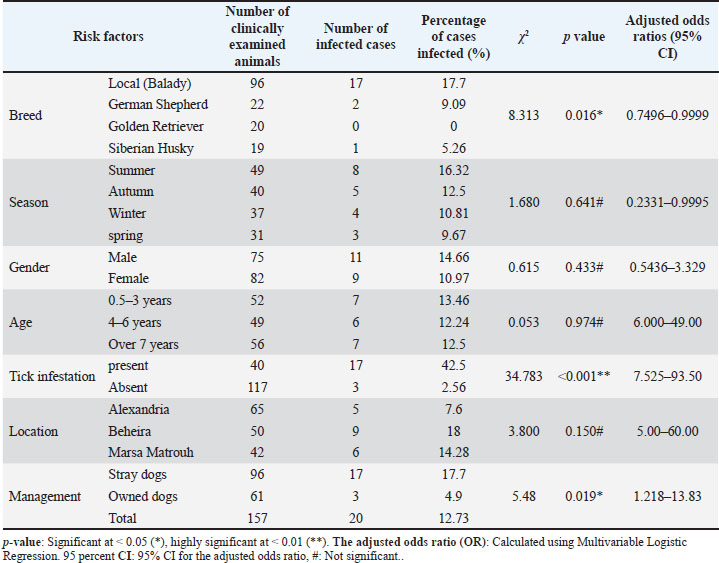
Risk factors associated with Ehrlichia sp. infectionThe local (Balady) breed recorded the highest significant (p=0.016, 95% CI=0.7496–0.9999) infection rate (17.7%), whereas the Golden Retriever did not. Although the highest infection rate was found in the Summer (16.32%), no significant differences were observed with the other seasons (p=0.641, 95% CI=0.2331 - 0.9995). Infected males (14.66%) were found to be insignificantly higher than females (10.97%) (p=0.433, 95% CI=0.5436 - 3.329). Animals at the age of 4–6 years were less infected (12.24%) than the younger animals up to 3 years old (13.46%) (p=0.974, 95% CI=6.000 - 49.00). The presence of ticks was found to be highly significant (p < 0.001, 95% CI= 7.525 - 93.50) related to infection (42.5%) compared with those having no external tick infestation (2.56%). Animals from the Alexanderia governorate (7.16%) were less infected compared with those from Marsa Matrouh (14.28%) and Behira (18%), respectively (p < 0.15, 95% CI=5.00–60.00). Interestingly, owned dogs (4.9%) were found to be significantly less infected than stray dogs (17.7%) (p < 0.019, 95% CI=1.218–13.83) (Table 5). Table 5. Risk factors associated with Ehrlichia sp. infection in dogs.
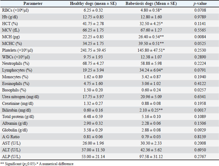
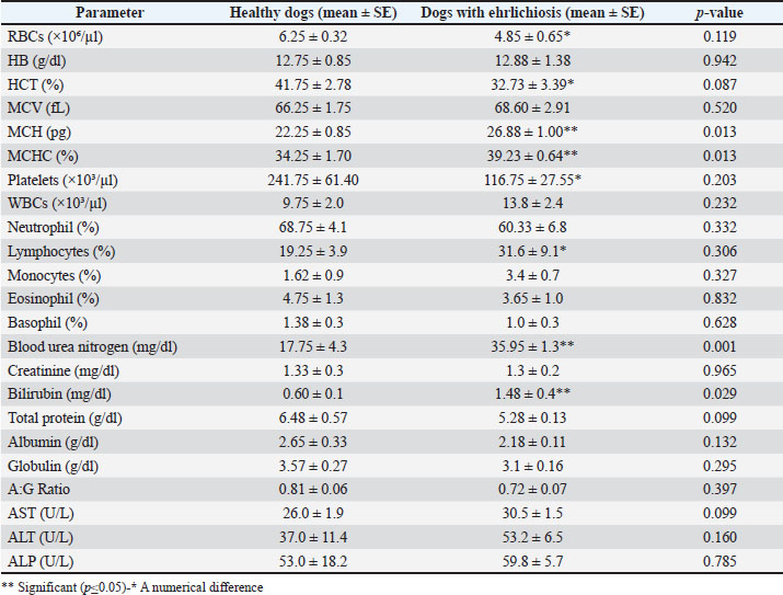
Hematological examinationInfected dogs showed a marked reduction in the RBC count, whereas the hemoglobin concentration remained within the normal reference range. Both mean corpuscular hemoglobin (MCH) and Mean Corpuscular Hemoglobin Concentration (MCHC) were significantly elevated, indicating hemoglobinemia secondary to RBC hemolysis. Although the RBC count and hematocrit (HCT) showed a numerical decrease in the infected group, the total Hb concentration remained stable (12.80 ± 1.60 g/dl). This discrepancy resulted in a significant increase in MCH (26.40 ± 0.54 pg; p= 0.008) and MCHC (39.50% ± 0.51%; p= 0.05). Such findings indicated intravascular hemolysis, where free plasma Hb contributed to the total Hemoglobin (Hb) measurement despite the loss of intact erythrocytes. This interpretation is further supported by the significant elevation in serum bilirubin levels (2.10 ± 0.25 mg/dl; p=0.0017) observed in the infected dogs, confirming the occurrence of active hemolytic processes. Moreover, the data presented in Tables 6 and 7 demonstrated nonsignificant thrombocytopenia and lymphocytosis in both Babesia- and Ehrlichia-infected dogs. Ehrlichiosis was characterized by a significant elevation in BUN. Other hematological and biochemical parameters remained within normal limits in all infected dogs, comparable to those of healthy control dogs. Table 6. Comparison of hemato-biochemical parameters between healthy dogs and dogs with babesiosis (mean ± S.E).
Table 7. Comparison of hemato-biochemical parameters between healthy and dogs with ehrlichiosis. (mean ± S.E)
DiscussionCanine babesiosis and ehrlichiosis are vector-borne diseases that result in significant adverse effects on the health status of infected cases (Khalifa et al., 2025). These pathogens are usually determined through the microscopic examination of blood smears. Although this tool is less sensitive and requires highly skilled analysts, it is usually applied due to its applicability and low cost (Mahmoud et al., 2024). Microscopy of the tested blood smears revealed a low prevalence rate of canine ehrlichiosis compared with babesiosis. The same pattern was observed during molecular detection. Although the latter technique was more sensitive and resulted in a higher detection rate. This result is less than that previously detected at the local level (25.6%) (Zaki et al., 2021) and (36.67%) (Mobark et al., 2024), but it could be attributed to variant factors, including different sampling localities, animal age, or season. These results were also different from those recently recorded in Thailand, which had infection prevalences of 26.12% for Ehrlichia sp. and 4.48% for Babesia sp. (Prasitsuwan et al., 2025). However, it was more than that reported in India (Bhagwan et al., 2024). Many previous diagnostic studies were constructed based on microscopy as a simple approach for veterinarians in rural areas. It was concluded that this technique could be reasonably sensitive during acute, clinically significant diseases only, but not sensitive enough to low parasitemia (Bai et al., 2017; Bhagwan et al., 2024). There was a recommendation for improving sensitivity and accuracy using buffy smears and testing lymph node aspirates (Nair et al., 2016). Therefore, molecular detection using conventional PCR was performed on the DNA of the same samples. Our results supported the theory of higher sensitivity, as the recorded results were 17.83% and 12.73% against babesiosis and ehrlichiosis, respectively. A higher rate of E. canis was detected in 46.9% and 30% of dog blood samples in India, respectively (Bai et al., 2017). Conversely, the recorded prevalence rate was higher than that in Iraq (5.1%) (Selim et al., 2022) and Nigeria (10.8%) (Obeta et al., 2020). Interestingly, a previous local study in Egypt revealed that the infection rate could differ even within the same country, as the infection rate of B. canis vogeli from southern Egypt was 4%, while the rate was higher in northern Egypt (Mahmoud et al., 2024). The current study revealed the absence of co-infection at the same time. This could be attributed to the phenomenon of immunologic interference or cross-modulation, which occurs when an established parasite modifies the host’s immune system to inhibit others or influence susceptibility to infection with other pathogenic microorganisms (Mabbott, 2018). Although previous research confirmed the presence of 2 or more multiple species of infection, especially in cases of tick infestation (López-Valencia et al., 2024). However, lower co-infection was determined in 6.5% of the tested animals in Turkey (Beristain-Ruiz et al., 2022). This study was limited to a specific geographical area (only 3 governorates), which may not represent the overall epidemiological situation in the entire country. Therefore, the findings should be interpreted as a local snapshot rather than a national prevalence. The development of advanced molecular techniques and phylogenetic analysis not only helped in the optimum identification and classification of multiple pathogens but also resulted in the tracing of the infection source (El Shanawany et al., 2025; Ouda et al., 2025; Abd El-Aziz et al., 2026). This approach greatly helped in accurate diagnosis and vaccine development for effective control (Ata et al., 2020 a,b). Sequencing and phylogenetic analysis confirmed that the Babesia sp. was of the B. canis subspecies vogeli. E. canis was the only detected; the presence of protozoan parasites is correlated with the presence of different tick species, which are located in tropical areas. The highest prevalence of B. canis was recorded in Europe, B. rossi is commonly located in South Africa, and B. vogeli has been reported in tropical and subtropical regions (Abdoli et al., 2024). It was also previously recorded in Egypt (Mahmoud et al., 2024). It is worth noting that B. rossi. Is the most virulent type recorded (Leisewitz et al., 2023). B. vogeli may have the longest evolutionary association with domestic dogs, in comparison to B. canis and B. rossi, which may explain the mild or subclinical infections that occur in these animals (Zygner et al., 2023). The close genetic relatedness of the current detected strains in this study with those previously reported in far countries, such as Australia, Thailand, and Malaysia, raises the concern of transmission methods that require further investigation. Such observation was previously recorded in free countries, even with the absence of a natural tick vector (Karasová et al., 2022). Furthermore, transmission via biting was strongly recommended, especially in dogfighting breeds that could have previously visited or come from an endemic country (Víchová et al., 2016). Clinical investigation of the diseased animals cleared the absence of clear, specific clinical features of the animals suffering from babesiosis or ehrlichiosis infections. Although the most prominent sign was fever. A lower percentage of animals showed pale mucus membranes and jaundice. These signs have been previously reported (Preena et al., 2021; Chan et al., 2025). The clinical findings differ according to many factors, such as the presence of immunity, the virulence of the strain, and the presence of multiple coinfections (Aziz et al., 2022). It is worth noting that the resulting clinical signs were only observed in animals subsequently confirmed to be infected. The presence of fever in a high percentage of infected animals is a supportive feature of the host cytokine response, which is more common with blood parasites (Brown et al., 2015). Therefore, the expression level of IFN-γ and TNF-α genes was determined. The results showed that the infected cases had higher expression of these cytokines than the non-infected ones, especially TNF-α. These results were previously reported as the IL-10 and IFN-γ levels were raised 2–4 weeks post infection, and the increase in TNF-α might have a role in the pathogenesis of these pathogens, especially in the acute infection phase (Faria et al., 2011; Stanilov et al., 2024). Furthermore, eliciting cytokines, including IFN-γ and TNF-α levels, might contribute to temporarily suppressing or preventing the establishment of subsequent pathogens in the host environment (Cloete et al., 2024). This supported our previous results of having no mixed infection. Analysis of the identified babesiosis and ehrlichiosis-associated risk factors revealed a significant correlation with the presence of external tick infestation. The same results were previously reported (Obeta et al., 2020). The presence of abundant ticks, cohabitation, and contact with other animals were marked risk factors and increased the risk of disease occurrence (Navarrete et al., 2018). Notably, the abundant presence of R. sanguineus tick species was frequently correlated with high ehrlichiosis morbidity (Khalifa et al., 2025). The presence of ticks is usually correlated with the summer and humid conditions, which favor their multiplication. Although in our study, the summer season was insignificantly higher than other seasons, multiple earlier studies confirmed that this season had a high morbidity rate (Thuo et al., 2014). Furthermore, transovarial transmission of B. vogeli in the intermediate host (tick) resulted in the survival of the infection foci, as the infection may be transmitted by the different stages of the tick. In addition, subclinical infection in adult dogs might affect the prevalence of the disease, as these cases were not treated with the optimum medications (Zygner et al., 2023). As we proved, the presence of ticks is a major driver, but the study did not include direct screening of the collected vectors for these pathogens. Integrating vector-host-pathogen data would have strengthened the link between the environment and infection (Agany et al., 2020). The obtained data concluded the presence of a significant prevalence based on the dog breed. The highest rate was determined in the local (Balady) breed compared with the others. The same findings were previously reported (Khalifa et al., 2025). The higher prevalence of canine babesiosis in Balady dogs than in exotic breeds could be explained by the fact that this breed was a stray dog that had almost no care, whereas the owned breeds had special care and interventions (Jegede et al., 2015). This result not only magnifies the role of stray dogs as a reservoir or focal point for disease persistence but also necessitates the establishment of governmental strategies to control these animals for these diseases or other zoonotic diseases (Otranto et al., 2017). The clinicopathological alterations observed in dogs infected with babesiosis and ehrlichiosis are generally nonspecific, and the severity of infection and the host’s immune response often determine the clinical manifestations (Bilwal et al., 2017). Both pathogens are recognized as significant causes of hemolytic anemia in canine populations worldwide (Boozer and Macintire, 2003; Sainz et al., 2015). As shown in Tables 6 and 7, a numerical decrease in RBC count and HCT% was evident in infected dogs, indicating anemia. In babesiosis, anemia results primarily from antibody-mediated cytotoxic destruction of erythrocytes rather than direct parasitic damage (Boozer and Macintire, 2003). In contrast, E. canis exhibits tropism for vascular endothelial cells, leading to vasculitis and hemorrhagic manifestations (petechiae and ecchymoses), which contribute to blood loss and anemia (Sainz et al., 2015). In the present study, platelet counts did not differ significantly between infected and control dogs, a finding consistent with some previous reports (Sudhakara Reddy et al., 2016). However, thrombocytopenia has frequently been reported in both babesiosis and ehrlichiosis because of immune-mediated platelet destruction or consumption during hemorrhagic episodes (Singh et al., 2021). The total leukocyte and lymphocyte counts showed variable trends among infected dogs; leukocytosis and lymphocytosis were occasionally observed, but without statistical significance, aligning with earlier observations (Pinto-Ferreira et al., 2019). Biochemically, serum total bilirubin levels were significantly elevated in both Babesia- and Ehrlichia-infected dogs compared with healthy controls, consistent with previous findings (Shah et al., 2011; Nivy et al., 2023). Hyperbilirubinemia most likely reflects intravascular and extravascular hemolysis rather than hepatic dysfunction, cholestasis, or sepsis (Nivy et al., 2023; Zygner et al., 2023). Reductions in RBC counts were occasionally accompanied by hemoglobin concentrations within the normal range, resulting in increased MCH and MCHC. Such patterns indicate hemoglobinemia secondary to red cell hemolysis, as described previously (Buys and Craven, 1990). Regarding renal function, serum creatinine concentrations did not differ significantly between infected and healthy dogs, consistent with the findings of (Wadhwa et al., 2011), although other studies have reported elevated creatinine in babesiosis (Sudhakara Reddy et al., 2016). BUN levels were significantly higher in patients with ehrlichiosis than in controls, suggesting that renal involvement is possibly linked to immune complex–mediated glomerular injury (Crivellenti et al., 2021). Additionally, total protein and albumin values tended to decrease, likely due to vasculitis-induced plasma protein loss through vascular endothelial pores (Bhadesiya and Raval, 2015). Collectively, these hematobiochemical alterations, which are characterized by anemia, hyperbilirubinemia, mild thrombocytopenia, and variable changes in renal parameters, reflect the shared pathophysiological mechanisms of hemolysis and vascular injury in canine babesiosis and ehrlichiosis. The limitations of this study include its limitedness to only 3 Egyptian governorates. Molecular identification of the external ticks and their correlation with the circulating parasites might also be determined. Finally, different blood parasites could be identified. All these limitations were mainly attributed to resource and time limitations. Despite these limitations, this study provided valuable baseline data for molecular detection and studied the related risk factors using a standardized protocol, which can help in local control strategies. ConclusionThe tested animals showed a prevalence rate of babesiosis (17.83%) and ehrlichiosis (12.73%). Although microscopy of stained blood smears is applicable, it is less sensitive than molecular tools. B. canis subspecies vogeli and E. canis were the circulating types. The obtained strains were not only closely related to the previously detected strains but also to those from geographically distant countries. Necessitates further study of other unusual transmission routes. Despite the detection of both pathogens being confirmed, the absence of co-infection could be attributed to immunologic interference or cross-modulation. Although the confirmed cases showed many clinical symptoms, fever and the presence of ticks were prominent, with high expression of IFN-γ and TNF-α genes. The local stray dog breed and the presence of external ticks were highly correlated with disease occurrence. The principal hemato-biochemical alterations were hemolytic anemia, thrombocytopenia, lymphocytosis, and hyperbilirubinemia. Based on the results obtained, we recommend structuring more molecular surveys to establish effective strategies to control stray dogs and tick infestation. AcknowledgmentNot applicable. Conflict of interestThe authors declare that there is no conflict of interest. FundingThe authors declare that no funds, grants, or other support were received during the preparation of this manuscript. Authors' contributionsAll authors contributed to the conceptualization, design of this work, and sample collection. S. M. H., R. D., and N. South shared in the microscopical examination. R.D., N.S., and E.B.A. shared in molecular identification. E. B. A. and S. shared in phylogenetic analysis. R. D. and E. B. A. shared in cytokine expression. S. M. H. performed the hematological and biochemical examinations. All authors contributed to writing the original draft and reviewing the final manuscript and agreed to submit the manuscript. Data availabilityAll data related to this manuscript are available upon request. REFERENCESAbd El-Aziz, T.H., Ata, E.B., Helal, M.A., Abdel-Rahman, E.H. and Nasr, S.M. 2026. Seroprevalence and risk factors associated with calves’ cryptosporidiosis in Egypt. BMC Vet. Res. 22, 125; doi: 10.1186/s12917-025-05216-7 Abdoli, A., Olfatifar, M., Badri, M., Zaki, L., Bijani, B., Pirestani, M., Hatam‐Nahavandi, K., Eslahi, A.V. and Karanis, P. 2024. A global systematic review and meta‐analysis on the babesiosis in dogs with special reference to Babesia canis. Vet. Med. Sci. 10(3):e1427; doi:10.1002/vms3.1427 Agany, D.D.M., Pietri, J.E. and Gnimpieba, E.Z. 2020. Assessment of vector-host-pathogen relationships using data mining and machine learning. Comput. Struct. Biotechnol. J. 18, 1704–1721; doi:10.1016/j.csbj.2020.06.031 Aihara, Y., Kasuya, H., Onda, H., Hori, T. and Takeda, J. 2001. Quantitative analysis of gene expressions related to inflammation in canine spastic artery after subarachnoid hemorrhage. stroke 32, 212–217; doi:10.1161/01.STR.32.1.212 Ata, E., Nasr, S.M., Mohamed, A.M., El-Aziz, T.H.A.A., Fouad, E.A., Sedky, D., Nassar, S. A. and Ghazy, A.A. 2020a. Bacteriological, hematological and biochemical diagnostic studies on diarrheic Arabian horse foals caused by enterobacterial infections. Adv. Anim. Vet. Sci. 8(4), 258–260; doi:10.17582/journal.aavs/2020/8.4.412.421 Ata, E., Salama, A., Zaghawa, A., Ghazy, A.A.A., Elsify, A., Nayel, M., Hegazy, Y., Abdel-Rahman, E.H.H. and Warda, S. 2020b. Seroprevalence of equine herpes virus-1 in endemic area of Egypt with risk factors assessment. Bulg. J. Vet. Med. 23, 102–111; doi:10.15547/bjvm.2168 Ata, E.B., Abdel-Aziz, T.H., Abdel-Ghany, H.S.M., Elsawy, B.S.M., Abdullah, H.H.A.M., Abouelsoued, D., Ashry, H.M., Hassan, M.R., Shapaan, R.M., Nasr, S.M., Mahmoud, M.S., Abdel Megeed, K.N. and Abdel-Shafy, S. 2024. Molecular and serological diagnosis of the circulating Trypanosoma evansi in Egyptian livestock with risk factors assessment. Microb. Pathog. 197, 107073; doi:10.1016/j.micpath.2024.10707 Ata, E.B., Shaapan, R.M., Ghazy, A.A., Kandil, O.M. and Abou-Zeina, H.A.A. 2023a. Epidemiological aspects of some equine viral diseases. Iraqi J. Vet. Sci. 37, 121–127; doi:10.33899/ijvs.2022.133255.2195 Ata, E.B., Shaapan, R.M., Nasr, S. and Abdel-Shafy, S. 2023b. Role of evolutionary epidemiology in the determination of the risk factors associated with some equine viral diseases. Iraqi J. Vet. Sci. 37, 143–150; doi:10.33899/ijvs.2022.133433.2228 Aziz, M.U., Hussain, S., Song, B., Ghauri, H.N., Zeb, J. and Sparagano, O.A. 2022. Ehrlichiosis in dogs: a comprehensive review about the pathogen and its vectors with emphasis on South and East Asian Countries. Vet. Sci. 10, 21; doi:10.3390/vetsci10010021 Bai, L., Goel, P., Jhambh, R., Kumar, P. and Joshi, V.G. 2017. Molecular prevalence and haemato-biochemical profile of canine monocytic ehrlichiosis in dogs in and around Hisar, Haryana, India. J. Parasit. Dis. 41, 647–654; doi:10.1007/s12639-016-0860-8 Beristain-Ruiz, D., Garza-Hernández, J., Figueroa-Millán, J., Lira-Amaya, J., Quezada-Casasola, A., Ordoñez-López, S., Laredo-Tiscareño, S., Alvarado-Robles, B., Castillo-Luna, O., Floriano-López, A., Hernández-Triana, L., Martínez-Ibáñez, F., Rivera-Barreno, R. and Rodríguez-Alarcón, C. 2022. Possible association between selected tick-borne pathogen prevalence and Rhipicephalus sanguineus sensu lato infestation in dogs from Juarez City (Chihuahua), Northwest Mexico–US Border. Pathogens 11, 552; doi: 10.3390/pathogens11050552 Bhadesiya, C.M. and Raval, S.K. 2015. Hematobiochemical changes in ehrlichiosis in dogs of Anand region, Gujarat. Vet. World 8, 713–717; doi:10.14202/vetworld.2015.713-717 Bhagwan, J., Singh, Y., Jhambh, R., Chaudhari, M. and Kumar, P. 2024. Molecular and microscopic detection of haemoprotozoan diseases in dogs from Haryana, India. Parasitol. Res. 123, 354; doi:10.1007/s00436-024-08376-x Bilwal, A.K., Mandali, G.C. and Tandel, F.B. 2017. Clinicopathological alterations in naturally occurring Babesia gibsoni infection in dogs of Middle-South Gujarat, India. Vet. World. 10, 1227–1232; doi;10.14202/vetworld.2017.1227-1232 Boozer, A.L. and Macintire, D.K. 2003. Canine babesiosis. Vet. Clin. North Am. Small Anim. Pract. 33, 885–904; doi:10.1016/S0195-5616(03)00039-1 Brown, A.L., Shiel, R.E. and Irwin, P.J. 2015. Clinical, haematological, cytokine and acute phase protein changes during experimental Babesia gibsoni infection of beagle puppies. Exp. Parasitol. 157, 185–196; doi:10.1016/j.exppara.2015.08.002 Buys, S. and Craven, C. 1990. MCHC in intravascular hemolysis. Am. J. Hematol. 33, 282–283; doi:10.1002/ajh.2830330415 Chan, K., Bęczkowski, P.M. and Almendros, A. 2025. Clinical and clinicopathological features of 108 dogs infected with Babesia gibsoni in Hong Kong. Animals 15, 645; doi:10.3390/ani15050645 Cloete, H.P.P., Rautenbach, Y., Leisewitz, A.L., Mellanby, R.J., Thompson, P.N. and Schoeman, J.P. 2024. Prevalence of co-infections with Ehrlichia spp. or Theileria spp. in dogs naturally infected with babesiosis in the Eastern Cape province. Vet. Parasitol. Reg. Stud. Reports. 54, 101092; doi:10.1016/j.vprsr.2024.101092 Crivellenti, L.Z., Cintra, C.A., Maia, S.R., Silva, G.E.B., Borin-Crivellenti, S., Cianciolo, R., Adin, C.A., Tinucci-Costa, M., Pennacchi, C.S. and Santana, A.E. 2021. Glomerulotubular pathology in dogs with subclinical ehrlichiosis. PLoS One 16, e0260702; doi:10.1371/journal.pone.0260702 Davis, R.E. and Garcia, L.S. 2023. Detection of blood parasites. In Clinical Microbiology Procedures Handbook Parasitology. New York City, NY: Wiley, pp: 1–52; doi:10.1002/9781683670438.cmph0120 El-gbily, S., Eldokmak, M.M., Diabb, R., Abas, O.M., Ata, E.B., Kamal, S. and Hassan, S.M.H. 2025. Severe lamb diarrhea outbreak: clinical features, identification of the causative agent, and a prophylactic approach. Comp. Immunol. Microbiol. Infect. Dis. 118, 102318; doi: 10.1016/j.cimid.2025.102318 El Shanawany, E.E., Ata, E.B., Hassan, S.E. and Abdel-Rahman, E.H. 2025. Validation of an in-house Sarcosystis fusiformis Glycoprotein-based ELISA for the serodiagnosis of sarcocystosis in Buffaloes. Egypt. J. Vet. Sci. 56, 757–770; doi:10.21608/ejvs.2025.410864.3019 Faria, J.L.M., Munhoz, T.D., João, C.F., Vargas-Hernández, G. André, M.R., Pereira, W.A.B., Machado, R.Z. and Tinucci-Costa, M. 2011. E. canis (Jaboticabal strain) induces the expression of TNF-α in leukocytes and splenocytes of experimentally infected dogs. Rev. Bras. Parasitol. Veterinária. 20, 71–74; doi:10.1590/S1984-29612011000100015 Ferrolho, J., Antunes, S., Vilhena, H., Anastácio, S., Ramalho de Sousa, S., Frouco, G., Ferreira, B. and Domingos, A. 2025. The complexities of canine monocytic ehrlichiosis: insights into Ehrlichia canis and its vector Rhipicephalus sanguineus. Microbiol. Res. (Pavia). 16, 85; doi:10.3390/microbiolres16040085 Gal, A., Loeb, E., Yisaschar-Mekuzas, Y. and Baneth, G. 2008. Detection of Ehrlichia canis by PCR in different tissues obtained during necropsy from dogs surveyed for naturally occurring canine monocytic ehrlichiosis. Vet. J. 175, 212–217; doi:10.1016/j.tvjl.2007.01.013 Gboeloh, L., Wagbara, B. and Sunday, B. 2023. Prevalence of Babesia Spp. in presumably healthy dogs and associated risk fators in OBIO/AKPOR local government area, Rivers State, Nigeria. J. Appl. Vet. Sci. 89–97; doi : 10.21608/javs.2023.188493.1211 Hassan, N.M., Helal, M.A., Elsawy, B.S., El Shanawany, E.E., Abu EL Ezz, N.M., Shalaby, H.A., El Namaky, A.H., Kandil, O.M., Mohamed, M.S., Desouky, H.M. and Ata, E.B. 2025. Current epidemiological and molecular patterns of haemonchosis in Cairo and Giza governorates, Egypt. Iraqi J. Vet. Sci. 39, 419–429; doi:10.33899/ijvs.2025.153449.3873 Jegede, O., Obeta, S. and Faisal, B. 2015. Infection of dogs with Babesia canis in Gwagwalada metropolis of Federal Capital Territory, Abuja, Nigeria. Sokoto J. Vet. Sci. 12, 37; doi:10.4314/sokjvs.v12i3.7 Karasová, M., Tóthová, C., Grelová, S. and Fialkovičová, M. 2022. The etiology, incidence, pathogenesis, diagnostics and treatment of canine babesiosis caused by Babesia gibsoni infection. Animals 12, 739; doi:10.3390/ani12060739 Khalifa, M.M., Salem, M.A., Fouad, E.A., Bakry, N.M., Kamel, M.S., El-Bahy and M.M., Ramadan, R.M. 2025. Vector-borne pathogens in dogs in Egypt: molecular and immunological insights. Res. Vet. Sci. 189, 105629; doi:10.1016/j.rvsc.2025.105629 Kumar, S., Stecher, G. and Tamura, K. 2016. MEGA7: molecular evolutionary genetics analysis version 7.0 for bigger datasets. Mol. Biol. Evol. 33, 1870–1874; doi:10.1093/molbev/msw054 Leisewitz, A.L., Mrljak, V., Dear, J.D. and Birkenheuer, A. 2023. The diverse pathogenicity of various Babesia parasite species that infect dogs. Pathogens 12, 1437; doi:10.3390/pathogens12121437 López-Valencia, G., Meza-Silva, K.M., Haro-Álvarez, A.P., Trasviña-Muñoz, E., Garcia-Reynoso, I.C., Herrera-Ramírez, J.C. and Gómez-Gómez, S.D. 2024. Prevalence, risk factors, and hematologic changes in dogs from Baja California with presence of Ehrlichia spp., and coinfection with Anaplasma spp. Austral J. Vet. Sci. 75–84; doi:10.4206/ajvs.562.06 Mabbott, N.A. 2018. The influence of parasite infections on host immunity to co-infection with other pathogens. Front. Immunol. 9, 2579; doi:10.3389/fimmu.2018.02579 Mahmoud, H.Y.A.H., Shahat, M.S., Fereig, R.M., Ali, A.O., Emeish, W.F.A., Soliman, A.M., Khalifa, F.A. and Tanaka, T. 2024. Molecular detection and characterization of Anaplasma marginale and Babesia canis vogeli infecting dogs in Luxor, Egypt. Sci. Rep. 14, 16888; doi:10.1038/s41598-024-67009-6 Matjila, P.T., Carcy, B., Leisewitz, A.L., Schetters, T., Jongejan, F., Gorenflot, A. and Penzhorn, B.L. 2009. Preliminary evaluation of the Br EMA1 gene as a tool for associating Babesia rossi genotypes and clinical manifestation of Canine babesiosis. J. Clin. Microbiol. 47, 3586–3592; doi:10.1128/JCM.01110-08 Mehra, M., Singh, O. and Desai, J. 2024. Clinical examination and diagnostic perspectives in dogs and cats. In Introduction to diseases, diagnosis, and management of dogs and cats. Academic Press, Elsevier, pp: 47–54; doi:10.1016/B978-0-443-18548-9.00004-4 Mobark, D., Elbaz, E., Atwa, S., Eisa, M., Sebaey, A. and Selim, A. 2024. Molecular, epidemiological, and hematological evaluation in Ehrlichia canis infected dogs from an endemic region in Egypt. Open Vet. J. 14, 1819; doi:10.5455/OVJ.2024.v14.i8.10 Nair, A.D.S., Cheng, C., Ganta, C.K., Sanderson, M.W., Alleman, A.R., Munderloh, U.G. and Ganta, R.R. 2016. Comparative experimental infection study in dogs with Ehrlichia canis, E. chaffeensis, Anaplasma platys and A. phagocytophilum. PLoS One 11, e0148239; doi:10.1371/journal.pone.0148239 Navarrete, M.G., Cordeiro, M.D., Silva, C.B., Massard, C.L., López, E.R., Rodríguez, J.C.A., Ribeiro, C.C.D.U., Rodríguez, O.F. and Fonseca, A.H. 2018. Serological and molecular diagnosis of Ehrlichia canis and associated risk factors in dogs domiciled in western Cuba. Vet. Parasitol. Reg. Stud. Reports. 14, 170–175; doi:10.1016/j.vprsr.2018.10.005 Nivy, R., Sutton, G. and Bruchim, Y. 2023. Carboxyhemoglobin as a diagnostic and prognostic biomarker of hemolytic anemias in dogs. J. Vet. Intern. Med. 37, 110–116; doi:10.1111/jvim.16617 Obeta, Sylvester S., Ibrahim, B., Lawal, I.A., Natala, J.A., Ogo, N.I. and Balogun, E.O. 2020. Prevalence of canine babesiosis and their risk factors among asymptomatic dogs in the Federal Capital Territory, Abuja, Nigeria. Parasite Epidemiol. Control 11, e00186; doi:10.1016/j.parepi.2020.e00186 Otranto, D., Dantas-Torres, F., Mihalca, A.D., Traub, R.J., Lappin, M. and Baneth, G. 2017. Zoonotic parasites of sheltered and stray dogs in the era of the global economic and political crisis. Trends Parasitol. 33, 813–825; doi:10.1016/j.pt.2017.05.013 Ouda, S., Hassan, R., Ismaeel, E., Abo-Sakaya, R., Ata, E., Sedky, D., Kandil, M. and Shabiny, L. 2025. Molecular diagnosis of hemoplasma infection in cats and sheep with hematological studies. Open Vet. J. 15, 2823; doi:10.5455/OVJ.2025.v15.i6.51 Paladsing, Y., Khanh, B.M.T., Thinphovong, C., Ketwang, S., Chaisiri, K., Carcy, B., De Garine-Wichatitsky, M., Morand, S., Inpankaew, T. and Kritiyakan, A. 2024. Dog blood parasite infection in upland and lowland communities of northern Thailand: the role of environment and care of dog owners. Vet. Parasitol. Reg. Stud. Reports 51, 101024; doi:10.1016/j.vprsr.2024.101024 Pinto-Ferreira, F., Mitsuka-Breganó, R., Monica, T.C., Martins, F.D.C., Matos, R.L.N. de, Mareze, M., Nino, B. de S.L., Narciso, S.G., Freire, R.L. and Navarro, I.T. 2019. Investigation and environmental analysis of samples from outbreak of toxoplasmosis at research institution in Londrina, Paraná, Brazil, 2016. Rev. Bras. Parasitol. Veterinária. 28, 518–521; doi:10.1590/s1984-29612019044 Prasitsuwan, W., Suwannachote, T., Sumalai, T., Lertpatarakomol, R., Trairatapiwan, T. and Ruenphet, S. 2025. A comparison of diagnostic methods for canine Ehrlichiosis: microscopy and RNases hybridization-assisted amplification technology compared with the quantitative polymerase chain reaction. Vet. World. 1214–1223; doi:10.14202/vetworld.2025.1214-1223 Preena, P., Sarangom, S.B., Ramesh Kumar, K.V., Seeja, S. and Rajalekshmi, S. 2021. Hematological alterations in large Babesia species infection in dogs of Kannur District of Kerala. J. Parasit. Dis. 45, 1090–1095; doi:10.1007/s12639-021-01404-y Rzhetsky, A. and Nei, M. 1992. A simple method for estimating and testing minimum-evolution trees. Mol. Biol. Evol. 9, 945–967; doi:10.1093/oxfordjournals.molbev.a040771 Sainz, Á., Roura, X., Miró, G., Estrada-Peña, A., Kohn, B., Harrus, S. and Solano-Gallego, L. 2015. Guideline for veterinary practitioners on canine ehrlichiosis and anaplasmosis in Europe. Parasit. Vectors 8, 75; doi:10.1186/s13071-015-0649-0 Saitou, N. and Nei, M. 1987. The neighbor-joining method: a new method for reconstructing phylogenetic trees. Mol. Biol. Evol. 4, 406–25; doi:10.1093/oxfordjournals.molbev.a040454 Salem, N.Y. and Farag, H.S. 2014. Clinical, hematologic, and molecular findings in naturally occurring Babesia canis vogeli in Egyptian Dogs. Vet. Med. Int. 2014, 270345; doi:10.1155/2014/270345 Salim, B., Alanazi, A.D., Omori, R., Alyousif, M.S., Alanazi, I.O., Katakura, K. and Nakao, R. 2019. Potential role of dogs as sentinels and reservoirs for piroplasms infecting equine and cattle in Riyadh City, Saudi Arabia. Acta Trop. 193, 78–83; doi:10.1016/j.actatropica.2019.02.029 Schmitz, S., Garden, O.A., Werling, D. and Allenspach, K., 2012. Gene expression of selected signature cytokines of T cell subsets in duodenal tissues of dogs with and without inflammatory bowel disease. Vet. Immunol. Immunopathol. 146, 87–91; doi:10.1016/j.vetimm.2012.01.013 Selim, A., Megahed, A., Ben Said, M., Alanazi, A.D. and Sayed-Ahmed, M.Z. 2022. Molecular survey and phylogenetic analysis of Babesia vogeli in dogs. Sci. Rep. 12, 6988; doi:10.1038/s41598-022-11079-x Shah, S.A., Sood, N.K. and Tumati, S.R. 2011. Haemato-biochemical Changes in natural cases of Canine babesiosis. Asian J. Anim. Sci. 5, 387–392; doi:10.3923/ajas.2011.387.392 Singh, J., Singh, R.S., Singh, H., Gupta, D.K. and Randhawa, S.S. 2021. Clinical and haemato-biochemical observations in dogs naturally infected with Canine monocytic ehrlichiosis. Explor. Anim. Med. Res. 11, 214; doi:10.52635/EAMR/11.2.214-219 Stanilov, I., Gospodinova, K., Petrov, V., Miteva, L., Tsachev, I. and Stanilova, S. 2024. Enhanced production of IL-10 in PCR-Positive dogs infected with E. canis and A. phagocytophilum facilitate specific immune responses. Microorganisms 12, 2516; doi:10.3390/microorganisms12122516 Sudhakara Reddy, B., Sivajothi, S., Varaprasad Reddy, L.S.S. and Solmon Raju, K.G. 2016. Clinical and laboratory findings of Babesia infection in dogs. J. Parasit. Dis. 40, 268–272; doi:10.1007/s12639-014-0491-x Tamura, K., Nei, M. and Kumar, S. 2004. Prospects for inferring very large phylogenies by using the neighbor-joining method. Proc. Natl. Acad. Sci. 101, 11030–11035; doi:10.1073/pnas.0404206101 Thuo, J.K.N., Aleri, J.W., Kitaa, J.M.A. and Mulei, C.M. 2014. Seasonality and occurrence of canine babesiosis in Nairobi and its environs in changing climatic patterns. Kenya Vet. 38, 18–23; doi:10.4314/kenvet.v38i1. van Steenbeek, F.G., Van den Bossche, L., Grinwis, G.C.M., Kummeling, A., van Gils, I.H.M., Koerkamp, M.J.A.G., van Leenen, D., Holstege, F.C.P., Penning, L.C., Rothuizen, J., Leegwater, P.A.J. and Spee, B. 2013. Aberrant gene expression in dogs with portosystemic shunts. PLoS One 8, e57662; doi:10.1371/journal.pone.0057662 Víchová, B., Horská, M., Blaňarová, L., Švihran, M., Andersson, M. and Peťko, B. 2016. First molecular identification of Babesia gibsoni in dogs from Slovakia, central Europe. Ticks Tick. Borne. Dis. 7, 54–59; doi:10.1016/j.ttbdis.2015.08.004 Wadhwa, D.R., Pal, B., Mandial, R.K., Kumar, A. and Agnihotri, R.K. 2011. Clinical, haemato-biochemical and therapeutic studies on canine babesiosis in Kangra Valley of Himachal Pradesh. J. Vet. Parasitol. 25(1), 39–41. Yuan, J.S., Reed, A., Chen, F. and Stewart, C.N. 2006. Statistical analysis of real-time PCR data. BMC Bioinformatics 7, 85; doi:10.1186/1471-2105-7-85 Zaki, A.A., Attia, M.M., Ismael, E. and Mahdy, O.A. 2021. Prevalence, genetic, and biochemical evaluation of immune response of police dogs infected with Babesia vogeli. Vet. World 14, 903–912; doi:10.14202/vetworld.2021.903-912 Zygner, W., Gójska-Zygner, O., Bartosik, J., Górski, P., Karabowicz, J., Kotomski, G. and Norbury, L.J. 2023. Canine babesiosis caused by large Babesia species: global prevalence and risk factors: a review. Animals 13, 2612; doi:10.3390/ani13162612 Zygner, W., Gójska-Zygner, O., Bąska, P. and Długosz, E. 2014. Increased concentration of serum TNF alpha and its correlations with arterial blood pressure and indices of renal damage in dogs infected with Babesia canis. Parasitol. Res. 113, 1499–503; doi:10.1007/s00436-014-3792-1 | ||

| How to Cite this Article |
| Pubmed Style Hassan SMH, Diabb R, Satour NS, Ata EB. Circulating Babesia sp. and Ehrlichia sp. in Egyptian dogs: Molecular detection, risk assessment, and hematobiochemical parameters. Open Vet. J.. 2026; 16(6): 3731-3748. doi:10.5455/OVJ.2026.v16.i6.45 Web Style Hassan SMH, Diabb R, Satour NS, Ata EB. Circulating Babesia sp. and Ehrlichia sp. in Egyptian dogs: Molecular detection, risk assessment, and hematobiochemical parameters. https://www.openveterinaryjournal.com/?mno=316713 [Access: June 26, 2026]. doi:10.5455/OVJ.2026.v16.i6.45 AMA (American Medical Association) Style Hassan SMH, Diabb R, Satour NS, Ata EB. Circulating Babesia sp. and Ehrlichia sp. in Egyptian dogs: Molecular detection, risk assessment, and hematobiochemical parameters. Open Vet. J.. 2026; 16(6): 3731-3748. doi:10.5455/OVJ.2026.v16.i6.45 Vancouver/ICMJE Style Hassan SMH, Diabb R, Satour NS, Ata EB. Circulating Babesia sp. and Ehrlichia sp. in Egyptian dogs: Molecular detection, risk assessment, and hematobiochemical parameters. Open Vet. J.. (2026), [cited June 26, 2026]; 16(6): 3731-3748. doi:10.5455/OVJ.2026.v16.i6.45 Harvard Style Hassan, S. M. H., Diabb, . R., Satour, . N. S. & Ata, . E. B. (2026) Circulating Babesia sp. and Ehrlichia sp. in Egyptian dogs: Molecular detection, risk assessment, and hematobiochemical parameters. Open Vet. J., 16 (6), 3731-3748. doi:10.5455/OVJ.2026.v16.i6.45 Turabian Style Hassan, Shahenaz M. H., Rasha Diabb, Neveen S. Satour, and Emad Beshir Ata. 2026. Circulating Babesia sp. and Ehrlichia sp. in Egyptian dogs: Molecular detection, risk assessment, and hematobiochemical parameters. Open Veterinary Journal, 16 (6), 3731-3748. doi:10.5455/OVJ.2026.v16.i6.45 Chicago Style Hassan, Shahenaz M. H., Rasha Diabb, Neveen S. Satour, and Emad Beshir Ata. "Circulating Babesia sp. and Ehrlichia sp. in Egyptian dogs: Molecular detection, risk assessment, and hematobiochemical parameters." Open Veterinary Journal 16 (2026), 3731-3748. doi:10.5455/OVJ.2026.v16.i6.45 MLA (The Modern Language Association) Style Hassan, Shahenaz M. H., Rasha Diabb, Neveen S. Satour, and Emad Beshir Ata. "Circulating Babesia sp. and Ehrlichia sp. in Egyptian dogs: Molecular detection, risk assessment, and hematobiochemical parameters." Open Veterinary Journal 16.6 (2026), 3731-3748. Print. doi:10.5455/OVJ.2026.v16.i6.45 APA (American Psychological Association) Style Hassan, S. M. H., Diabb, . R., Satour, . N. S. & Ata, . E. B. (2026) Circulating Babesia sp. and Ehrlichia sp. in Egyptian dogs: Molecular detection, risk assessment, and hematobiochemical parameters. Open Veterinary Journal, 16 (6), 3731-3748. doi:10.5455/OVJ.2026.v16.i6.45 |


